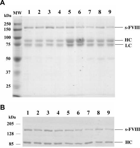
Figure 1.
SDS-PAGE and Western blot analysis of factor VIII mutants and WT factor VIII. (A) Purified WT and mutant factor VIII proteins (0.77 μg) after SDS-PAGE on 8% polyacrylamide gels were visualized by GelCode. (B) Purified WT and mutant factor VIII proteins (0.34 μg) were electrophoresed on 8% polyacrylamide gels, transferred to PVDF membranes, and probed by biotinylated R8B12 antibody. Bands were visualized by chemifluorescence. WT (lane 1), Glu272Ala (lane 2), Glu272Val (lane 3), Asp519Ala (lane 4), Asp519Val (lane 5), Glu665Ala (lane 6), Glu665Val (lane 7), Glu1984Ala (lane 8), and Glu1984Val (lane 9). MW indicates molecular weight marker; sFVIII, single chain form factor VIII; HC, heavy chain; LC, light chain. An apparent stoichiometry ratio of single chain form to heterodimer of WT and mutant factor VIII forms were 0.96 (WT), 0.64 (Glu272Ala), 0.92 (Glu272Val), 0.74 (Asp519Ala), 0.8 (Asp519Val), 0.64 (Glu665Ala), 0.63 (Glu665Val), 0.91 (Glue1984Ala), and 0.5 (Glu1984Val).

