Figure 1.
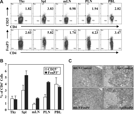
Human regulatory T cells (Treg cells) in DKO-hu HSC mice. Lymphoid organs from each DKO-hu HSC mouse were individually analyzed for development of human Treg cell by flow cytometry analysis at 12 to 40 weeks after transplantation. Experiments were repeated using 5 independent cohorts of DKO-hu HSC mice (generated using 5 different human donor fetal liver tissues). (A) Human CD4 T cells (mCD45−hCD45+CD3+CD4+) in different lymphoid organs of one representative DKO-hu HSC mouse were stained with CD25 or FoxP3 antibodies (see Figure S1 for FoxP3 vs CD25 plots). Numbers on plots are percentages of double-positive cells from the mCD45−huCD45+ CD3+CD4+ gated cells. (B) Bar graphs represent percentages of CD3+CD4+CD25+ or CD3+CD4+FoxP3+ Treg cells of total CD3+CD4+ T cells in different lymphoid organs. Data are summarized from DKO-hu mice derived from 5 different human fetal liver donor tissues (n > 30 DKO-hu mice). Error bars display standard deviations. (C) Human FoxP3+ Treg cells in lymphoid tissues of DKO-hu HSC mice were detected by immunostaining. Immunohistochemistry (IHC) staining was performed with anti–human FoxP3 antibody on paraffin sections of mesenteric lymph node (mLN) and spleen from DKO-hu HSC mice. Control nonspecific hybridoma supernatant was used as negative control.

