Abstract
Syncope is a commonly encountered problem in the emergency department (ED). Its causes are many and varied, some of which are potentially life threatening. A review was carried out of relevant papers in the available literature, and this article attempts to assimilate current evidence relating to ED management. While the cause of syncope can be identified in many patients, and life threatening conditions subsequently treated, a risk stratification approach should be taken for those in whom a cause is not identified in the ED. Aspects of the history and examination that may help identify high risk patients are explored and the role of investigations to aid this stratification is discussed. Identifying a cardiac cause for syncope is a poor prognostic indicator. Patients with unexplained syncope who have significant cardiac disease should therefore be investigated thoroughly to determine the nature of the underlying heart disease and the cause of syncope, although presently there is little evidence that this improves their dismal prognosis. This risk stratification approach has led to the development of several clinical decision rules, which are discussed along with current international guidelines on syncope management. This review suggests that presently the American College of Emergency Physicians guidelines are the most useful aids specific to the management of syncope in the ED; however, the Osservatorio Epidemiologico sulla Sincope nel Lazio (OESIL) score may also be a useful ED risk stratification tool
Keywords: Diagnostic guidelines, emergency department, evaluation, syncope
Syncope is a transient loss of consciousness with an inability to maintain postural tone followed by a spontaneous recovery.1 The word derives from a Greek term meaning "to cut short" and may have been first described by Hippocrates.2 Syncope accounts for approximately 3% of emergency department (ED) visits and between 1 and 6% of acute hospital medical admissions, affecting 6 per 1000 people per year.2,3 Clinical assessment of syncope is challenging, owing to the heterogeneous nature of underlying causes, ranging from benign neurocardiogenic syncope to potentially fatal dysrhythmias and pulmonary embolism.
There is some evidence of suboptimal clinical management of patients with syncope. Thakore et al in 1999 looked at practice in one UK ED and showed that few patients had relevant syncope symptoms documented and 25% of patients did not have an electrocardiogram (ECG) recorded. In addition, 28% of patients with an abnormal ECG and 40% with a history of organic heart disease were sent home from the ED.4
The aim of this article was to review and assimilate all available evidence for the management of adult patients presenting to the ED with syncope.
METHODS
Search strategy
To address the aim, a search strategy was devised, using the search terms [syncope, vaso‐vagal/ or syncope/ or syncope.mp AND emergency service, hospital/ or emergency department.mp. or emergency medical services/,] b. The search was applied via the OVID interface, to MedLine (1966 to October 2005 week 2), EMBASE (1980 to 2005 week 42) and the Cochrane Database of Systematic Reviews. All articles relevant to the management of adult patients with syncope in the ED were included. Any articles that did not focus on the management of adult syncope within the ED were rejected.
In total, 292 articles were identified from the search strategy, of which 82 were thought to be relevant. To prevent selection bias, no limits were placed on the search. The abstracts of all papers identified were read to determine relevance. The full texts of relevant articles were then obtained and read to determine if they should be included in the review. The references of all papers designated for review inclusion were also hand searched to identify further suitable studies.
RESULTS AND DISCUSSION
History
In treating syncope, a history of transient loss of consciousness followed by spontaneous recovery must be elicited. A thorough history and physical examination is able to determine the reason for syncope in approximately 40% of patients.5,6,7,8 Most patients do not remember their syncopal episode. Some patients can recall the event as it may terminate just prior to the loss of consciousness ("presyncope"). It is important to identify features in the history that may point to seizure activity, the most important of which is the presence of a post‐ictal phase. While confusion may be present immediately after syncope, this should not last for more that a minute.1 Other discriminators such as tonic‐clonic activity, incontinence, and tongue biting may help, but do not in isolation rule out syncope if a period of cerebral anoxia has occurred.9 Seizure activity that is thought not to be primarily due to a period of cerebral anoxia (that is, epilepsy) should not be classified as syncope.
The presence of presyncopal symptoms such as nausea, diaphoresis, dizziness, and a feeling of warmth may suggest vaso‐vagal syncope.7,10,11 Precipitant factors (micturition and coughing) may suggest situational syncope, and a positional aspect (syncope precipitated by rising from a sitting position) may suggest orthostatic syncope. Kapoor et al found that vaso‐vagal syncope, orthostatic hypotension, and situational syncope were the diagnoses most commonly made on the basis of history and examination alone, and accounted for 30% of syncope presentations.6
Other important symptoms prior to the syncopal event include chest pain, sudden onset of headache or dyspnoea, palpitations, back pain, or focal neurological deficits. The presence of any of these may suggest an alternative serious cause. A brief or absent presyncopal period may be associated with syncope of a cardiac nature, especially a dysrhythmia.11 Here, an average length of presyncopal symptoms of 3 seconds has been reported.10 Syncope associated with neurocardiogenic (vaso‐vagal) syncope has been reported to last an average of 2.5 minutes.1,10 Recurrent episodes of syncope, while leading to an increased likelihood of injury, are not associated with major morbidity. Mortality decreases with increasing syncope frequency.6,12 Calkins et al found that patients experiencing syncope secondary to dysrhythmias were more likely to be male, aged >54 years, to have less than 5 seconds of presyncope warning, and less likely to have had previous syncope episodes, compared to those patients with neurocardiogenic syncope. This latter group were more likely to have palpitations, blurred vision, and feelings of nausea, warmth and light headedness prior to the syncope episode, and feelings of nausea, warmth, dizziness, and fatigue afterwards.11
A witness history should be sought and a drug history taken to identify the use of antihypertensive or other cardiac medication, and drugs that cause bradycardia, hypotension, or prolong the QT interval (erythromycin, quinine and major tranquilisers). Nitrate use immediately prior to the syncopal episode is associated with glyceryl trinitrate syncope. A menstrual history should also be taken in women of childbearing age, as syncope is a not uncommon presentation of ectopic pregnancy. In addition, neurocardiogenic syncope is relatively common in early pregnancy.
Some patients presenting with syncope may be under the influence of alcohol or recreational drugs, making a thorough history difficult. While these substances may lead to collapse, syncope is unlikely to occur as a direct consequence of either alcohol or recreational drugs. These patients should be assessed at the time of presentation with a thorough examination and ECG; however, subsequent assessment of risk and additional investigations may need to wait until the patient is more compliant.
Finally, a family history of cardiac disease or sudden unexplained family death or history of syncope precipitated by exercise raise the possibility of hypertrophic cardiomyopathy, Brugada's syndrome, or pre‐excitation disorders such as congenital long QT syndrome, and arrhythmogenic right ventricular dysplasia, which can be precipitated by a sympathetic surge.
Examination
A detailed physical examination should be performed, vital signs obtained, and a point of care blood glucose measurement taken. The cardiovascular system should be specifically examined, looking for a postural drop (a fall of ⩾20 mmHg, or a fall to <90 mmHg after standing for at least 3 minutes), a displaced apex, valve lesions, the presence of cardiac failure, carotid bruits, and a ventricular pause of >3 seconds precipitated by carotid sinus massage.1 This final test is diagnostic for carotid sinus hypersensitivity, and should be performed if syncope may have been precipitated by neck movements or pressure on the neck. It is important to first exclude the presence of a carotid bruit and to be aware of the risk of precipitating a prolonged sinus pause or an episode of hypotension. Patients should also have intravenous access and be in an area where resuscitation equipment is available if required. Neurological examination should attempt to identify signs suggestive of seizure activity pointing towards a primary neurological seizure rather than true syncope. Finally, evidence of related trauma should be sought and a rectal examination performed to identify gastrointestinal haemorrhage if suggested by the history.
Oh et al7 prospectively studied 497 patients with syncope to determine whether symptoms and comorbidities predicted adverse outcome. History and physical examination identified a cause in 222 patients (47%). In the remaining patients, the absence of presyncopal nausea and vomiting (odds ratio (OR) = 7.1) and the presence of ECG abnormalities (OR = 23.5) were predictors of dysrhythmic syncope.
Investigations
Despite full blood count and urea and electrolyte estimation seeming reasonable investigations in syncope, laboratory investigations have not been shown to discriminate in the management of syncope,10,14,15 except for a profoundly low haematocrit,13 and current guidelines do not recommend routine testing.16,17 In one study of syncopal patients, two of 134 patients were found to be hypoglycaemic,10 and one, later diagnosed with diuretic induced orthostatic hypotension, was hyponatremic.8 Of 134 patients with syncope secondary to gastrointestinal haemorrhage, four had an abnormal haematocrit that dropped with rehydration;1 however, on each occasion the diagnosis was suspected on clinical grounds. A urinary β‐HCG test should be considered in all women of childbearing age to rule out an ectopic pregnancy.
The only studies that have shown brain computed tomography and electroencephalogram to be helpful have included primary neurological seizures as a cause of syncope. All other studies have shown no benefit in performing these or any radiological investigations in the management of syncope.6,10,18,19,20
Electrocardiogram
A standard 12 lead ECG is warranted in all cases of syncope unless the history and physical examination reveal an obvious non‐cardiac cause. This initial ECG is normal in most patients with syncope.5,6,10,19,20,21 Martin et al suggested that the ECG is diagnostic in only 2% of patients,10 while Kapoor et al found that 28 of 433 patients (6%) had a diagnostic initial ECG.6 Martin et al also found that the presence of an abnormal ECG (defined as any abnormality of rhythm or conduction, ventricular hypertrophy, or evidence of prior myocardial infarction, but excluding non‐specific ST segment and T wave changes) was a multivariate predictor for dysrhythmia or death within one year of syncope.8 A further study showed that an abnormal ECG, defined as rhythm or conduction abnormality, atrioventricular block, signs of an old myocardial infarction (MI), left or right ventricular hypertrophy or frequent premature ventricular contractions (PVCs), was a predictor for dysrhythmic syncope.7 Equally, a normal ECG is associated with negative electrophysiology studies,6 and a low risk for syncope secondary to a cardiovascular cause.8,16,22,23 The ECG also allows assessment of the QT interval and may suggest disorders such as Wolff‐Parkinson‐White syndrome.24
The current European Society of Cardiology syncope guidelines16 document the ECG abnormalities that increase the risk of a syncope secondary to dysrhythmia: bifascicular block, QRS >0.12 seconds, Mobitz second degree atrioventricular block, sinus bradycardia (<50 bpm), sinoatrial block, sinus pause >3 seconds, pre‐excited QRS complexes, prolonged QT interval, signs of Brugada syndrome (right bundle branch block, ST segment elevation in leads V1–V3) or arrhythmogenic right ventricular dysplasia (epsilon wave or localised QRS >110 ms in V1–V3, or inverted T waves in V2 and V3 without right bundle branch block), and Q waves suggesting MI. It is suggested that patients with these abnormalities should be admitted for monitoring and be investigated for dysrhythmic syncope. There is no evidence that any of these findings are associated with an early adverse outcome and no studies have been powered to assess the prognostic value of ECG abnormalities.
Other cardiac investigations
For patients considered at risk of having an arrhythmic cause for their syncope, longer electrocardiogram assessment in the form of 24 hour tape monitoring and loop recording may be considered on either an inpatient or outpatient basis. These investigations have good sensitivity; however, patients experiencing arrhythmias may not demonstrate abnormalities during the monitoring period. While arrhythmias demonstrated during routine ED monitoring are obviously diagnostic, more prolonged monitoring does not form part of ED investigation.
Echocardiography is also considered part of syncope investigation. There is no evidence yet that ED echocardiography is able to aid ED risk stratification; however, early echocardiography may prove helpful in the future.
Cardiac markers
The routine measurement of cardiac markers in adult patients presenting to the ED with syncope has a diagnostic yield for acute MI of <1%.25,26,27 This may be higher in elderly patients who are more likely to present with atypical symptoms of MI such as syncope.28 Even in this group, the number of patients who do not have other features suggestive of MI is small.25 Other groups prone to "silent" MI such as those with diabeties have not been investigated. There is no evidence that raised cardiac markers have any prognostic value.27,29
Diagnosis of syncope
In the 1980s, the commonest underlying diagnosis of syncope was vaso‐vagal syncope (37–40%).5,6,10,14,18,19,30 Other diagnoses included dysrhythmia (8–20%), orthostatic hypotension (8–10%), situational syncope (3–8%), organic heart disease (4–8%) and carotid sinus syncope (1%). In 31–47% of patients, no cause of syncope was found.5,6,10,14,18,19,30 The underlying reason for syncope is now more likely to be elicited with increased availabilities of tilt testing, and 24 hour tape monitoring and loop recording; however, commonly it is not clear after initial ED assessment.31 The most recent study employing diagnostic algorithms and newer diagnostic modalities suggests that unexplained syncope still accounts for 14% of all patients (table 1).31
Table 1 Diagnosis of cause of syncope in 650 patients.
| Cause of syncope | n | % | ||
|---|---|---|---|---|
| Non‐cardiac causes | 456 | 70 | ||
| Vasodepressor syncope | 242 | 37 | ||
| Orthostatic hypotension | 158 | 24 | ||
| Neurological | 30 | 5 | ||
| Psychiatric | 11 | 2 | ||
| Other | 9 | 1.5 | ||
| Carotid sinus hypersensitivity | 6 | 1 | ||
| Unknown | 92 | 14 | ||
| Cardiac | 69 | 11 | ||
| Arrhythmias | 44 | 7 | ||
| Sinus bradycardia or pause | 15 | 2 | ||
| Atrioventricular block | 15 | 2 | ||
| Ventricular tachycardia | 9 | 1.5 | ||
| Supraventricular tachycardia | 4 | 0.5 | ||
| Pacemaker malfunction | 1 | 0.2 | ||
| Acute coronary syndrome | 9 | 1.5 | ||
| Aortic stenosis | 8 | 1 | ||
| Pulmonary embolism | 8 | 1 | ||
| Incompletely assessed | 33 | 5 |
Reprinted from Sarasin et al31 with permission from Excerpta Medica Inc.
Stratification by cause of syncope
In 1983, Kapoor et al19 published the first prospective study of 204 syncopal patients. A cardiovascular cause (dysrhythmia, aortic stenosis, MI, pulmonary embolus, dissecting aortic aneurysm) was determined in 53 patients, a non‐cardiovascular cause in 54, and in 97 patients no cause was identified. At 12 months, mortality was 14%. Mortality was greater in the patients in whom a cardiovascular cause had been identified (30%) than in the patients in whom a non‐cardiovascular cause had been identified (12%), or in those in whom no cause had been found (6.4%). Sudden death (defined as death within 24 hours of the onset of symptoms) was found to be greater in the patients in whom a cardiovascular cause had been identified (24%) compared with a non‐cardiovascular cause (4%) and an unknown cause (3%). This study was the first to highlight the greater risk to a patient whose syncope is due to a cardiac cause.
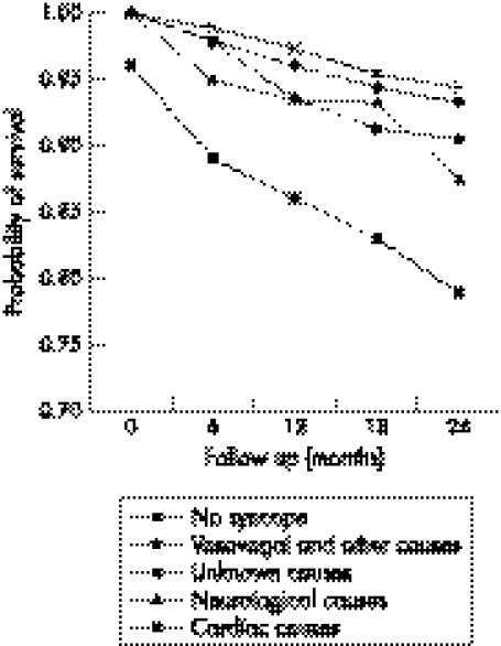
Soteriades et al32 studied 7814 participants of the Framlingham heart study. Of these, 822 had syncope in the 17 years of follow up (6.2 per 1000 person years). Vaso‐vagal syncope, the most common cause (21.2%), was not associated with any increased risk of death; however, a cardiac cause for syncope, found in 9.5%, was associated with a two fold increase in death, and a 6 month mortality rate exceeding 10% (fig 1). Getchell et al studied elderly hospitalised patients (mean age 73 years) presenting with syncope, and showed that mortality was not associated with a cardiac cause for syncope, but rather with age and comorbid illnesses.33
Figure 1 Overall survival of participants with syncope according to cause, and participants without syncope, among 7814 participants of the Framlingham heart study. Adapted from Soteriades et al,3 with permission from the publishing division of the Massachusetts Medical Society.
Subsequent studies controlling for cardiac mortality have showed that the higher mortality in patients with syncope due to a cardiovascular cause is largely related to underlying cardiovascular disease.7,21,34 A study comparing patients with and without syncope, who were matched for cardiac disease, showed that syncope itself was not a significant predictor of 1 year survival,21 however male sex, age >55 years and congestive heart failure were significant predictors. Middlekauff et al in 1993 studied 491 patients with advanced cardiac failure, 60 of whom had an episode of syncope. The 1 year mortality was greater in the patients with cardiac failure who had a history of syncope, compared to a matched group of patients with cardiac failure and without a syncope history (45% versus 12%). The major predictor of sudden death, however, was poor left ventricular function, not whether the cause of the syncope was cardiac or not.34 This study demonstrated syncope itself to be a good predictor of mortality. Whether these results are applicable to other patient populations is unclear.
It therefore seems it is the presence of significant underlying heart disease that is associated with a poor prognosis in syncope. It is likely that the presence of cardiac failure, commonly secondary to coronary artery disease, predisposes the patient to dysrhythmias and consequent syncope or sudden death. Patients with syncope and with signs of cardiac failure should be notionally high risk patients and therefore should be investigated to delineate underlying heart disease and the cause of syncope, in an attempt to reduce mortality.21,35
Clinical decision rules
Martin et al prospectively developed and validated a risk stratification system for patients presenting to the ED with syncope.8 In total, 252 patients were enrolled into a derivation cohort and 374 into a validation group. Four factors were predictive of 1 year mortality or dysrhythmia occurrence. These were abnormal presenting ECG findings (rhythm abnormalities, frequent PVCs, conduction disorders, left or right ventricular hypertrophy, short PR interval, evidence of an old MI, and atrioventricular block), a history of ventricular dysrhythmias, a history of congestive cardiac failure, and age >45 years. The 1 year mortality and dysrhythmia risk in patients with none of the four risk factors was 4.4–7.3%, increasing to 57.6–80.4% in patients with three risk factors.
Emphasis subsequently moved from the importance of making an underlying diagnosis in syncopal patients to risk stratification of patients into groups correlating with mortality. As the underlying conditions associated with short term mortality in syncope are related to structural cardiac disease and dysrhythmias, the rationale behind risk stratification is to focus resources into monitoring and investigating these high risk patients to reduce mortality.
Oh et al7 found that history and physical examination was able to determine a cause in 47% of patients. The only independent predictor of 1 year mortality was the presence of underlying cardiac disease (defined as coronary artery disease, valvular disease, cardiomyopathy, congestive cardiac failure, or other organic heart disease found clinically or during investigations). Crane36 conducted the only UK ED study of syncope outcome. This retrospective study of 210 patients presenting during an 8 week period showed that it was possible to stratify UK ED patients with syncope according to the American College of Physicians (ACP) guidelines.37,38 Patients in the ACP group 1 (high risk), had a 1 year mortality rate of 36%, compared to patients assigned to ACP group 2 (intermediate risk) (14%), and ACP group 3 (low risk) (0%).
Shen et al39 showed that patients in an intermediate risk group can be investigated in a ED based syncope unit, leading to an increased diagnostic yield, reduced hospital admission, and length of hospital stay, without increasing mortality.
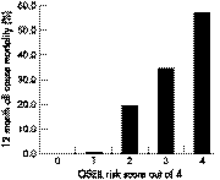
Colivicchi et al40 performed a six centre study that recruited 270 patients into a derivation study and 328 into a validation group. They developed a risk score (Osservatorio Epidemiologico sulla Sincope nel Lazio (OESIL) score) based on four characteristics: age >65 years, a clinical history of cardiovascular disease, syncope without prodromal symptoms, and an abnormal ECG. The presence of each characteristic scored 1 point. The authors found that 1 year mortality increased with increasing risk score and suggested that the tool could therefore be used in the assessment of ED patients with syncope (fig 2).
Figure 2 Rates of 12 month, all cause mortality according to the OESIL score in the derivation cohort. Reprinted from Colivicchi F et al,40 with permission from Oxford University Press.
Sarasin et al41 prospectively recruited 175 Swiss patients with unexplained syncope after ED investigation into a derivation study, and 269 similar US patients into a validation group. They found that predictors for dysrhythmic syncope were abnormal ECG, a history of congestive cardiac failure, and age >65 years. Risk of dysrhythmia (diagnosed by defined 24 hour Holter or loop recorder abnormalities) rose from 0–2% in patients with no risk factors to 6–17% in patients with one risk factor, 35–41% in those with two, and 27–60% in those with all three risk factors. They concluded that a risk score based on clinical and ECG factors is able to identify patients in the ED at risk of dysrhythmia.
The most recent and largest derivation study on syncope risk stratification focused on short term risk (probably more relevant to ED practice) and was performed by Quinn et al.13 They prospectively studied 684 patients who presented to a US ED with syncope, 79 of whom experienced a serious 7 day outcome. Of the 50 studied predictor variables, 26 were associated with a serious outcome. A clinical decision rule (the San Francisco syncope rule) was devised using five risk factors: abnormal ECG, anaemia (haematocrit <30%), complaint of shortness of breath, systolic hypotension (<90 mmHg), and a history of congestive cardiac failure. This rule was found to be 96% sensitive and 62% specific at predicting serious short term outcome, and if applied to the derivation cohort, would have decreased hospital admissions by 10%. This group has not yet prospectively validated their rule; however, two other studies have attempted to do so.42,43 Sensitivity in both studies was much lower than in the derivation cohort of Quinn et al (52% v 91%), in one study missing 26 of the 50 patients who had a serious outcome.42 An attempt has also been made to validate the rule for long term (1 year) mortality with a sensitivity of 88% and specificity of 56% in 658 ED attendees.44
With underlying cardiac failure being associated with a poor prognosis in syncope, clinical decision rules utilising biochemical markers of cardiac failure severity (C‐reactive protein or brain natriuretic peptide)45,46,47,48 may prove useful in the future. As yet, these have not been studied.
Guidelines
The ACP guidelines of 199737,38 reviewed all existing literature in order to provide guidelines on diagnosing syncope. They included guidance on which patients with unexplained syncope should be admitted to hospital, and divided patients into groups depending on the apparent risk of adverse outcome. Three main groups were identified. High risk patients in whom admission was indicated were those with a history of coronary artery disease, congestive cardiac failure (CCF) or ventricular tachycardia, those with accompanying symptoms of chest pain, those with physical signs of CCF, significant valve disease, stroke or focal neurology, and patients with ECG findings of ischemia, dysrhythmia (serious bradycardia or tachycardia), long QT interval, or bundle branch block.
The second group identified were those in whom they felt admission was often indicated. This "intermediate risk" group included patients with a sudden loss of consciousness with injury, tachycardia, or exertional syncope, those with frequent episodes (which lead to an increased likelihood of injury but are not associated with an increased mortality), those with a suspicion of coronary heart disease or dysrhythmia, moderate to severe postural hypotension, and those aged >70 years.
A third "low risk" group was defined as those who do not fall into either of the above groups. These patients may be discharged with or without outpatient follow up. Thakore et al showed that adherence to these guidelines in their UK ED population, would have increased hospital admissions by 38–58%.4
While other guidelines have followed,16,17 none have been prospectively validated. All guidelines include history, examination and investigation of syncopal patients, however only the American College of Emergency Physicians (ACEP) guidelines have focused directly on ED investigations and management.49 These suggest admission for patients with a history of congestive heart failure or ventricular dysrhythmias, associated chest pain or other symptoms compatible with acute coronary syndrome, evidence of significant congestive heart failure or valvular heart disease on physical examination, or ECG findings of ischemia, dysrhythmias, prolonged QT interval, or bundle branch block.
The ACEP guidelines also suggest that admission should be considered for patients with syncope who are older than 60 years, have a history of coronary artery or congenital heart disease, have a family history of unexpected sudden death, or in younger patients who present with exertional syncope without an obvious benign aetiology. Presently it is unclear whether either the application of guidelines to syncope management or the practice of admitting patients with syncope to hospital has any impact on patient outcome. No such benefits have ever been demonstrated.
CONCLUSIONS
Identifying a cardiac cause for syncope is a poor prognostic indicator for ED patients presenting with syncope. This is related to the severity of the patient's underlying cardiac disease rather than the syncopal event itself. Patients presenting with syncope who have significant cardiac disease should be investigated thoroughly to determine the nature of the underlying heart disease and the cause of syncope. At present however, there is little evidence that this improves their dismal prognosis (>30% 1 year mortality).
There are five small risk stratification studies on syncope in the ED.7,8,29,40,41 All five used different characteristics and outcome measures in their risk stratification tools. Only two were prospective and had mixed results.29,40 None have been examined in a UK population.
Presently the ACEP guidelines49 are the most useful aids to the management of syncope in the ED; however, the OESIL score40 may be a useful ED risk stratification tool.
Abbreviations
ACEP - American College of Emergency Physicians
ACP - American College of Physicians
artery disease - CCF, congestive cardiac failure
ECG - electrocardiogram
ED - emergency department
MI - myocardial infarction
OESIL - Osservatorio Epidemiologico sulla Sincope nel Lazio
PVC - premature ventricular contraction
Footnotes
Competing interests: there are no competing interests
References
- 1.Morag R. Syncope. In: Peak DA, Talavera F, Halamka J, et al. eMedicine on line emergency medicine textbook. www.emedicine.com/emerg/topic876.htm
- 2.Maisel W H, Stevenson W G. Syncope: getting to the heart of the matter. N Engl J Med 2002347931–933. [DOI] [PubMed] [Google Scholar]
- 3.Soteriades E S, Evans J C, Larson M G.et al Incidence and prognosis of syncope. N Engl J Med 2002347878–885. [DOI] [PubMed] [Google Scholar]
- 4.Thakore S B, Crombie I, Johnston M. The management of syncope in a British Emergency Department compared to recent American guidelines. Scot Med J 199944155–157. [DOI] [PubMed] [Google Scholar]
- 5.Day S C, Cook E F, Funkenstein H.et al Evaluation and outcome of emergency room patients with transient loss of consciousness. Am J Med 19827315–23. [DOI] [PubMed] [Google Scholar]
- 6.Kapoor W. Evaluation and outcome of patients with syncope. Medicine (Baltimore) 199069160–175. [DOI] [PubMed] [Google Scholar]
- 7.Oh J H, Hanusa B H, Kapoor W N.et al Do Symptoms Predict Cardiac Arrhythmias and Mortality in Patients With Syncope? Arch Intern Med 1999159375–380. [DOI] [PubMed] [Google Scholar]
- 8.Martin T P, Hanusa B H, Kapoor W N. Risk stratification of Patients with Syncope. Annals of Emergency Medicine 199729459–466. [DOI] [PubMed] [Google Scholar]
- 9.Dunn M J G, Breen D P, Davenport R J.et al Early management of adults with an uncomplicated first generalised seizure. Emerg Med J 200522237–242. [DOI] [PMC free article] [PubMed] [Google Scholar]
- 10.Martin G J, Adams S L, Martin H G.et al Prospective evaluation of syncope. Ann Emerg Med 198413499–504. [DOI] [PubMed] [Google Scholar]
- 11.Calkins H, Shyr Y, Frumin H.et al The value of the clinical history in the differentiation of syncope due to ventricular tachycardia, atrioventricular block, and neurocardiogenic syncope. Am J Med 199598365–373. [DOI] [PubMed] [Google Scholar]
- 12.Kapoor W, Peterson J, Wieand H.et al Diagnostic and prognostic implications of recurrences in patients with syncope. Am J Med 198783700–707. [DOI] [PubMed] [Google Scholar]
- 13.Quinn J V, Stiell I G, McDermott D A.et al Derivation of the San Francisco syncope rule to predict patients with short‐term serious outcomes. Ann Emerg Med 200443224–232. [DOI] [PubMed] [Google Scholar]
- 14.Lipitz L A, Wei J Y, Rowe J W. Syncope in an elderly institionalized population: prevalence, incidence, and associated risk. Q J Med 19855545–55. [PubMed] [Google Scholar]
- 15.Junaid A, Dubinsky I L. Establishing an approach to syncope in the emergency department. J Emerg Med 199715593–599. [DOI] [PubMed] [Google Scholar]
- 16.Brignole M, Alboni P, Benditt D G.et al Task Force on Syncope, European Society of Cardiology. Guidelines on management (diagnosis and treatment) of syncope–update 2004. Executive summary. Eur Heart J 2004252054–2072. [DOI] [PubMed] [Google Scholar]
- 17.Brignole M, Alboni P, Benditt D.et al Task Force on Syncope, European Society of Cardiology. Guidelines on management (diagnosis and treatment) of syncope. Eur Heart J 2001221256–1306. [DOI] [PubMed] [Google Scholar]
- 18.Silverstein M D, Singer D E, Mulley A G.et al Patients with syncope admitted to medical intensive care units. JAMA 19822481185–1189. [PubMed] [Google Scholar]
- 19.Kapoor W N, Karpf M, Wieand S.et al A prospective evaluation and follow‐up of patients with syncope. N Engl J Med 1983309197–204. [DOI] [PubMed] [Google Scholar]
- 20.Eagle K, Black H. The impact of diagnostic tests in evaluating patients with syncope. Yale J Biol Med 1983561–8. [PMC free article] [PubMed] [Google Scholar]
- 21.Kapoor W N, Hanasu B H. Is syncope a risk factor for poor outcomes? Comparison of patients with and without syncope. Am J Med 1996100646–655. [DOI] [PubMed] [Google Scholar]
- 22.Krol R B, Morady F, Flaker G C.et al Electrophysiologic testing in patients with unexplained syncope: Clinical and non‐invasive predictors of outcome. J Am Coll Cardiol 198710358–363. [DOI] [PubMed] [Google Scholar]
- 23.Denes P, Vretz E, Ezr M D.et al Clinical predictors of electrophysiologic findings in patients with syncope of unknown origin. Arch Intern Med 19881481922–1928. [PubMed] [Google Scholar]
- 24.Klitzner T S. Sudden cardiac death in children. Circulation 199082629–632. [DOI] [PubMed] [Google Scholar]
- 25.Link M S, Lauer E P, Homoud M K.et al Low yield of rule‐out myocardial infarction protocol in patients presenting with syncope. Am J Cardiol 200188706–707. [DOI] [PubMed] [Google Scholar]
- 26.Grossman S A, Van Epp S, Arnold R.et al The value of cardiac enzymes in elderly patients presenting to the emergency department with syncope. J Gerontol A Biol Sci Med Sci 2003581055–1058. [DOI] [PubMed] [Google Scholar]
- 27.Hing R, Harris R. Relative utility of serum troponin and the OESIL score in syncope. Emerg Med Australas 20051731–38. [DOI] [PubMed] [Google Scholar]
- 28.Bayer A J, Chadha J S, Farag R R.et al Changing presentation of myocardial infarction with increasing old age. J Am Geriatr 198334263–266. [DOI] [PubMed] [Google Scholar]
- 29.Lipsitz L A, Pluchino F C, Wei J Y. The prevalence and prognosis of minimally elevated creatine kinase‐myocardial band activity in elderly patients with syncope. Arch Intern Med 19871471321–1323. [PubMed] [Google Scholar]
- 30.Ben‐Chetrit E, Flugelman M, Eliakim M. Syncope: a retrospective study of 101 hospitalized patients. Isr J Med Sci 198521950–953. [PubMed] [Google Scholar]
- 31.Sarasin F P, Louis‐Simonet M, Carballo D.et al Prospective evaluation of patients with syncope: A population‐based study. Am J Med 2001111177–184. [DOI] [PubMed] [Google Scholar]
- 32.Soteriades E S, Evans J C, Larson M G.et al Incidence and prognosis of syncope. N Engl J Med 2002347878–885. [DOI] [PubMed] [Google Scholar]
- 33.Getchell W S, Larsen G C, Morris C D.et al Epidemiology of syncope in hospitalized patients. J Gen Intern Med 199914677–687. [DOI] [PMC free article] [PubMed] [Google Scholar]
- 34.Middlekauff H, Stevenson W, Stevenson L.et al Syncope in advanced heart failure: high risk of sudden death regardless of origin of syncope. J Am Coll Cardiol 199321110–116. [DOI] [PubMed] [Google Scholar]
- 35.Kapoor W. Current evaluation and management of syncope. Circulation 20021061606–1609. [DOI] [PubMed] [Google Scholar]
- 36.Crane S D. Risk Stratification of patients with syncope in an accident and emergency department. Emerg Med J 20021923–27. [DOI] [PMC free article] [PubMed] [Google Scholar]
- 37.Linzer M, Yang E H, Estes NA I I I.et al Diagnosing syncope. 1. Value of history, physical examination, and electrocardiography: Clinical Efficacy Assessment Project of the American College of Physicians, Ann Intern Med 1997126989–996. [DOI] [PubMed] [Google Scholar]
- 38.Idem Diagnosing syncope. 2. Unexplained syncope: Clinical Efficacy Assessment Project of the American College of Physicians, Ann Intern Med 199712776–86. [DOI] [PubMed] [Google Scholar]
- 39.Shen W K, Decker W W, Smars P A.et al Syncope evaluation in the emergency department (SEEDS). Circulation 20041103636–3645. [DOI] [PubMed] [Google Scholar]
- 40.Colivicchi F, Ammirati F, Melina D.et al Development and prospective validation of a risk stratification system for patients with syncope in the emergency department: the OESIL risk score. European heart journal 200324811–819. [DOI] [PubMed] [Google Scholar]
- 41.Sarasin F P, Hanusa B H, Perneger T.et al A Risk score to predict arrhythmias in patients with unexplained syncope. Acad Emerg Med 2003101312–1317. [DOI] [PubMed] [Google Scholar]
- 42.Fischer C M, Shapiro N I, Lipsitz L.et al External validation of the San Francisco syncope rule. Acad Emerg Med 200512(suppl)127 [Google Scholar]
- 43.Stracner D L, Kass L. Validation of the San Francisco syncope rule. Acad Emerg Med 200512(suppl)87–88. [Google Scholar]
- 44.Quinn J V, McDermott D A, Kohn M.et al Death rates of emergency department patients with syncope: Can the San Francisco syncope rule predict long‐term mortality? Acad Emerg Med 200512(suppl)8 [Google Scholar]
- 45.Doust J A, Glasziou P P, Pietrzak E.et al A systematic review of the diagnostic accuracy of natriuretic peptides for heart failure. Arch Intern Med 20041641978–1984. [DOI] [PubMed] [Google Scholar]
- 46.Doust J A, Pietrzak E, Dobson A.et al How well does B‐type natriuretic peptide predict death and cardiac events in patients with heart failure: systematic review. BMJ 2005330625–627. [DOI] [PMC free article] [PubMed] [Google Scholar]
- 47.Tanimoto K, Yukiiri K, Mizushige K.et al Usefulness of brain natriuretic peptide as a marker for separating cardiac and noncardiac causes of syncope. Am J Cardiol 200493228–230. [DOI] [PubMed] [Google Scholar]
- 48.Alonso‐Martinez J L, Llorente‐Diez B, Echegarey‐Agara M.et al C‐reactive protein as a predictor of improvement and readmission in heart failure. Eur J Heart Fail 20024331–336. [DOI] [PubMed] [Google Scholar]
- 49.Molzen G W, Suter R E, Whitson R. American College of Emergency Physicians: Clinical Policy: critical issues in the evaluation and management of patients presenting with syncope. Ann Emerg Med 200137771–776. [DOI] [PubMed] [Google Scholar]


