Abstract
Recent modifications in total knee prosthesis design theoretically better accommodate the anatomy of the female femur and thereby have the theoretical potential to improve clinical results in TKA by more accurately restoring femoral posterior condylar offset, reducing femoral notching, reducing femoral component flexion, and reducing component overhang. First, we radiographically evaluated whether a contemporary unisex prosthesis would accommodate female anatomy equally as well as male anatomy. Next, we radiographically evaluated female knees in which a gender-specific prosthesis was used. Pre- and postoperative radiographs of 122 knees (42 female unisex, 41 male unisex, 39 female gender-specific) were reviewed. In the unisex groups, there were no differences in femoral notching or femoral component flexion. Posterior femoral offset increased in both groups. However, femoral component overhang was worse in female knees (17%) than in male knees (0%). In the gender-specific female group, the incidence of component overhang was similar to that in the unisex female group. Unisex femoral components of this specific design do not equally match the native anatomy male and female knees. In some women, a compromise was required in sizing.
Introduction
The concepts of sexual dimorphism in humans and anatomic variations in various ethnic groups are well known and supported by anatomic and radiographic studies [6, 10–12, 16]. In general, the distal femurs of females are not only smaller, but have different shapes with a narrower medial-lateral (ML) diameter for any given anteroposterior (AP) distance than in males [6, 10, 12]. The recent introduction and marketing of gender-specific knee arthroplasty implants was a new approach to the ongoing trend across TKA systems to offer more sizing options and is based on the anatomic differences between male and female femurs [4, 9]. Rather than simply offering more sizes with similar AP to ML ratios, the gender-specific component is designed to better accommodate the anatomic differences noted in females with a narrower ML dimension for any given AP dimension [4, 9]. In addition, the angle of the trochlear groove was increased and the anterior flange thickness was reduced to better match the native female anatomy [4, 9].
The introduction of the gender-specific components, optimized for the female anatomy, raised the question of whether the unisex components in this system equally matched the native anatomy of both male and female patients in TKA or whether they better matched one gender. Theoretically, intraoperative problems such as overresection of the posterior femoral condyles, femoral notching, excessive femoral component flexion, or acceptance of component overhang may be caused when trying to accommodate the native female anatomy with the standard unisex femoral component sizes that are based on mean anatomic measurements from males and females. In circumstances in which a female knee requires a larger size component in the AP dimension than it can accommodate in the ML dimension, the surgeon has four choices, all of which may contribute to clinical problems, including (1) overresection of the posterior femoral condyles, which can reduce postoperative flexion [3]; (2) increased femoral component flexion, which can lead to cam-post impingement in a posterior stabilized knee [1, 5, 7]; (3) femoral notching with potential for supracondylar fracture [1, 8, 14]; and (4) femoral component overhang, which may create soft tissue irritation. Anecdotal intraoperative observations suggested the operating surgeon (HDC) tended to downsize the femoral component in some female patients because of difficulties accommodating the ML dimension of the prosthesis that was optimal in the AP dimension.
We therefore posed the following hypotheses: (1) unisex femoral components do not adequately match the native femoral anatomy of female knees and thereby use of these implants in female patients will result in higher rates of undesirable radiographic outcomes, including a reduction of femoral posterior condylar offset, increased femoral component flexion, increased femoral notching, and increased component overhang versus male patients with a unisex component; and (2) use of gender-specific femoral components in female patients will eliminate the increased rates of undesirable radiographic outcomes hypothesized in female patients with a unisex device.
Materials and Methods
We retrospectively reviewed 39 females (42 knees) with a unisex femoral component, 38 males with a unisex femoral component (41 knees), and 35 females (39 knees) with a gender-specific femoral component who had undergone primary TKA from January 2005 to April 2007. All patients had received either a NexGen® Legacy® Posterior Stabilized prosthesis (Zimmer, Inc, Warsaw, IN) or a Gender Solutions™ NexGen® Legacy® Posterior Stabilized prosthesis (Zimmer, Inc). Additional inclusion criteria were absence of periarticular bony deformities or ligamentous insufficiency and availability of true lateral radiographs of the femur both preoperatively and postoperatively. During the same time period, we performed 263 primary TKAs. Of these, 13 were excluded because they required a constrained implant for bony or ligamentous deficiencies and 12 had received a primary implant from a different manufacturer. A further 116 knees were excluded because they lacked a true lateral radiograph or adequate AP radiograph either preoperatively or postoperatively. The study protocol was approved by the Institutional Review Board at our institution.
All operations were performed by the same surgeon (HDC) using the same instruments. Bone cuts were performed in a standard manner with the distal femoral and proximal tibial bone cuts perpendicular to the mechanical axis in the coronal plane. Also, to reduce the risk of femoral notching, 3° of flexion was incorporated into the distal femur in the sagittal plane. The proximal tibial cut incorporated a limited posterior slope of approximately 3° in the sagittal plane. Femoral component sizing was performed with a posterior referencing instrument that sets the resection of the posterior condyles equal to the thickness of the component to attempt to accurately restore the posterior condylar offset. This guide also incorporates an anterior boom that allows secondary referencing of the anterior cortex that helps prevent notching. The anterior cortex of the midpoint of the femur was used as the point marking the depth of the anterior femoral resection. The femoral component rotation was set parallel to the transepicondylar axis. If downsizing was required based on intraoperative assessment that the desired femoral component was too wide, additional posterior resection was performed. The other alternative was to accept some medial or lateral overhang. In most cases, some compromise was required and included the surgeon’s assessment of the extent of the overhang, flexion and extension gap balance, and remaining anterior bone that could be resected before producing a notch. However, because this was a retrospective review, none of the details of this customization process were recorded.
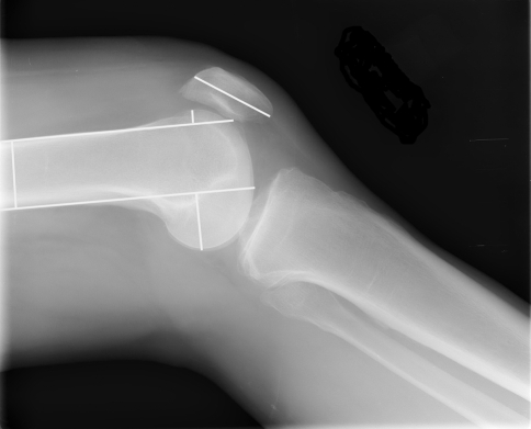
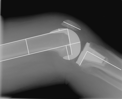
The pre- and postoperative radiographs were reviewed by the treating surgeon (HDC) (Figs. 1, 2). On each occasion, a standing AP view and Merchant view of both knees were obtained along with a true lateral view of the operative knee. Each radiograph was performed according to a standard protocol. These images were evaluated with standard digital imaging software and the same measurements were performed in each case. Posterior condylar offset measurements were performed according to the techniques previously published by Bellemans et al. [3]. Preoperative measurements included AP diameter of the femur 10 cm from the joint line on the lateral view, maximum length of the patella on the lateral view, posterior condylar offset on the lateral view, and anterior femoral offset on the lateral view (Fig. 1). Postoperative measurements included the same four measurements as previously stated and four additional measurements: femoral component flexion relative to the anterior cortex on the lateral view, notching of the anterior cortex on the lateral view, medial or lateral overhang of the femoral component at the distal joint line on the standing AP view, and posterior slope of the tibial component on the lateral view (Fig. 2). The posterior tibial slope measurement was added as a reference measurement, because we believed this measure independent of the femoral component sizing or positioning and gender. On both the pre- and postoperative radiographs, the diameter of the femur 10 cm proximal to the joint line was used to standardize any magnification differences on the two sets of radiographs according to the previously described technique by Bellemans et al. [3]. In addition, the length of the patella was also measured for similar purposes to see if this would be an alternative to the femoral diameter measurement.
Fig. 1.
A true lateral preoperative radiograph demonstrates the four measurements recorded in each case: anteroposterior diameter of the femur 10 cm from the joint line, maximum length of the patella, posterior condylar offset, and anterior femoral offset.
Fig. 2.
A true lateral postoperative radiograph demonstrates seven of the eight measurements recorded in each case: the same four measurements recorded preoperatively plus femoral component flexion relative to the anterior cortex, notching of the anterior cortex, and posterior slope of the tibial component. The eighth postoperative measurement was medial or lateral overhang of the femoral component at the distal joint line taken on the standing anteroposterior view.
The primary outcome measures were the change from baseline posterior condylar offset, femoral component flexion, whether the patient had any femoral notch, whether the patient had any medial or lateral femoral component overhang, and change from baseline anterior offset. The primary outcome measures and methods of analysis were specified before analyzing the data. Two knees from the same patient were treated as independent observations. For offset measurements, we used the thickness of the femur to measure the followup radiograph in the same scale as the baseline radiograph. That is, offset measurements on the followup radiograph were multiplied by the ratio of the preoperative femoral thickness to the postoperative femoral thickness. The diameter of the femur was used to standardize the magnification rather than the length of the patella because patellar resurfacing made it difficult to accurately perform this measurement on the postoperative radiographs.
Mean posterior offset change, femoral flexion, anterior offset change, and tibial slope were compared among the groups by using the two-sample t test. Assumptions for the test were verified by inspecting the distributions of the residuals. The incidence of notch and overhang were compared among the groups by using the Pearson chi square test.
Results
The unisex femoral components did not match the native female anatomy. Although there were no differences in mean femoral component flexion (95% confidence interval [CI], –1.6 to 0.3) or the incidence of notching (95% CI, −0.15 to 0.19) between the female patients with the unisex components and the male group, the incidence of medial or lateral femoral component overhang was higher (p = 0.006) in females than in males (seven knees or 17% versus none in the males) (Table 1). In addition, the difference in the mean change in posterior condylar offset was lower (p = 0.01) in female than male patients with the unisex devices (Table 1). However, in both groups, the change was a slight increase in thickness postoperatively (female 1.3 mm, male 2.8 mm) rather than the decrease that was hypothesized would occur in the female group.
Table 1.
Radiographic measurements in unisex and gender-specific TKAs
| Radiographic measurement | Female gender-specific (n = 39 knees) | Female unisex (n = 42 knees) | Male unisex (n = 41 knees) | Female gender-specific versus female unisex | Female gender-specific versus male unisex | Female unisex versus male unisex | |||
|---|---|---|---|---|---|---|---|---|---|
| p | 95% CI | p | 95% CI | p | 95% CI | ||||
| Posterior offset change (mm)* | 2.5 (2.2) | 1.3 (2.8) | 2.8 (2.6) | 0.04 | 0.1 to 2.3 | 0.59 | –1.4 to 0.8 | 0.01 | –2.6 to –0.3 |
| Femoral flexion (º)* | 4.4 (1.9) | 3.7 (2.3) | 4.4 (2.2) | 0.16 | –0.3 to 1.6 | 0.99 | –0.9 to 0.9 | 0.18 | –1.6 to 0.3 |
| Notch, number | 7 (18%) | 8 (19%) | 7 (17%) | 0.90 | –0.18 to 0.16 | 0.92 | –0.16 to 0.18 | 0.82 | –0.15 to 0.19 |
| Medial or lateral femoral component overhang, number | 2 (5%) | 7 (17%) | 0 (0%) | 0.10 | –0.25 to 0.02 | 0.14 | –0.02 to 0.12 | 0.006 | 0.05 to 0.28 |
| Anterior offset change (mm)* | −4.5 (2.3) | –3.1 (2.7) | –4.8 (3.4) | 0.01 | –2.6 to –0.3 | 0.66 | –1.0 to 1.6 | 0.01 | 0.4 to 3.1 |
| Tibial slope (º)* | 3.6 (1.6) | 3.3 (2.0) | 3.2 (2.1) | 0.57 | –0.6 to 1.0 | 0.40 | –0.5 to 1.2 | 0.78 | –0.8 to 1.0 |
*Values for variable expressed as means with standard deviations in parentheses; 95% CI = 95% confidence interval.
Use of the gender-specific prostheses in female patients did not eliminate the increased incidence of detrimental radiographic findings noted in the female versus male unisex groups. Specifically, in the female patients with the gender-specific components, 5% (two knees) demonstrated medial or lateral overhang versus 17% (five knees) in the female unisex group (Table 1). However, the margin of error was large for a sample of this size (95% CI, −0.25 to 0.02). There were also no differences in the mean femoral component flexion (95% CI, −0.3 to 1.6) or incidence of notching (95% CI, −0.18 to 0.16) between the female-specific and female unisex groups (Table 1). Finally, the unisex female group had less (p = 0.04; 95% CI, 0.1 to 2.3) mean posterior condylar offset change than the female gender-specific group, but both groups demonstrated an increase in posterior condylar offset rather than a detrimental decrease (female-specific 2.5 mm, female unisex 1.3 mm).
Discussion
The recent introduction and marketing of the gender-specific total knee implants is a new approach in the ongoing trend over the past 30 years to offer more prosthetic sizing options in TKA [4, 9] This theoretically allows surgeons to better match the prosthesis size with the patient anatomy. In most systems, the increase in size options has occurred by progressively reducing the step between sizes in the AP and ML dimensions. However, the introduction of the gender-specific implants is based on trying to better match the anatomic differences of female femurs versus male femurs [4, 6, 10, 12]. Prior anatomic studies have demonstrated female distal femurs not only are smaller, but have a different shape than male femurs with a narrower ML diameter for any given AP distance [6, 9, 12]. Theoretically, intraoperative sizing problems may be encountered when a unisex device that does not adequately match the female anatomy is used in female patients. These sizing difficulties have been reported to contribute to the occurrence of postoperative clinical problems in prior studies. Specifically, if the ML width of the female femur is too narrow to accommodate the prosthesis of a given AP dimension, downsizing to a smaller size may result in undesirable outcomes: overresection of the posterior femoral condyles by even 1 mm has been previously associated with reduced postoperative flexion [3]; increased femoral component flexion may lead to cam-post impingement and polyethylene wear in a posterior stabilized knee [5, 7]; and femoral notching has been associated with increased risk of supracondylar fracture [1, 8, 14]. Alternatively, use of the larger component may require acceptance of ML femoral component overhang that may create soft tissue irritation. We examined the use of a unisex device in male and female patients to see if the sizing options resulted in higher rates of undesirable radiographic findings in the female patients as compared with the male patients and whether use of the gender-specific components in female patients eliminated the increased rates of undesirable radiographic results that were hypothesized to occur in female patients with a unisex device.
As a result of the retrospective and purely radiographic study design, there are many limitations that should be considered when interpreting the current data. Our findings pertain only to a single implant system. At this time, the gender bias favoring the unisex femoral components better matching the male anatomy only pertains to the specific design studied; this may simply represent an isolated deficiency of this particular product. Additional studies of the femoral components in other knee systems are needed before this conclusion can be applied to other designs. However, because the current system is the only one that offers a gender-specific component, the clinical value of identifying this potential failing in other systems is limited because no current alternative exists. A second concern regarding this study is the fact that all surgeries were performed by only one surgeon. A systematic bias may have produced the noted findings and the results may not be applicable to other surgeons. However, the theoretical argument remains that if the components do not adequately match the native anatomy, one of the four detrimental radiographic outcomes will likely result. Therefore, although another surgeon may not have a higher rate of medial or lateral component overhang in female patients than in male patients, they likely would have a higher rate of a different undesirable radiographic finding; furthermore, of the four possible outcomes, medial or lateral overhang is probably the least likely to be associated with potential clinical problems. Therefore, the findings in this study may indeed represent a best case scenario. Another limitation of the study is interpretation of radiographs is dependent on the technical quality of the radiograph. This potentially interfered with our ability to accurately perform measurements. Attempts to minimize this factor included standard protocols for the radiographs; furthermore, only patients with perfectly positioned lateral views with overlapping condyles were included (Figs. 1, 2). Exclusion of some patients based on inadequate radiographs may therefore have introduced selection bias. However, this technical error was likely random and it is unlikely patients with one radiographic characteristic consistently had radiographs in which poor technical quality was accepted by the technician. Another major limitation of this study is no clinical outcomes were correlated with the radiographic findings. However, previous work has raised concerns about the potential clinical effects of each radiographic variable studied, except overhang, to support further efforts to eliminate these radiographic outliers. Data in the literature to condemn overhang are lacking, but prominent hardware is a clinically accepted cause of irritation after orthopaedic surgery in general, and because there is no theoretical advantage to overhang, this should probably be avoided if possible at reasonable cost and presuming the effects are clinically important. Moreover, although the clinical effects of these radiographic findings may be small, it is possible they contribute to the lower range of motion, outcome scores, and postoperative satisfaction scores noted in female patients when reviews of very large total knee registries are performed [2, 13, 15]. This study, which was only ever conceived as a radiographic study, will hopefully serve as the basis for larger studies with adequate power to identify any small clinical differences that may result. Finally, no cost data were examined in this study. Therefore, it is impossible to perform an economic analysis to determine the financial impact of the new component options. Again, financial analysis should be part of future studies that attempt to correlate changes in clinical outcomes with the use of the new prosthetic options to determine the costs of any changes noted.
In a study of 200 consecutive knees (100 male and 100 female), Chin et al. [6] reported the mean AP to ML ratio of the knee for the entire population to be 0.8 [6]. In comparison, this mean ratio was 0.82 in the female knees versus 0.79 in the male knees [6]. These general findings are supported by the report from Hitt et al. [10], who measured the intraoperative anatomy of the distal femur in a cohort of 337 adult male and female patients and then compared these measurements with the geometries of modern total knee prostheses [10]. Although it was clear the systems that were evaluated approximated the mean AP to ML ratios of the entire population, the prosthesis did not as accurately match the subpopulation of female patients [10]. In support of these prior publications, we found that although two of our four primary outcome measures were similar between the male and female patients in whom a unisex prosthesis had been implanted (degree of femoral component flexion and rate of femoral notching), differences were identified that supported our primary hypothesis that the unisex implants do not adequately match the female anatomy. Medial or lateral overhang occurred more frequently in the female patients. Furthermore, changes in mean posterior condylar offset were also less in the female compared to the male patients. It is interesting to note in both male and female patients with a unisex prosthesis, rather than identifying a reduction in mean posterior condylar offset, a slight increase occurred in both groups. It is possible this increase may reflect a failure of the preoperative radiographic measurement to account for the residual thickness of posterior cartilage that is included in the resection thickness using the intraoperative cutting guide. In distinction, on the postoperative radiograph, the entire thickness of the metal component is easily measured. Therefore, this slight increase may not represent a true increase in the posterior condylar offset. The fact that the increase in posterior condylar offset was considerably less in the female unisex group than in the male group (1.3 mm versus 2.8 mm) may therefore also support the primary hypothesis that the unisex implants in this system do not adequately match native female anatomy. In prior work by Bellemans et al. [3], even a 1-mm decrease in offset reduced postoperative flexion by more than 5°. Therefore, the potential for this radiographic finding of approximately 1.5 mm producing a potentially important clinical issue remains open for further study.
In distinction to supporting the primary hypothesis that unisex implants do not adequately match the native female anatomy, the results of this study do not unequivocally support the secondary hypothesis regarding the gender-specific implants in females eliminating the increased rates of undesirable radiographic findings noted in female patients with a unisex implant. Specifically, because the gender-specific femoral component is narrower in the ML dimension for a given AP dimension, use of this implant would have been expected to reduce the increased incidence of medial or lateral overhang noted in female patients with a unisex implant. We found the incidence of medial or lateral overhang in the female knees with the gender-specific component was 5% versus 17% in the female unisex group but this difference was not significant and therefore did not support the secondary hypothesis. In distinction, in the female group with the gender-specific components, the increase in mean posterior offset was equal to that in the male group (2.5 mm versus 2.8 mm) and it was greater than the female unisex group. This provides some secondary evidence that the gender-specific components better match the female femoral anatomy. Based on these two contradictory findings in the gender-specific group, the question remains whether the changes made to the current gender-specific implant are extensive enough to accurately match female femoral anatomy or whether additional geometric modifications would provide better radiographic results.
Our data support the primary hypothesis that unisex femoral components in the NexGen® Legacy® Posterior Stabilized system are gender-biased and do not accurately match female anatomy. Use of these unisex components in female patients results in the need for accepting a compromise that is radiographically evident in some patients; in this study, the compromise was to accept some medial or lateral overhang of the femoral component. In distinction, we were unable to confirm use of the gender-specific femoral component available in this system was associated with better radiographic results. However, because each of the potential compromises associated with use of a unisex device has been associated with potentially negative clinical effects, use of the gender-specific component that does not have any apparent theoretical risks appears reasonable when using this particular knee system in female patients.
Footnotes
Each author certifies that he or she has no commercial associations (eg, consultancies, stock ownership, equity interest, patent/licensing arrangements, etc) that might pose a conflict of interest in connection with the submitted article.
Each author certifies that his or her institution has approved the human protocol for this investigation, that all investigations were conducted in conformity with ethical principles of research, and that informed consent for participation in the study was obtained.
References
- 1.Aaron RK, Scott R. Supracondylar fracture of the femur after total knee arthroplasty. Clin Orthop Relat Res. 1987;219:136–139. [PubMed]
- 2.Baker PN, Van der Meulen JH, Lewsey J, Gregg PJ. The role of pain and function in determining patient satisfaction after total knee replacement. J Bone Joint Surg Br. 2007;89:893–900. [DOI] [PubMed]
- 3.Bellemans J, Banks S, Victor J, Vandenneucker H, Moemans A. Fluoroscopic analysis of the kinematics of deep flexion in total knee arthroplasty: influence of posterior condylar offset. J Bone Joint Surg Br. 2002;84:50–53. [DOI] [PubMed]
- 4.Booth RE. The gender specific (female) knee. Orthopedics. 2006;29:768–769. [DOI] [PubMed]
- 5.Callaghan JJ, O’Rourke MR, Goetz DD, Schmalzried TP, Campbell PA, Johnston RC. Tibial post impingement in posterior-stabilized total knee arthroplasty. Clin Orthop Relat Res. 2002;404:83–88. [DOI] [PubMed]
- 6.Chin KR, Dalury DF, Zurakowski D, Scott RD. Intraoperative measurements of male and female distal femurs during primary total knee arthroplasty. J Knee Surg. 2002;15:213–217. [PubMed]
- 7.Clarke HD, Math KR, Scuderi GR. Polyethylene post failure in posterior stabilized total knee arthroplasty. J Arthroplasty. 2004;19:652–657. [DOI] [PubMed]
- 8.Culp RW, Schmidt RG, Hanks G, Mak A, Esterhai JL Jr, Heppenstall RB. Supracondylar fracture of the femur following prosthetic knee arthroplasty. Clin Orthop Relat Res. 1987;222:212–222. [PubMed]
- 9.Greene KA. Gender-specific design in total knee arthroplasty. J Arthroplasty. 2007;22(Suppl 3):27–31. [DOI] [PubMed]
- 10.Hitt K, Shurman JR, Greene K, McCarthy J, Moskal J, Hoeman T, Mont MA. Anthropometric measurements of the human knee: correlation to the sizing of current knee arthroplasty systems. J Bone Joint Surg Am. 2003;85(Suppl 4):115–122. [PubMed]
- 11.Kwak DS, Surendran S, Pengatteeri YH, Park SE, Choi KN, Gopinathan P, Han SH, Han CW. Morphology of the proximal tibia to design the tibial component of total knee arthroplasty for the Korean population. Knee. 2007;14:295–300. [DOI] [PubMed]
- 12.Polivache P, Insall JN, Scuderi GR, Font-Rodriguez DE. Rotational landmarks of the distal femur in total knee arthroplasty. Clin Orthop Relat Res. 1991;331:35–46. [DOI] [PubMed]
- 13.Ritter MA, Harty LD, Davis KE, Meding JB, Berend ME. Predicting range of motion after total knee arthroplasty: clustering, log-regression, and regression tree analysis. J Bone Joint Surg Am. 2003;85:1278–1285. [DOI] [PubMed]
- 14.Ritter MA, Thong AE, Keating EM, Faris PM, Meding JB, Berend ME, Pierson JL, Davis KE. The effect of femoral notching during total knee arthroplasty on the prevalence of postoperative femoral fractures and on clinical outcomes. J Bone Joint Surg Am. 2005;87:2411–2414. [DOI] [PubMed]
- 15.Robertson O, Dunbar M, Pehrsson T, Knutson K, Lidgren L. Patient satisfaction after total knee arthroplasty: a report on 27372 knees operated on between 1981 and 1995 in Sweden. Acta Orthop Scand. 2000;71:262–267. [DOI] [PubMed]
- 16.Vaidya SV, Ranawat CS, Aroojis A, Laud NS. Anthropometric measurements to design total knee prostheses for the Indian population. J Arthroplasty. 2000;15:79–85. [DOI] [PubMed]


