Abstract
Fifty-eight osteoarthritic and thirty-one rheumatoid patients underwent modular total knee-replacement arthroplasty. The major indication for the operation was relief of pain. Contraindications to this resurfacing arthroplasty included varus-valgus instability of over 20 degrees, combined varus-valgus instability with flexion contracture of over 40 degrees, marked recurvatum, and predominant patellofemoral symptoms. In 59 per cent of the osteoarthritic and 58 per cent of the rheumatoid patients, complete relief of pain was evident when they were evaluated twenty-four months after surgery, while another 35 per cent of each group had only mild pain related to inclement weather. Their ability to walk long distances without support or limp was increased. Range of motion and ability to climb stairs were not significantly improved.
Introduction
The major aims of reconstructive surgery in the arthritic knee are to relieve pain, restore stability, and permit a functional range of motion. In the past, procedures to achieve these aims have not been as successful as was hoped for in the majority of cases. While the solidly arthrodesed knee was indeed painless, the functional disability was often unacceptable. The Magnuson and Pridie debridement procedures temporarily relieved pain, but did not restore motion or stability. Osteotomy of the proximal end of the tibia was extremely successful in aligning the knee with varus deformity caused by osteoarthritis, thereby relieving pain and instability. In the knee with valgus deformity, however, it was less effective, and in neither did it improve motion. It was ineffective in the rheumatoid knee. Hemiarthroplasty of the femoral or tibial surface alone gave short-term relief of pain, at best.
The technical knowledge gained from a decade of total hip-replacement arthroplasty has allowed the development of several technical variations in replacement arthroplasty for the knee joint. Ideally, each variation should serve best according to the specific disability for which the model of prosthesis was designed.
The present models for total knee-replacement arthroplasty fall into three basic types:
The constrained type: This imparts stability to the knee by virtue of an articulation, either hinged (Walldius, Guepar, Shiers, Young) or ball-in-socket (Herbert). Ex-tensive resection of bone is required for implantation of these models.
The non-constrained, partially stabilizing type: The models called total condylar, Geomedic, and University of California (Irvine) are examples of this group. The amount of bone resection varies, but is less than for the constrained models. Stability is imparted to the knee by means of tracks or interdigitated components.
The resurfacing type: This has been advocated for knees in which there is minimum instability. Little or no bone resection is needed to implant this type of prosthesis. The Marmor modular and Savastano models are examples.
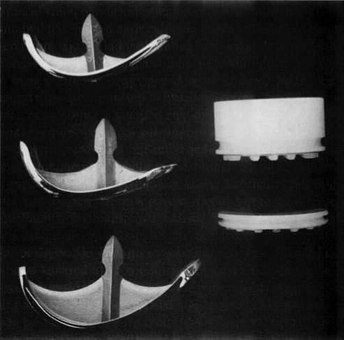
For the past two and one-half years I have used the Marmor modular prosthesis (Fig. 1) for total knee arthroplasty in patients with destruction of the articular surfaces of the knee joint and only mild instability. The present report describes eighty-nine of these patients, each of whom was followed for at least two years. The population base for this study was the first 100 consecutive patients on whom total knee-replacement arthroplasty was performed with the modular prosthesis. Eleven patients who had only eighteen months of follow-up were not included.
Fig. 1.
The femoral components (left) are available in three sizes. The choice of component is determined by template fit at the time of surgery. The tibial components (right) are available in thicknesses of from six to twenty-one millimeters, in increments of three millimeters.
Surgical Technique and Postoperative Care
The basic technique was described by Marmor. Certain technical points warrant emphasis. In nearly all of our patients we used a single, relatively straight medial parapatellar incision carried directly down to the joint without developing skin flaps. Proximally, the incision separated the interval between the rectus femoris and the vastus medialis muscles. Distally, 20 per cent of the insertion of the patellar tendon into the tibial tubercle was elevated sharply. This permitted easy lateral displacement of the patella and wide exposure of the knee joint. With the knee hyperflexed, a wedge of the popliteal surface of each femoral condyle five millimeters thick can be removed to give wide exposure to the entire posterior aspect of the tibial plateaus. The Hall II Pneumatic Instrument, fitted with a contra-angle handpiece and a large, sharp cutting burr, facilitates the preparation of the tibial plateau. The anterior portion of the lateral femoral implant is countersunk into the femoral condyle to prevent possible patellar impingement during flexion. Anchoring holes three centimeters deep are made in the proximal surface of the tibia and are directed downward and anteriorly. After all prosthetic components have been implanted and the methylmethacrylate has set, the tourniquet is released and hemostasis is obtained. The patella should be replaced in the trochlear groove and free motion verified during passive flexion and extension of the knee. In patients with a preoperative valgus deformity, it often is necessary to release the lateral patellar retinaculum and imbricate the medial retinaculum to restore patellofemoral alignment. The capsule of the knee is closed with the knee in flexion, while the remaining layers are closed with the knee fully extended. Closed Hemovac drainage is maintained for forty-eight hours. At that point the postoperative Robert Jones dressing and the drains are removed, and a gentle range of motion is begun. The patients are allowed to bear weight on the third postoperative day and walk with either a walker or crutches as tolerated. Subsequently a cane is used for a minimum of four weeks. Quadriceps-setting isometric exercises are begun preoperatively and continued through the entire postoperative period. All patients are started on intravenous cephalosporin two hours prior to surgery, and this is maintained intravenously for forty-eight hours after surgery. In the initial portion of the study, no specific antithromboembolic medications were used. For the past year, however, all patients were started on aspirin, 600 milligrams orally twice a day, beginning the day before surgery and continuing until the day of discharge from the hospital.
Modular components fabricated by the Richards Manufacturing Company were implanted in the first thirty patients. All subsequent procedures were performed using the Zimmer compartmental type of modular knee prosthesis.
Clinical Material
During the study period, eleven patients underwent unicompartmental tibiofemoral replacement for “apparently” localized degenerative arthritis involving either the medial or the lateral compartment. Since the biomechanical implications and, indeed, the rationale for unicompartmental replacement differ from those for bicompartmental tibiofemoral replacement, these eleven patients were not included in the series. We also eliminated from the series two osteoarthritic and five rheumatoid patients who underwent bilateral modular knee replacement, because of the difficulty of comparing functional results in patients with bilateral disease with results in those who have undergone unilateral total knee replacement. At times, functional results are difficult to assess in the presence of disabling problems in other weight-bearing joints of the same or the contralateral extremity. This problem existed to some extent in the osteoarthritic patients, but was more manifest in the rheumatoid patients. In the latter group, functional ability was correlated as well as possible with the involved knee only. Relief of pain was relatively simple to correlate with the affected joint; however, the need for support and the ability to walk distances were greatly influenced by disease of adjacent joints.
The series under discussion comprises eighty-nine patients, fifty-eight with osteoarthritis and thirty-one with rheumatoid arthritis. All rheumatoid patients were seropositive, and in those with a proliferative invasive synovium an anterior synovectomy was performed at the time of joint replacement. All patients who had been taking systemic corticosteroids at any time during the twelve months prior to surgery were begun on steroid medication preoperatively and maintained on it during the operation and for seventy-two hours thereafter. Subsequent steroid requirements as well as general medical management of the patients were variable and no standard regimen was followed.
There were thirty-one male and twenty-seven female osteoarthritic patients, 79 per cent of whom were over the age of sixty. Only two of the thirty-one rheumatoid patients were men, and the greatest number of patients (seventeen) were between fifty and sixty years old.
In this series, the major purpose of total knee replacement was to relieve pain. Eighty-six per cent of the osteoarthritic patients and 78 per cent of the rheumatoid patients had pain which was rated as moderate, severe, or disabling. In each group there was one patient who did not complain of any pain but who was essentially bedridden, with severe limitation of joint motion. The purpose of surgery in these two patients was to regain motion for adequate nursing care and chair-to-bed transfer. There were seven rheumatoid patients (23 per cent) whose major complaint was recurrent swelling associated with pain and limitation of motion. At surgery, large areas of cartilage erosion were visualized, and despite the presence of minimum roentgenographic changes I thought that synovectomy alone would he inadequate. Therefore. bicompartmental replacement was also performed in these knees.
The patients were evaluated and rated preoperatively and at three, six, twelve, eighteen, and twenty-four months postoperatively. It was noted early in the study that the great majority of patients achieved a plateau in terms of functional and numerical rating by the third postoperative month, and that further improvement beyond this point was minimum. No deterioration of results was encountered during follow-up.
All patients were operated on by myself and my two associates (Dr. M. A. Gruber and Dr. A. J. Zimmerman) or by senior resident orthopaedic surgeons under our direction. I personally performed all evaluations of the patients. Questions regarding pain and function were asked in a standardized manner. The evaluation of gems varum or valgum was made on the basis of weight-bearing anteroposterior roentgenograms made with the knee in maximum extension. The degree of flexion contracture or recurvatum, when present, was measured on lateral roentgenograms with the patient recumbent and the leg suspended by the ankle so that maximum knee extension was achieved.
A rating system similar to that described by Ranawat and Shine was used. Out of a total of 100 points, thirty were allocated to pain, thirty-five to function, and ten to muscle strength. Static passive range of motion (five points), flexion deformity (ten points), instability (ten points), and extensor lag (up to five negative points) were less important in the rating, although these parameters obviously influenced both the patient’s function and the initial choice as to whether a resurfacing implant would be the best prosthesis for the reconstruction.
After evaluation, we did not accept for operation any patient in whom varus or valgus instability exceeded 20 degrees, or in whom the sum of the degrees of instability and flexion contracture or recurvatum exceeded 40 degrees. Recurvatum of over 15 degrees was thought to indicate absence of the posterior cruciate ligament and was a contraindication for resurfacing arthroplasty. A history of prior intro-articular infection also was an absolute contraindication.
Results
Postoperatively 94 per cent of the patients (fifty-four of fifty-eight in the osteoarthritic group and twenty-nine of thirty-one in the rheumatoid arthritis group) had no pain or only slight discomfort with inclement weather (Table 1). In each group over half of the patients were pain-free at all times. Eighty-five per cent of the osteoarthritic patients and 67 per cent of the rheumatoid patients had no swelling or only slight swelling when evaluated twenty-four months postoperatively.
Table 1.
| Osteoarthritic patients | Rheumatoid arthritis patients | |||
|---|---|---|---|---|
| Preop. | 24 Mos. Postop. | Preop. | 24 Mos. Postop | |
| Pain | ||||
| None | 1 | 34 | 1 | 18 |
| Sight | 7 | 20 | 6 | 11 |
| Moderate | 43 | 2 | 13 | 1 |
| Severe | 7 | 2 | 11 | 1 |
| Range of motion | ||||
| Over 100 degrees | 3 | 6 | 1 | 2 |
| 80 to 100 degrees | 16 | 21 | 5 | 12 |
| 60 to 80 degrees | 32 | 27 | 15 | 15 |
| Less than 60 degrees | 7 | 4 | 10 | 2 |
| Limp | ||||
| None | 3 | 20 | 1 | 13 |
| Mild | 32 | 32 | 8 | 8 |
| Moderate | 15 | 5 | 11 | 7 |
| Marked | 8 | 1 | 11 | 3 |
| Support needed | ||||
| None | 20 | 34 | 3 | 8 |
| Cane | 32 | 21 | 17 | 16 |
| Walker or crutches | 5 | 2 | 8 | 5 |
| Non-ambulatory | 1 | 1 | 3 | 2 |
| Walking distance | ||||
| Unlimited | 0 | 22 | 0 | 3 |
| Over 5 blocks | 2 | 34 | 5 | 14 |
| 1 to 5 blocks | 18 | 9 | 5 | 9 |
| Less than 1 block | 38 | 3 | 21 | 5 |
| Extensor lag | ||||
| None | 18 | 44 | 4 | 24 |
| Up to 10 degrees | 32 | 13 | 20 | 6 |
| Over than 1 block | 8 | 1 | 7 | 1 |
| Genu valgum* | ||||
| Less than 5 degrees | 8 | 22 | 3 | 16 |
| 5 to 10 degrees | 14 | 9 | 14 | 5 |
| 10 to 20 degrees | 10 | 1 | 5 | 1 |
| Genu varum* | ||||
| Less than 5 degrees | 9 | 21 | 1 | 8 |
| 5 to 10 degrees | 2 | 5 | 4 | 1 |
| 10 to 20 degrees | 15 | 0 | 4 | 0 |
| Flexion contracture* | ||||
| None | 9 | 14 | 2 | 6 |
| Less than 15 degrees | 25 | 42 | 14 | 21 |
| Over 15 degrees | 24 | 2 | 15 | 4 |
* As measured on roentgenograms.
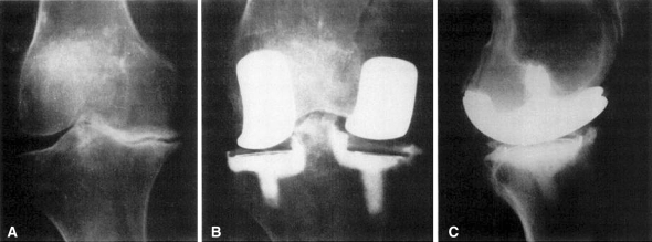
Limitation of motion was not a major preoperative indication for surgery in either group. Preoperatively 88 per cent of the osteoarthritic patients had over 60 degrees of knee motion, and 33 per cent had over 80 degrees. In the rheumatoid group 73 per cent had greater than 60 degrees of motion and 19 per cent, over 80 degrees. When evaluated postoperatively, the total range did not change significantly in either group. In many of the rheumatoid patients, however, the arc of motion shifted so that extension was gained and flexion was lost. By trimming the anterior part of the tibia it was possible to obtain full extension in almost all patients. Postoperative flexion beyond 80 degrees was obtained in 46 per cent of the osteoarthritic patients, and 10 per cent could flex the knee beyond 100 degrees. In the rheumatoid patients, 45 per cent of the patients flexed the knee over 80 degrees and 6 per cent could flex beyond 100 degrees (Fig. 2A–C).
Fig. 2A–C.
A seventy-two-year-old woman with marked osteoarthritic changes of the right knee. The preoperative point rating was thirty-six: the patient had disabling pain and could walk only half a block. When evaluated eighteen months after surgery, she had a rating of eighty-seven. She had no pain and could walk an unlimited distance. (A) Preoperative standing roentgenogram, revealing a 15-degree varus deformity and loss of joint height medially. (B) Postoperative anteroposterior roentgenogram. (C) Postoperative lateral roentgenogram.
Midway through the study we began to perform capsular closures with the knee flexed. Patients treated with this method were found to regain active flexion postopera-tively more rapidly than those in whom the capsule was closed in extension. This was most marked if the patients were evaluated two weeks postoperatively, but the results tended to equalize by about the second postoperative month. Beyond the third month there was little further increase in flexion.
We noted that despite an adequate range of both passive and active motion postoperatively, most of our patients continued to walk with limited knee flexion during the stance phase. The reason for this oft-reported abnormality [7] has not been determined.
Walking ability was increased in both groups, mainly because of relief of pain. Many activities of daily living for our patients required the ability to walk for up to five blocks. Preoperatively, only 4 per cent of the osteoarthritic and 16 per cent of the rheumatoid patients could walk that far. When evaluated twenty-four months after surgery, 79 per cent of the osteoarthritic patients and 55 per cent of the rheumatoid patients could walk five blocks. Preoperatively, most patients could ascend stairs only one at a time, at best. Of the thirty-five patients who postoperatively achieved the theoretical 95 degrees of motion required to ascend stairs foot over foot, only ten could actually walk up stairs in this manner; the other twenty-five, all of whom were completely pain-free and whose knees were stable to medial and lateral stress, climbed stairs one step at a time. Almost all of them had less than 2.5 centimeters of quadriceps atrophy as compared with the opposite side. When they were questioned directly regarding their inability to ascend stairs, the commonest explanation given was fear of falling.
The mild varus or valgus preoperative instability in some of our patients usually was attributed to the asymmetrical loss of the spacer effect of normal articular carti-lage, rather than to lesions of the collateral ligaments. By restoring the “height of the cartilage” we tried to restore stability without the need for ligament procedures [3]. At the time of surgery, the height of the plateau was adjusted so as to achieve the minimum possible laxity on varus or valgus stress. For a proper test the knee had to be fully extended, the patella replaced in the trochlear notch, and the medial retinaculum temporarily approximated by means of towel clips. If this was not done, the tendency always was to insert a tibial component that was too thick, and flexion and extension of the knee would cause a rocking motion rather than normal gliding of femoral and tibial surfaces. Although the tibial plateaus of the modular prosthesis are available only in thicknesses differing by three millimeters, smaller differences in height could be compensated for by adjusting the depths of the trough in the tibial plateau. Postoperatively, when roentgenographic measurements of standing tibial and femoral alignment were made, over 70 per cent of the patients had less than 5 degrees of genu varum or genu valgum remaining. All knees were clinically stable to varus or valgus stresses postoperatively, and the patients walked without medial or lateral thrust. Although it has been emphasized that an attempt should always be made to obtain a slight valgus position, we sought to achieve functional stability rather than a preconceived ideal alignment. In three knees residual varus deformity remained, which was considered to be of cosmetic rather than functional importance. It mainly was due to varus deformity of the tibial shaft, and even with restoration of alignment between the femur and the tibial metaphysis the patient still had a bowed leg. This result was not considered a complication or failure as regards the use of the modular knee prosthesis. Plateau units of standard width were used routinely, except in the four rheumatoid patients of short stature in whom the small-diameter implants were needed. The choice of diameter of the femoral implant was made at the time of surgery on the basis of the fit of the template on the femoral condyle. By deepening the channel in the femoral condyle either anteriorly or posteriorly, an adequate fit between the template and the femoral condyle could be achieved. The anterior leading edge of the lateral condylar implant was routinely embedded in the femur to prevent possible impingement by the patella.
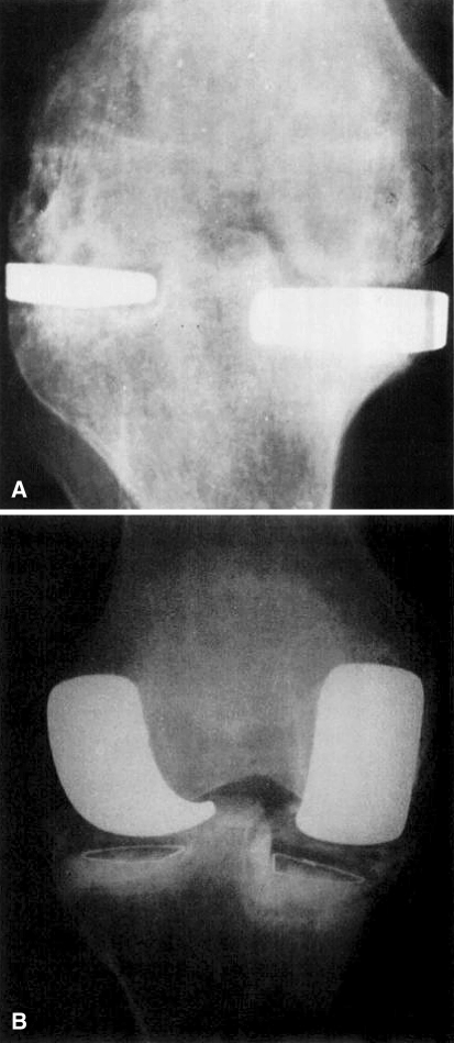
Six rheumatoid patients had undergone prior synovectomy of the knee and one had had a previous MacIntosh arthroplasty (Fig. 3A and B). Eight osteoarthritic patients had had Magnuson joint debridement. There was no significant difference in the eventual outcome after surgery in these fifteen patients as compared with the other seventy-four in the series.
Fig. 3A–B.
A fifty-two-year-old woman with rheumatoid arthritis who had undergone prior tibial-plateau replacement with a MacIntosh prosthesis. Pain recurred one year after the initial surgery and was disabling; the patient could walk only a few steps. She had a 20-degree flexion contracture and no angular deformity. When evaluated eighteen months after operation, she could walk ten blocks and had some discomfort related to inclement weather. (A) Preoperative anteroposterior roentgenogram. (B) Postoperative roentgenogram, after insertion of modular total knee replacement.
The redevelopment of quadriceps strength and range of motion postoperatively demanded a considerable degree of cooperation and motivation on the part of the patient. For this reason, a psychological assessment of each candidate was made in consultation with the patient’s rheumatologist or internist prior to operation, and any patient who was poorly motivated, not fully cooperative, or over-reactive was not chosen for surgery.
Twenty-four months postoperatively a roentgenographic evaluation was done to determine wear and settling of the prosthesis [1, 4, 10]. We measured changes in the distance between the lowest portion of the femoral condylar implant and the marking wires on the tibial components; the changes were evaluated, correcting for magnification, with the width of the femoral condylar implant as a guide. However, minor variations in position of the knee in maximum extension produced totally unreliable measurements, and this method of evaluating wear had to be abandoned.
Evaluation of settling was done by measuring the distance between the marking wire in each tibial component and the furthest superior bone portion of the tibial plateau after correction for magnification, again using the width of the femoral implant as a reference. Slight changes in knee flexion during roentgenography did not significantly affect the measurements.
Settling of over one millimeter occurred in only 5 per cent of the osteoarthritic patients and 8 per cent of the rheumatoid patients. In only two patients was there settling of over 1.5 millimeters: one was the only patient with deep infection, and the other had a non-infected knee, to be described.
Extensor lag is defined as the inability to extend the knee actively to the limits of its passive extensions [8]. Preoperatively, 69 per cent of the osteoarthritic patients and 87 per cent of the rheumatoid patients had extensor lag. The usual range was up to 10 degrees, although a few patients in each group had greater lag. Postoperatively, 76 per cent of the osteoarthritic and 77 per cent of the rheumatoid patients did not have any extensor lag at all. In the modular arthroplasty, the patella and patellar tendon are not displaced anteriorly, and therefore the diminution in extensor lag in these patients was probably due to the restoration of strength to the quadriceps muscle. It would be of interest to see if extensor lag could be eliminated in even more patients if the modular arthroplasty included anterior displacement of the patella. Convery and Beber, in evaluating their cases of Geomedic total knee replacement, noted that there was extensor lag in almost all of their patients three months postoperatively. They felt that stretching of the patellar ligament from a long-standing flexion deformity was its major cause, and discussed tibial tubercleplasty [6] or patellar-tendon advancement as possible solutions. Bearing this in mind, we compared preoperative flexion deformities with residual extensor lag, and with the exception of two patients who had preoperative flexion contractures of 30 and 35 degrees, we found no correlation. The two patients cited each had an extensor lag of approximately 10 degrees postoperatively, which persisted even with intensive physiotherapy.
Despite the fact that at surgery all peripheral patellar osteophytes were removed, and the patella was recentered in the trochlear notch, no attempt was made at resurfacing the articulating surfaces of the patellofemoral compartment. Therefore, many patients postoperatively had crepitus on retropatellar pressure, but no patient complained of more than mild discomfort behind the patella with walking or stair-climbing. Although patellofemoral pain did not appear to be a significant problem in our patients after bicompartmental replacement, this may be a reflection of our patient selection. No patient was included in our series in whom the predominant site of pain was retropatellar, particularly related to stair-climbing. When this pattern prevails, even if the knee is fairly stable, we prefer to use the total condylar knee prosthesis, in order to replace the patellofemoral articulation as well as the femorotibial articulation.
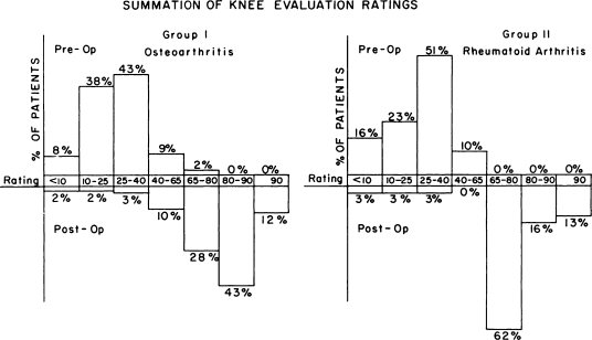
The evaluation ratings of the osteoarthritic patients preoperatively showed a mean of thirty points. Only 11 per cent had over forty points (Chart 1). After follow-up the mean point rating was seventy-nine and only 10 per cent scored less than fifty. In the rheumatoid patients the preoperative mean was twenty-eight, with 10 per cent scoring over forty. Twenty-four months later, the mean was seventy-five and only 9 per cent scored less than sixty-five. Of the total of eighty-nine patients, thirty had final ratings in the eighties and eleven had ratings in the nineties.
Chart 1.
In interpreting these data, it should be borne in mind that the patients selected for modular arthroplasty were carefully chosen so as to exclude those with marked patellofemoral abnormalities or significant instability.
Complications
No patient had clinical signs of thrombophlebitis [5] or of pulmonary embolus, although some patients were maintained on salicylates and others were not. No patient who received salicylates in our series had gastrointestinal bleeding. There were no cases of fat embolism. One patient had a deep infection (Staphylococcus aureus) which required debridement and closed suction-irrigation. This patient had rheumatoid arthritis and had been taking steroids for fourteen years prior to surgery. At final follow-up she had no drainage, although she continued to have disabling pain with walking. Roentgenograms revealed three millimeters of settling. Three other patients had superficial infections: culture of fluid from one revealed Pseudomonas aeruginosa: from one, Staphylococcus epidermidis: and one culture was sterile. The drainage in these patients ceased spontaneously and did not recur. All three of these patients also had rheumatoid arthritis and had been taking steroid medication for a long time. In four other rheumatoid patients, erythema developed around the operative wound during the first postoperative week; these lesions subsided without drainage. None of these patients was febrile, nor did they manifest a leukocytosis. One rheumatoid patient had slight skin necrosis which healed spontaneously. There was one case of capsule rupture that occurred approximately two months after surgery. This patient was osteoarthritic and had previously undergone two unsuccessful tibial osteotomies for genu valgum. Two months after the arthroplasty, she had massive swelling of the knee and an obvious rupture of the sutured capsule. When the knee was re-explored, an exuberant amount of synovial hypertrophy was found covering the articular surfaces and the implants. She underwent synovectomy and reapproximation of the capsule. Microscopic examination of the synovium revealed a nonspecific synovitis without lymphoid nodules. There was no evidence of foreign-body reaction to the cement or of particulate matter in the tissue. The synovial fluid and the synovium were cultured for aerobic, anaerobic, fungal, and mycobacterial organisms, and all cultures were sterile. The exact cause of the complications in this patient could not be determined. She died of renal disease before the eighteen-month evaluation, and therefore is not included in Table 1. Had she survived, she probably would have had a poor result, considering both function and relief of pain.
No cases of frank loosening of the prosthesis occurred in this series. One of our patients with a unicompartmental replacement did have complete loosening of the tibial unit at the cement-bone interface.
Marked dysesthesia developed on the sole of the foot in one osteoarthritic patient immediately postoperatively. This decreased somewhat but did not completely disappear. Despite the fact that this patient had no pain in the knee and had an arc of motion of 110 degrees, the result was classified as poor because of the disabling foot pain. There were no other neurological problems in the series.
Although the modular arthroplasty is completely unconstrained (there are no tracks in the polyethylene of the tibial components), and although some latitude in orientation of the implants is permissible, stresses applied to the implant by improper placement may lead to early failure. One patient in the series had a valgus deformity of the knee preoperatively, associated with a moderate flexion contracture. In this patient the operative exposure was extremely difficult, despite the use of a second incision. Postoperative roentgenograms revealed that both tibial implants were severely angulated in the coronal plane. Although there was initial relief of pain, there was recurrence of pain and limitation of motion in the knee by the sixth postoperative week, and the prosthesis showed settling of up to three centimeters. The patient had no systemic or local signs of infection, but by the third month she had become so disabled that revision of the arthroplasty was required. We did a bone resection and implanted a total condylar knee prosthesis, and after nine months she was completely free of pain.
Conclusion
In this series of patients, the surgery achieved a striking postoperative relief of pain in both the osteoarthritic and the rheumatoid patients. This improvement was well maintained for two years in almost all patients. The problem of implant loosening, which was cited by some surgeons, did not occur, possibly because of the use of cement in anchoring holes. Additionally, the lack of a significant incidence of settling in our series may have been due to our limitation of use of the prosthetic implant to those patients in whom significant preoperative instability did not exist, and in whom, therefore, large asymmetrical postoperative stresses were avoided.
Footnotes
The Classic Article is © 1976 by the Journal of Bone and Joint Surgery, Inc. and is reprinted with permission from Laskin RS. Modular total knee-replacement arthroplasty. A review of eighty-nine patients. J Bone Joint Surg Am. 1976;58:766–773.
Richard A. Brand MD ✉ Clinical Orthopaedics and Related Research, 1600 Spruce Street, Philadelphia, PA 19103, USA e-mail: dick.brand@clinorthop.org
References
- 1.Charnley, John: The Long-Term Results of Low-Friction Arthroplasty of the Hip Performed as a Primary Intervention. J. Bone and Joint. Surg., 54-B: 61–76, Feb. 1972. [PubMed]
- 2.Convery, F. R., and Beber, C. A.: Total Knee Arthroplasty Indications, Evaluation and Postoperative Management. Clin. Orthop., 94: 42–49, 1973. [PubMed]
- 3.Coventry, M. J.; Upshaw, J. E.; Riley, L. H.; Finerman, G. A. M.; and Turner, R. H.: Geometric Total Knee Arthroplasty. I: Conception, Design, Indications, and Surgical Technic. Clin. Orthop., 94: 171–176, 1973. [PubMed]
- 4.Ewald, F. C.: Metal to Plastic Total Knee Replacement. Orthop. Clin. North America, 6: 811–821, 1975. [PubMed]
- 5.Harris, W. R.; Salzman, E. W.; Athanasoulis, Christos; Waltman, A. C.; Baum, Stanley; and Desanctis, R. W.: Comparison of Warfarin, Low-Molecular-Weight Dextran, Aspirin, and Subcutaneous Heparin in Prevention of Venous Thromboembolism following Total Hip Replacement. J. Bone and Joint Surg., 56-A: 1552–1562, Dec. 1974. [PubMed]
- 6.Kaufer, Herbert: Mechanical Function of the Patella. J. Bone and Joint Surg., 53-A: 1551–1560, Dec. 1971. [PubMed]
- 7.Kettelkamp, D. B., and Nasca, Richard: Biomechanics and Knee Replacement Arthroplasty. Clin. Orthop., 94: 8–14, 1973. [DOI] [PubMed]
- 8.Lieb, F. J., and Perry, Jacquelin: Quadriceps Function. An Anatomical and Mechanical Study Using Amputated Limbs. J. Bone and Joint Surg., 50-A: 1535–1548, Dec. 1968. [PubMed]
- 9.Marmor, Leonard: The Modular Knee. Clin. Orthop., 94: 242–248, 1973. [DOI] [PubMed]
- 10.Potter, T. A.; Weinfeld, M. S.; and Thomas, W. H.: Arthroplasty of the Knee in Rheumatoid Arthritis and Osteoarthritis. A Follow-up Study after Implantation of the McKeever and MacIntosh Prostheses. J. Bone and Joint Surg., 54-A: 1–24, Jan. 1972. [PubMed]
- 11.Ranawat, C. S., and Shine, J. J.: Duo-Condylar Total Knee Arthroplasty. Clin. Orthop., 94: 185–195, 1973. [DOI] [PubMed]


