Abstract
Knee forces directly affect arthroplasty component survivorship, wear of articular bearing surfaces, and integrity of the bone-implant interface. It is not known which activities generate forces within a range that is physiologically desirable but not high enough to jeopardize the survivorship of the prosthetic components. We implanted three patients with an instrumented tibial prosthesis and measured knee forces and moments in vivo during exercise and recreational activities. As expected, stationary bicycling generated low tibial forces, whereas jogging and tennis generated high peak forces. On the other hand, the golf swing generated unexpectedly high forces, especially in the leading knee. Exercise on the elliptical trainer generated lower forces than jogging but not lower than treadmill walking. These novel data allow for a more scientific approach to recommending activities after TKA. In addition, these data can be used to develop clinically relevant structural and tribologic testing, which may result in activity-specific knee designs such as a knee design more tolerant of golfing by optimizing the conflicting needs of increased rotational laxity and conformity.
Introduction
Knee forces directly affect arthroplasty component survivorship, wear of articular bearing surfaces, and integrity of the bone-implant interface. Excessive knee forces have been implicated in the breakdown of the cement interface or in the collapse of underlying bone. Knee forces along with component design also determine the contact stresses on the bearing surfaces. Contact stresses have been correlated with the magnitude and distribution of wear.
Forces in the physiological range are essential to preserve the health of the bones and adjacent joints of the implanted knee. In addition, current health and lifestyle requirements include regular exercise and recreational activities to maintain body weight, muscle tone, and cardiovascular fitness. The lengthening life expectancy and higher overall fitness of the age group of patients undergoing knee arthroplasty continues to raise the bar on durability and survival of the prosthetic components.
It is not known which activities generate forces within a range that is physiological desirable but not high enough to jeopardize the survivorship of the prosthetic components. The conventional wisdom is low-impact activities (such as biking, walking, and golf) are relatively safer than high-impact activities (such as jogging and tennis). However, scientific justification of these recommendations requires quantitative measurements of knee forces in vivo during these activities.
Almost all studies relating to knee forces have involved either in vitro measurements [6, 12, 23] or estimates using mathematical models [15, 20, 22, 25]. We previously reported the first direct measurement of knee forces in vivo after TKA [3, 4]. The tibial component used in that study was instrumented with four load cells that measured the axial components of load on the four quadrants of the tibial tray. This instrumented implant measured the total axial force and the location of the center of pressure. However, shear and moments, which are also important components of knee forces, could not be measured in that design. In collaboration with Zimmer, Inc (Warsaw, IN), we developed a second-generation, force-sensing device that measured all components of tibial forces [13]. The stem of this design was instrumented with strain gauges that measured all six components of force. We earlier reported the first in vivo measurements of shear and moments in the knee after TKA [2] for four activities: walking, stair climbing, chair rise, and squat. Since then, we have implanted a total of three patients with the second-generation device (Fig. 1). For this study, we measured knee forces during recreational activities such as tennis, golf, and biking and during use of exercise equipment such as the treadmill, elliptical trainer, step treadmill, leg press, knee extension, and rowing machines.
Fig. 1.

Postoperative radiograph of one of the subjects displays the location of the strain gauges (S), microprocessor (M), internal induction coil (C), and transmitting antenna.
Many patients want to return to an active lifestyle after TKA. However, as a result of the lack of objective data, surgeons’ recommendations are based on subjective opinions and are not always consistent [10, 19]. The purpose of this study was to measure knee forces during exercise and recreational activities. These results could prove important in defining scientific guidelines for advising patients regarding the safety and benefits of various postarthroplasty activities.
Materials and Methods
A custom tibial component was manufactured by Zimmer, Inc, based on the Natural Knee® II (NK-II) tibial tray design. The tray and locking mechanism were identical to the standard design for implantation with a standard insert. The stem was instrumented with strain gauges to measure three orthogonal forces and three moments. The stem also housed a microtransmitter, which performed analog-to-digital conversion, filtering, and multiplexing before transmitting data through a hermetic glass, feed-through tantalum antenna. External coil induction was used to power the implant. Details of the implant design, strain gauges, microtransmitter, telemetry system, and accuracy have been previously reported [13].
The direction of forces and moments were computed in the coordinate system of the tibial tray, which was implanted at 90° to the intramedullary axis of the tibia in the coronal and the sagittal planes. For example, the shear generated at the tibial tray by the femoral component moving in the anterior direction was denoted as anterior shear. Similarly, the moment generated at the tibial tray by adduction of the knee was termed adduction moment.
Three subjects, two men and one woman, received the implants. The men were aged 83 and 81 years with body weights of 75 kg and 70 kg, respectively. The woman was 67 years old (body weight, 89 kg). Appropriate Institutional Review Board approval and patient consents were obtained. A standard polyethylene insert (NK-II CR Congruent; Zimmer, Inc) and a posterior cruciate-retaining femoral component (NK-II CR; Zimmer, Inc) were implanted using a standard anteromedial approach. The tibial bone cut was made at 90° to the long axis in the coronal plane (0° varus) and at 90° in the sagittal plane (0° posterior slope). The distal femoral cut was made at 6° valgus to the anatomic axis of the femur. The posterior femoral cut was made at 3° external rotation with reference to the posterior surface of the posterior condyles. Intramedullary alignment was used for femoral and tibial bone preparation. The patella was resurfaced with a standard dome-shaped, all-polyethylene component. All components were cemented. All patients underwent routine postoperative rehabilitation as per a standard primary TKA.
We earlier reported in vivo measurements of shear and moments in the knee in our first subject 3 months after TKA [2] (for walking, stair climbing, chair rise, and squat). Since then, we have implanted a total of three patients with the second-generation device (Fig. 1). For this study, tibial forces and moments were measured 1 year postoperatively for the following activities: walking on a treadmill (Star Trac TR 4500; Star Trac, Irvine, CA) at speeds ranging from 1 to 4 miles/hour; jogging on a treadmill (5 miles/hour) as well as on the laboratory floor (subject-selected speed); using a rowing machine (Indoor Rower; Concept II, Morrisville, VT), a stair-climbing machine (StairMaster® 4000 PT; Nautilus Inc, Vancouver, WA), an elliptical trainer (9500 HR; Life Fitness, Schiller Park, IL), a leg-press machine (MD-122 Super Leg Press; Body Masters, Rayne, LA), and knee extension against resistance (S-109 Super Leg Extension; Body Masters). The resistance on the leg-press machine was altered to generate a net reaction force of 50%, 40%, or 20% of body weight under the foot on the instrumented side. The resistance on the knee extension machine was set to 9.1 kg (20 lbs) or 18.2 kg (40 lbs).
Stationary bicycling (Lifecycle® HR; Lifecycle Inc, Franklin Park, IL) was analyzed at various levels of difficulty and speeds ranging from 60 to 90 revolutions per minute (rpm). During this activity, the subjects were instructed to maintain the desired pedaling rate using a digital readout for feedback. For analysis, six cycles on the instrumented side were selected for each level and speed; the biking speed (rpm) was confirmed by measuring the cycle peak-to-peak duration.
Golfing was analyzed in the laboratory (golf swing with a club but no golf ball), at the TaylorMade Performance Lab, Carlsbad, CA (golf swing with a club and a golf ball against a net), as well as on a driving range (Torrey Pines Golf Course, La Jolla, CA). All three subjects were right-handed golfers; two subjects had the left (leading) knee instrumented, whereas the third had the right (trailing) knee instrumented. In addition, one of the subjects (with a left-sided implant) could also drive left-handed. Therefore, knee forces were monitored for the leading knee in two subjects and for the trailing knee in two subjects. Tennis was analyzed in the laboratory (simulated tennis strokes without a ball) as well on the tennis courts at the La Jolla Beach and Tennis Club during actual play. Under both conditions, knee forces generated during the serve, forehand, and backhand strokes were analyzed.
Results
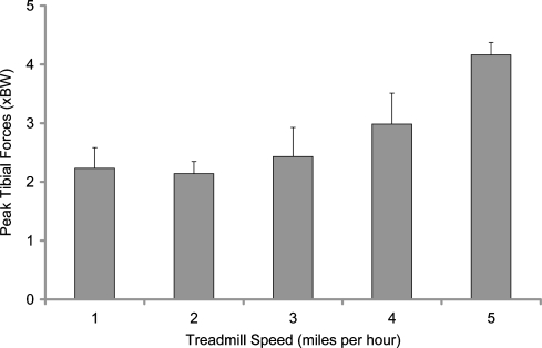
In walking, there was some variation among subjects, but peak forces remained in the range of 1.8 to 2.5 times body weight for treadmill walking at the speeds tested (Fig. 2). Treadmill speed during comfortable walking (range, 1–3 miles per hour) had no effect on peak tibial forces. In general, knee forces during treadmill walking (2.05 ± 0.20 times body weight) were lower than those measured during level walking on a laboratory floor (2.6 times body weight). A speed comparable to power walking (4 miles per hour) generated forces on the treadmill (2.80 ± 0.43 times body weight) similar to the forces generated during walking on the laboratory floor.
Fig. 2.
Peak forces were in the range of 1.8 to 2.5 times body weight (BW) for treadmill walking at the speeds tested. Treadmill speed during comfortable walking (range, 1–3 miles per hour) had no effect on peak tibial forces. A speed comparable to power walking (4 miles per hour) generated higher forces on the treadmill. Peak forces recorded during jogging were even higher than those recorded during power walking.
During jogging, peak forces recorded were even higher than those recorded during power walking (Fig. 2). Differences between peak forces generated on the treadmill (at 5 miles per hour) and those generated during jogging on the laboratory floor (subject-selected speed) were minimal. Peak ground reaction forces increased by a mean of 40% relative to walking on the laboratory floor.
During stationary bicycling, the seat height was adjusted to achieve a knee flexion angle of 90° with the pedal at the highest position. Actual measured peak flexion during stationary bicycling was 94.3°± 1.2°. Overall, tibial forces peaked at 1.03 ± 0.20 times body weight. Increasing the speed of bicycling from 60 to 90 rpm did not affect peak tibial forces but did affect the flexion angle at which tibia force peaked (Fig. 3). At 60 rpm, tibial forces peaked earlier at 74°± 4°; at 90 rpm, tibial forces peaked later at 37°± 2°. In general, anterior shear was low (overall average, 0.21 ± 0.01 times body weight).
Fig. 3.
Increasing the speed of bicycling from 60 to 90 rpm or increasing the resistance up to Level 3 did not substantially affect peak tibial forces. BW = body weight.
High tibial forces were generated during the golf swing (Fig. 4A–B). Much higher forces were generated in the leading knee relative to the trailing knee. Forces generated during simulated swinging in the laboratory (without a ball) were similar to those generated on the driving range. Anterior tibial shear (0.34 ± 0.01 times body weight) and axial tibial torque (13.0 ± 0.33 N-m) were in the moderate range.
Fig 4A–B.
High tibial forces were generated during the golf swing. (A) Much higher forces were generated in the leading knee relative to the trailing knee. (B) A golf swing with a driver tended to generate higher forces than a sand wedge. BW = body weight.
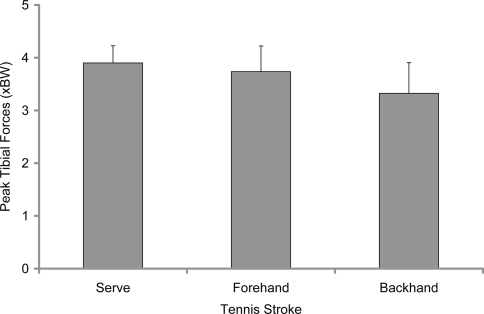
Mean peak forces generated in tennis during the serve and during a forehand return were higher than those generated during the backhand return (Fig. 5). Anterior shear was moderate (0.28 ± 0.12 times body weight). Forces measured during actual play on the tennis courts were on average 12% higher than those generated during simulated strokes in the laboratory.
Fig. 5.
Mean peak forces generated during the serve and during a forehand return were higher than those generated during the backhand return. BW = body weight.
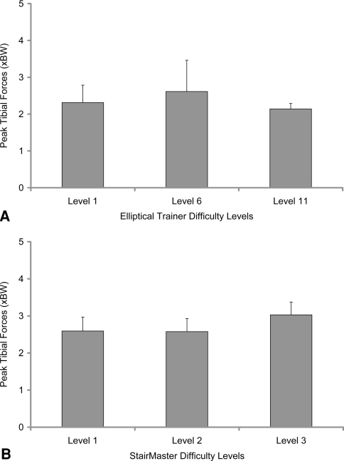
Rowing generated a peak mean tibial force of 0.85 ± 0.08 times body weight at a maximum flexion angle 90° ± 1°. Exercising on the elliptical trainer generated mean peak tibial force of 2.24 ± 0.22 times body weight, which remained unchanged with increasing levels of difficulty (Fig. 6A). Anterior tibial shear forces were very low (0.15 ± 0.06 times body weight). Exercising on the StairMaster generated similar forces at low levels of intensity but increased to over 3 times body weight at higher levels (Fig. 6B). The knee flexion angle at peak tibial forces also increased with increasing intensity.
Fig. 6A–B.
(A) Exercising on the elliptical trainer generated mean peak tibial forces, which remained largely unchanged with increasing levels of difficulty. (B) Exercising on the StairMaster generated similar forces at low levels of intensity but increased to over 3 times body weight (BW) at higher levels.
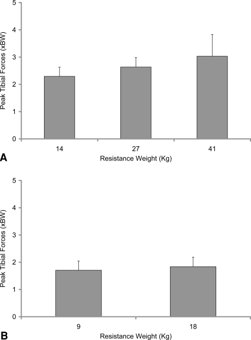
Tibial forces increased with increasing resistance during the leg-press activity but did not change during the knee extension activity (Fig. 7A). In the leg-press activity, the tibial forces peaked at decreasing knee flexion angle with increasing resistance. Conversely, in the knee extension activity (Fig. 7B), the tibial forces peaked at increasing knee flexion angle with increasing resistance. Both the leg-press and the knee-extension activities generated similar magnitudes of peak anteroposterior shear (0.24–0.35 times body weight). However, peak shear was directed anteriorly during the leg-press activity and directed posteriorly during the knee-extension activity.
Fig. 7A–B.
(A) Peak tibial forces increased with increasing resistance during the leg-press activity but (B) did not change during the knee-extension activity. BW = body weight.
Discussion
Improvements in surgical technique, biomaterials, and implant design, together with the increase in life expectancy and the present focus on physical fitness, have made it desirable for patients to return to relatively active lifestyles after TKA. Because polyethylene wear has been linked to increased activity, the general tendency is for surgeons to be conservative in their recommendations for post-TKA exercise, sports, and recreational activity. Of even greater concern are activities that generate high impact because of the potential for irreversible damage to the bearing material. However, the results of clinical outcomes are mixed, with some studies reporting increased component failure in more active patients, whereas others report no difference [5, 11, 14, 17–19]. We therefore directly measured tibial forces in vivo during exercise and various recreational activities.
In this study, data were collected in only three patients and this cohort is too small to cover the entire spectrum of patients undergoing knee arthroplasty. These results may apply only to one type of knee arthroplasty design, moderately conforming cruciate-retaining prostheses. Generalization of these force values to all knee arthroplasty designs is therefore not recommended. Finally, the level of intensity of these activities (particularly the recreational activities) was patient-selected and force magnitudes are likely to change with varying levels of intensity and skill.
Treadmill walking at all speeds up to 3 miles per hour generated lower peak tibial forces relative to walking on the laboratory floor. Because the treadmill was not instrumented with a force plate, we could not directly determine whether this was the result of the better shock absorption provided by the treadmill surface or the result of decreased muscle activity during pushoff. Power walking (at 4 miles per hour) generated higher peak tibial forces, but remained in the range of those generated while walking on the laboratory floor. These results indicate treadmill walking is a relatively safe activity (even safer than regular walking) and power walking on a treadmill can be recommended to improve cardiovascular function without substantial increase in knee forces.
Jogging is generally considered a high-impact force with various estimates of knee forces ranging from 7 times body weight [8] up to 22 times body weight [9]. In two subjects implanted with instrumented distal femoral tumor replacement prosthesis, jogging generated peak forces of 3.6 times body weight [24]. Our subjects were implanted with a primary knee arthroplasty design and generated higher forces (consistently greater than 4 times body weight), most likely because the musculature around the knee was intact (relative to the more extensive tissue resection of a tumor replacement surgery). Increase in knee forces can be the result of increased ground reaction force as well as increased extensor muscle activity. The knee is flexed more during the stance phase of jogging (15°–45°) than during walking (5°–25°) resulting in a fivefold increase in the flexion moment about the knee [1]. Because we recorded minimal difference between treadmill jogging and jogging on the laboratory floor, the increased knee moment appears the dominant contribution. This is also supported by the modest increase in peak ground reaction force (40%) relative to the 100% increase in peak knee forces when jogging relative to walking on the laboratory floor.
Stationary bicycling is frequently recommended for patients recovering from knee injury and surgery, because it is considered a low-impact activity that exercises the knee through a large range of flexion. Overall, stationary bicycling generated even lower knee forces than walking. Knee joint moments increase with pedaling rate when the power is kept constant [21]. However, peak knee forces remained largely unchanged between pedaling rates of 60 and 90 rpm. Knee forces at low speeds (60 rpm) peaked earlier at high flexion angles (approximately 75°), whereas forces at high speeds (90 rpm) peaked later at lower flexion angles (approximately 35°), perhaps indicating a compensatory mechanism that prevented knee moments from increasing substantially. Increase in the level of difficulty increased peak knee forces but by a modest amount. The measured peak tibial forces (1.03 times body weight) were slightly lower than those estimated with dynamic modeling (1.2 times body weight), but measured anterior shear (0.21 times body weight) was higher than estimated (0.05 times body weight) [7].
Similar to jogging, tennis is also considered a high-impact activity. As expected, playing tennis also generated high knee forces (in the same range as jogging). The tennis serve and forehand stroke generated greater forces than the backhand stroke. Surgeons are reluctant to permit patients to play tennis after TKA [19]. The Knee Society consensus recommendations permit doubles tennis but do not allow singles tennis [10]. However, the revision rates at intermediate-term followup in a cohort of patients who played tennis at a fairly high level (mean National Tennis Player Rating greater than 4) were not higher those reported for less active patients [19]. Our results rank tennis in the same category as jogging in terms of peak knee force magnitude.
Surprisingly, the golf swing generated high peak tibial forces with magnitudes in the leading knee approaching those generated during jogging. These forces were high during a simulated golf swing without a ball as well as striking a ball on a driving range. Patients undergoing TKA experience mild knee pain during or after golfing [16]. This pain is more common in the leading knee and has been attributed to increased torque. Our results indicate increased loading in the leading knee during golf swing may also be a contributing factor.
Elliptical trainers are frequently recommended as a low-impact exercise machine for patients who experience knee pain during higher-impact jogging. Peak tibial forces were similar to those measured during treadmill walking and anterior shear forces were low. Using the elliptical trainer may be recommended as an alternative to jogging, but in the context of reducing knee forces, elliptical training is no better than treadmill walking and does not appear as safe as stationary bicycling.
Rowing machines are often recommended to increase knee flexion. However, surgeons are cautious about recommending rowing after TKA [10]. Rowing generated the lowest peak knee forces of all activities reported here, less than stationary bicycling, active knee extension, or treadmill walking. The leg-press machine exercises similar muscles as the squat. One advantage of the leg-press machine is that the resistance can be reduced to below body weight. The knee extension exercise against resistance is a safe method of strengthening the quadriceps muscle without generating high tibial forces. Both the leg-press (against a resistance force equal to body weight) and the squat generated the same peak tibial forces. The leg-press machine can therefore be recommended to build up to a squat if a full weightbearing squat is not possible.
As expected, stationary bicycling generated low tibial forces, whereas jogging and tennis generated high peak forces. On the other hand, the golf swing generated unexpectedly high forces, especially in the leading knee. Exercise on the elliptical trainer generated lower forces than jogging, but not lower than treadmill walking. These results allow for a more scientific approach to recommending activities after TKA. Tibial forces comprise only one of the factors that may contribute to the potential for prosthetic wear and damage. Other factors include the kinematics (which affect contact area) and the number of cycles of the activity. For example, the golf swing generated forces similar in magnitude to jogging. However, the number of cycles during which the knee is exposed to high forces during a golf game are fewer than the number of cycles during jogging. Further analysis of structural and tribologic studies using these results may enhance knee design as well as result in activity-specific knee designs such as a knee design more tolerant of golfing. Optimizing the conflicting needs of increased rotational laxity and conformity can now be accomplished using in vivo knee forces to support computational methods.
Acknowledgments
We thank Juan Hermida, MD, for his contribution to the in vitro testing of the implants, Zachary Dooley, MS, for assistance with data collection and analysis, and Judy Blake for manuscript formatting and copyediting.
Footnotes
Each author certifies that he or she has no commercial associations (eg, consultancies, stock ownership, equity interest, patent/licensing arrangements, etc) that might pose a conflict of interest in connection with the submitted article.
Each author certifies that his or her institution has approved the human protocol for this investigation, that all investigations were conducted in conformity with ethical principles of research, and that informed consent for participation in the study was obtained.
References
- 1.Biewener AA, Farley CT, Roberts TJ, Temaner M. Muscle mechanical advantage of human walking and running: implications for energy cost. J Appl Physiol. 2004;97:2266–2274. [DOI] [PubMed]
- 2.D’Lima DD, Patil S, Steklov N, Chien S, Colwell C Jr. In vivo knee moments and shear after total knee arthroplasty. J Biomech. 2007;40:S11–S17. [DOI] [PubMed]
- 3.D’Lima DD, Patil S, Steklov N, Slamin JE, Colwell CW Jr. The Chitranjan Ranawat Award: in vivo knee forces after total knee arthroplasty. Clin Orthop Relat Res. 2005;440:45–49. [DOI] [PubMed]
- 4.D’Lima DD, Patil S, Steklov N, Slamin JE, Colwell CW Jr. Tibial forces measured in vivo after total knee arthroplasty. J Arthroplasty. 2006;21:255–262. [DOI] [PubMed]
- 5.Diduch DR, Insall JN, Scott WN, Scuderi GR, Font-Rodriguez D. Total knee replacement in young, active patients. Long-term follow-up and functional outcome. J Bone Joint Surg Am. 1997;79:575–582. [DOI] [PubMed]
- 6.Ellis MI, Seedhom BB, Wright V. Forces in the knee joint whilst rising from a seated position. J Biomed Eng. 1984;6:113–120. [DOI] [PubMed]
- 7.Ericson MO, Nisell R. Tibiofemoral joint forces during ergometer cycling. Am J Sports Med. 1986;14:285–290. [DOI] [PubMed]
- 8.Glitsch U, Baumann W. The three-dimensional determination of internal loads in the lower extremity. J Biomech. 1997;30:1123–1131. [DOI] [PubMed]
- 9.Harrison RN, Lees A, McCullagh PJ, Rowe WB. A bioengineering analysis of human muscle and joint forces in the lower limbs during running. J Sports Sci. 1986;4:201–218. [DOI] [PubMed]
- 10.Healy WL, Iorio R, Lemos MJ. Athletic activity after joint replacement. Am J Sports Med. 2001;29:377–388. [DOI] [PubMed]
- 11.Jones DL, Cauley JA, Kriska AM, Wisniewski SR, Irrgang JJ, Heck DA, Kwoh CK, Crossett LS. Physical activity and risk of revision total knee arthroplasty in individuals with knee osteoarthritis: a matched case-control study. J Rheumatol. 2004;31:1384–1390. [PubMed]
- 12.Kaufman KR, Kovacevic N, Irby SE, Colwell CW. Instrumented implant for measuring tibiofemoral forces. J Biomech. 1996;29:667–671. [DOI] [PubMed]
- 13.Kirking B, Krevolin J, Townsend C, Colwell CW Jr, D’Lima DD. A multiaxial force-sensing implantable tibial prosthesis. J Biomech. 2006;39:1744–1751. [DOI] [PubMed]
- 14.Lavernia CJ, Sierra RJ, Hungerford DS, Krackow K. Activity level and wear in total knee arthroplasty: a study of autopsy retrieved specimens. J Arthroplasty. 2001;16:446–453. [DOI] [PubMed]
- 15.Lu TW, O’Connor JJ, Taylor SJ, Walker PS. Validation of a lower limb model with in vivo femoral forces telemetered from two subjects. J Biomech. 1998;31:63–69. [DOI] [PubMed]
- 16.Mallon WJ, Callaghan JJ. Total knee arthroplasty in active golfers. J Arthroplasty. 1993;8:299–306. [DOI] [PubMed]
- 17.Mintz L, Tsao AK, McCrae CR, Stulberg SD, Wright T. The arthroscopic evaluation and characteristics of severe polyethylene wear in total knee arthroplasty. Clin Orthop Relat Res. 1991;273:215–222. [PubMed]
- 18.Mont MA, Marker DR, Seyler TM, Gordon N, Hungerford DS, Jones LC. Knee arthroplasties have similar results in high- and low-activity patients. Clin Orthop Relat Res. 2007;460:165–173. [DOI] [PubMed]
- 19.Mont MA, Rajadhyaksha AD, Marxen JL, Silberstein CE, Hungerford DS. Tennis after total knee arthroplasty. Am J Sports Med. 2002;30:163–166. [DOI] [PubMed]
- 20.Morrison JB. The mechanics of the knee joint in relation to normal walking. J Biomech. 1970;3:51–61. [DOI] [PubMed]
- 21.Redfield R, Hull ML. On the relation between joint moments and pedalling rates at constant power in bicycling. J Biomech. 1986;19:317–329. [DOI] [PubMed]
- 22.Seireg A, Arvikar. The prediction of muscular lad sharing and joint forces in the lower extremities during walking. J Biomech. 1975;8:89–102. [DOI] [PubMed]
- 23.Singerman R, Berilla J, Archdeacon M, Peyser A. In vitro forces in the normal and cruciate-deficient knee during simulated squatting motion. J Biomech Eng. 1999;121:234–242. [DOI] [PubMed]
- 24.Taylor SJ, Walker PS. Forces and moments telemetered from two distal femoral replacements during various activities. J Biomech. 2001;34:839–848. [DOI] [PubMed]
- 25.Taylor WR, Heller MO, Bergmann G, Duda GN. Tibio-femoral loading during human gait and stair climbing. J Orthop Res. 2004;22:625–632. [DOI] [PubMed]


