Abstract
Group A streptococcus (GAS) causes variety of diseases ranging from common pharyngitis to life-threatening severe invasive diseases, including necrotizing fasciitis and streptococcal toxic shock-like syndrome. The characteristic of invasive GAS infections has been thought to attribute to genetic changes in bacteria, however, no clear evidence has shown due to lack of an intriguingly study using serotype-matched isolates from clinical severe invasive GAS infections. In addition, rare outbreaks of invasive infections and their distinctive pathology in which infectious foci without neutrophil infiltration hypothesized us invasive GAS could evade host defense, especially neutrophil functions. Herein we report that a panel of serotype-matched GAS, which were clinically isolated from severe invasive but not from non-invaive infections, could abrogate functions of human polymorphnuclear neutrophils (PMN) in at least two independent ways; due to inducing necrosis to PMN by enhanced production of a pore-forming toxin streptolysin O (SLO) and due to impairment of PMN migration via digesting interleukin-8, a PMN attracting chemokine, by increased production of a serine protease ScpC. Expression of genes was upregulated by a loss of repressive function with the mutation of csrS gene in the all emm49 severe invasive GAS isolates. The csrS mutants from clinical severe invasive GAS isolates exhibited high mortality and disseminated infection with paucity of neutrophils, a characteristic pathology seen in human invasive GAS infection, in a mouse model. However, GAS which lack either SLO or ScpC exhibit much less mortality than the csrS-mutated parent invasive GAS isolate to the infected mice. These results suggest that the abilities of GAS to abrogate PMN functions can determine the onset and severity of invasive GAS infection.
Introduction
Streptococcus pyogenes (group A streptococcus; GAS) is one of the most common human pathogens. It causes a wide variety of infections ranging from uncomplicated pharyngitis and skin infections to severe and even life-threatening manifestations, such as necrotizing fasciitis (NF) and streptococcal toxic shock-like syndrome (STSS) [1], [2], with high mortality rates ranging from 20% to 60% [3]. Several streptococcal virulence factors, including streptolysin, and M protein, have been reported to be involved in these diseases, by genetic studies or animal-passaged models [1], [2], [4]–[6]. However, which of factors are involved in pathogenesis mediated by clinically isolated severe invasive GAS remains obscure.
The strains of emm1 genotype, among more than 100 emm genes encoding the serotype-determinant M protein, are the predominant cause of severe GAS infections in Japan [7]. Recently, GAS with diverse emm genotypes, especially, emm49-genotype, have been isolated from patients of severe invasive GAS infections since 2000; however, these genotypes were not isolated before 1999 in Japan [8]. Therefore, emm49 GAS isolated from invasive infections seems to acquire the novel or altered virulence factors by mutations, genomic additions, or deletions.
Epidemiological and pathological findings, including sporadic incidents of severe invasive GAS infections [9], high frequency of severe invasive infections in immunocompromised host [9], and aggregation of bacteria and a paucity of polymorphnuclear neutrophils (PMN) in foci of invasive GAS infection [10] suggest that host defense factors play an important role in the onset of invasive infections. These findings led us to postulate that invasive GAS infections hampered host innate immune defense, especially on PMN, providing the front-line defense against GAS infection by quick recruitment to infection site and clearance of bacteria following phagocytosis [11], [12]. So far, using animal-passaged GAS mutants, gene-manipulated GAS, many virulence-associated molecules are pointed out to play some roles in the bacterial evasion from phagocytic ingestion by neutrophils [13]. However, restricted availability of clinical isolates with the same serotypes fail to elucidate direct relationship between definitive genetic changes in clinically isolated severe invasive GAS and the lack of PMN at the site of bacterial growth.
In the present study, we aimed to explore the crucial factors in the pathogenesis of severe invasive GAS infections in the context of PMN-GAS relationship, using a panel of emm49 clinical isolates from patients with or without severe invasive infection, and their gene-manipulated mutants. We now show a direct and previously unrecognized link between functional loss of a factor CsrS of two-component sensor/regulator system (CsrS/CsrR: also known as CovS/CovR) and escape from killing by PMN via inducing necrosis to them and digesting IL-8, a PMN-attracting chemokine. We further determined CsrS mutations in the severe invasive GAS was essential to control the expression of various virulence genes and contributed to the in vivo virulence and disease-specific pathophysiology in a mouse model. These data may participate in prediction of GAS potential for future invasive infection as well as risk assessment of patients by measuring PMN function.
Results
Group A streptococcus isolates from severe invasive infections is resistant to killing by human PMN
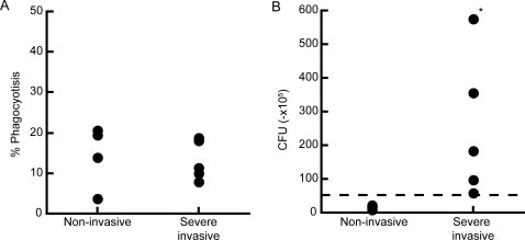
To examine whether emm49 GAS isolated from severe invasive infection might alter human PMN function, we performed phagocytosis assay in vitro. As non-opsonized GAS was resistant to the phagocytosis by PMN [14], we opsonized GAS with human plasma in advance to the assay. As shown in Figure 1A, there was no significant difference between GAS that were isolated from non-invasive and severe invasive infections in phagocytosis by PMN (p = 0.5556). However, as shown in Figure 1B, in vitro killing assay revealed that PMN killed non-invasive GAS, resulting in 15–42% of initial number of bacteria, but not invasive GAS (p = 0.019). The similar results were obtained when opsonized with either FCS or human serum regardless of complements immobilization (data not shown). These results were common among all PMN donors. These data indicated that clinically isolated severe invasive GAS were phagocytosed, but escaped from killing by human PMN.
Figure 1. Severe invasive GAS evade killing activity of PMN.
(A) Phagocytosis activity of PMN to emm49 GAS. Four non-invasive strains and 5 severe invasive strains labeled with Alexa-488, followed by incubation with PMN at MOI 10 for 60 min. The proportion of GAS-phagocytosed PMN was then analyzed using flow cytometry. (B) Severe invasive or non-invasive strains were incubated with PMN at MOI 10. After 2 hours, the killing capacity of PMN was estimated by counting the number of bacteria colonies. The dotted line indicates the number of bacteria applied to the culture.
Severe invasive GAS rapidly induce necrosis to human PMN
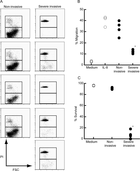
In an acute bacterial infection, PMN were quickly recruited at the site of infectious foci according to the gradient of chemoattractants. Therefore, we examined whether GAS clinically isolated from severe invasive infections could affect the migration ability of PMN in response to chemokines. As a model of local infection of the initial phase, we utilized a transwell system and added IL-8 and GAS in culture medium within the lower wells. PMN were applied in the upper wells and subsequently incubated for 90 min. As shown in Figure 2A and 2B, a substantial number of PMN, consisted largely of viable cells, was detected in the lower wells consisted of IL-8 and non-invasive GAS, as a control. Contrarily, number of PMN was significantly low in the presence of severe invasive GAS (p = 0.016) compared to that of control culture. Flow cytometry analysis suggested that although PMN was detected in the lower well consisted of IL-8 and severe invasive GAS, but most of them were dead as defined by propidium iodine staining (p = 0.016) (Figure 2A and 2C) demonstrating that severe invasive GAS affected survival of PMN and its migration activity in a transwell system.
Figure 2. Severe invasive GAS kill human PMN and impair migration ability in response to IL-8.
(A) Flow cytometry profiles of migrated PMN in response to 100 nM IL-8 plus GAS. PMN were applied into the upper well (5×10⁵ cells) and those migrated into the lower well in a transwell system in response to IL-8 in the presence of GAS (5×10⁶ CFU) were stained with propidium iodine and were analyzed using flow cytometry. Each panel represents migrated PMN encountered with individual clinically isolated GAS strain. The representative data are shown. (B) The proportion of PMN that migrated into lower wells in response to IL-8, in the presence (closed circles) or absence (open circles) of invasive or non-invasive GAS strains. Total cell numbers consisted of both viable and dead cells were estimated 60 minutes after incubation. (C) The proportion of live PMN that migrated into lower wells in response to IL-8 alone (open circles) or IL-8 in the presence of severe invasive or non-invasive GAS strains (closed circles). Viable cell numbers were analyzed at 60 minutes incubation. *p<0.05 estimated by Mann-Whitney's U test.
PMN were killed by streptolysin O (SLO) from severe invasive GAS
PMN death was induced shortly after encounter with severe invasive GAS, and PMN were not in apoptotic death because of low frequency of cells positive for annexin V (data not shown), which was seen in the case of cytolysin-dependent cell injury [15]. GAS produce two cytolysins that may contribute to pathogenesis. Streptolysin S (SLS) is an oxygen-stable β-hemolysin and Streptolysin O (SLO) is a pore-forming cholesterol-binding toxin [16]. Therefore, to know the mechanism underlying GAS-mediated killing of PMN, we investigated whether SLO produced by invasive GAS affect survival of PMN in an in vitro migration assay system.
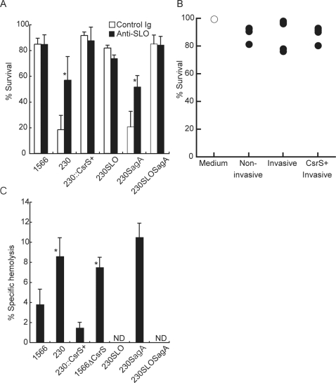
Figure 3A shows that PMN killing by invasive GAS was blocked by anti-SLO Ab in culture medium within lower wells of a transwell system (p = 0.018 compared with control Ig), at similar extent by adding free cholesterol in the medium (data not shown). Furthermore, an SLO deficient mutant from a STSS isolate NIH230 (NIH230slo) lost the killing activity for PMN (Figure 3A), thereby, strongly suggesting that SLO is a key element for PMN killing mediated by invasive GAS. Contrarily, SLS-deficient mutant from NIH230 strain (NIH230sagA) killed PMN, as efficiently as did parent strain, indicating that SLS is dispensable for killing of PMN mediated by invasive GAS. SLO and SLS double mutant GAS from NIH230 strain (NIH230slosagA) displayed the killing activity indistinguishable from that of NIH230slo, confirming the primarily role of SLO for GAS-mediated PMN killing. As shown in Figure 3B, incubation of PMN with supernatant from co-culture of IL-8 and invasive or non-invasive GAS did not affect PMN viability, suggesting that severe invasive GAS causes PMN killing following encounter with bacteria in a contact-dependent manner.
Figure 3. Severe invasive GAS killed PMN by SLO in a contact-dependent manner.
(A) The viability of viable PMN that migrated in lower wells of transwell system was estimated as Figure 2. To investigate the role of SLO in PMN survival, lower wells consisted of IL-8 in the presence of either polyclonal rabbit anti-SLO antibodies (25 µg/ml at a final concentration, closed column) or control rabbit IgG (open column), together with either non-invasive GAS (1566), invasive GAS (NIH230), an csrS-transduced NIH230 (NIH230::csrS+), an SLO deficient NIH230 mutant (NIH230slo), an SLS deficient NIH230 mutant (NIH230sagA), or SLO and SLS double mutant (NIH230slosagA). Values are mean±SD. *p<0.05 estimated by Student's t-test. (B) No substances secreted from GAS reduced PMN viability. PMN was incubated for 2 hours with supernatants from co-culture of IL-8 and invasive or non-invasive GAS and live cell number was examined as described in Figure 2C. (C). SLO-specific hemolytic activity for sheep erythrocytes in supernatants from overnight culture of invasive or non-invasive GAS as listed in (A). Forty-five minutes after incubation, the absorbance of culture supernatants was measured at 540 nm and the SLO-specific hemolytic activity was calculated as described in methods and presented as the mean±SD. The results represent one of two independent experiments. *p<0.05 significantly higher than non-invasive strain 1566 estimated by ANOVA. ‘ND’ represents less than 0.5% hemolysis.
To confirm that SLO activity is increased in invasive GAS strain, we measured SLO hemolytic activity of GAS strains used in this study. As shown in Figure 3C, SLO activity of severe invasive isolate NIH230 is increased as twice as that of non-invasive strain 1566 (p = 0.017).
Impaired migration of PMN is due to degradation of IL-8 by serine proteinase ScpC
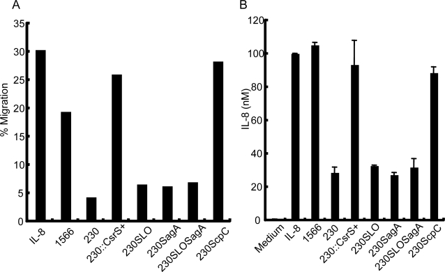
Although NIH230slo lost the killing activity for PMN, migration of PMN in response to IL-8 in a transwell system was not restored in the presence of this mutant (Figure 4A), thereby, suggesting that severe invasive GAS blocks PMN migration by influence on IL-8 activity. Therefore, we quantified the amount of IL-8 in culture before and after co-culture with clinically isolated GAS or its mutants. Figure 4B shows that the amount of IL-8 was significantly reduced by 60-min co-culture with NIH230, as well as NIH230slo, but not with non-invasive GAS 1556. As previous reports suggested that the GAS envelope peptidase ScpC (also known as SpyCEP) degrades the CXC chemokines, such as human IL-8, Groα, murine KC and MIP-2 [17]–[19], we established a NIH230 mutant deficient with ScpC (NIH230scpC) and analyzed the property in a PMN migration assay. The results showed that NIH230scpC neither digested IL-8, like 1566 strain, (Figure 4B) nor abrogated the migration of PMN in response to IL-8, comparable to 1566 strain (Figure 4A), whereas the mutant killed the migrated PMN as well as the parent strain NIH230 (data not shown). These results demonstrate that clinically isolated invasive GAS impaired PMN recruitment and its survival, as a result of productions of ScpC and SLO, respectively.
Figure 4. Severe invasive GAS degrade IL-8 by serine protease ScpC, resulting in impaired PMN migration.
(A) Migration abilities of PMN in response to IL-8 in the presence of non-invasive and invasive GAS, as listed in Figure 3A plus ScpC deficient NIH230 mutant (NIH230scpC). PMNs that migrated into the lower well in a transwell system were estimated by flow cytometry. (B). IL-8 was added into the culture medium (100 nM at a final concentration) and incubated with invasive or non-invasive GAS as listed in (A). Sixty minutes after incubation, the amount of IL-8 in triplicates was measured by sandwich ELISA and presented as the mean±SD. The results represent one of two independent experiments.
Enhanced expression of the slo and the scpC genes in severe invasive GAS is attributed to mutation of a transcriptional regulator CsrS
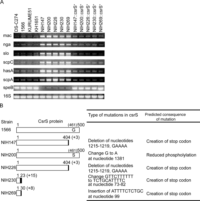
Although sequences of the slo gene and the scpC gene were identical among clinically isolated non-invasive and severe invasive GAS (data not shown), Figure 5A shows that the slo and the scpC genes were expressed in the severe invasive GAS greater in extent than those in the non-invasive GAS. The expression of the other virulence-associated genes, such as IgG degrading protease of GAS, Mac-1-like protein (mac), nicotine adenine dinucleotide glycohydrolase (nga), polysaccharide capsule production (hasA), and C5a peptidase (scpA), was also upregulted in the severe invasive GAS, greater than that detected in the non-invasive GAS (Figure 5A). Contrarily, the levels of streptococcal pyrogenic endotoxin (speB), SLS (sagA), and mitogenic factor (speF) genes were downregulated in the severe invasive GAS, compared to that found in the non-invasive GAS (Figure 5A and data not shown). These results demonstrate the prominent changes in the transcriptional profile of several virulence-associated genes, including the slo and the scpC, in the all severe invasive GAS.
Figure 5. Mutation of the csrS gene in the isolates of the patients with severe invasive infections is responsible for increased virulence of GAS.
(A) Expression of virulence-associated genes in non-invasive and invasive GAS isolates and mutants transduced with csrS, analyzed by RT-PCR. The expression of virulence-associated factors mRNA: IgG degrading protease of GAS, Mac-1-like protein (mac), nicotine adenine dinucleotide glycohydrolase (nga), slo, scpC, polysaccharide capsule production (hasA), C5a peptidase (scpA), and streptococcal pyrogenic endotoxin (speB), plus expression of 16S rRNA (16S) were shown. (B) The csrS mutations in GAS isolates from the patients with severe invasive streptococcal infections. The numbers at the end of the bars indicate the total amino acid residues of CsrS proteins from the start codon in non-invasive GAS (1566) and invasive Gas from the patients (NIH147, NIH200, NIH226, NIH230 and NIH 269). Solid boxes represent the newly created amino acids as a result of frameshift mutations, with length of amino acids ( “+ number” within parentheses). In the NIH200 strain, Ser replaced Gly at position 461 of the CsrS protein. Type of mutations are listed at the end of bars.
Mutation of csrR or csrS can cause significant alterations in virulence in mouse models of infection,either increasing lethality or the severity of localized soft tissue lesions [5], [6]. GAS isolates from mice with severe invasive disease had mutations in csrS, raising the notion that CsrR/S function is important in modulating gene expression during infection. Therefore, we analyzed the linkage between the csrS and/or csrR genes and the property of invasive GAS infection by sequencing these genes in the emm49 strains used in this study. The nucleotide sequence of the csrR gene was identical in all the isolates, and that of the csrS gene was identical among the all non-invasive GAS isolates (data not shown). However, as shown in Figure 5B, the csrS genes of all clinically isolated severe invasive GAS had a deletion, a point mutation, or an insertion, thereby, resulting in the creation of translational stop codons (NIH147, NIH226, NIH230, and NIH269) or in a mutation in the presumed kinase domain (NIH200). In order to clarify the role of CsrS regarding expression of the virulence-associated genes and resistance to PMN killing, we introduced the intact csrS gene of the 1566 strain into the severe invasive GAS (see Figure 5A). The csrS-introduced severe invasive GAS reduced the expression levels of the slo and the scpC genes, comparable to those detected in the non-invasive GAS. In contrast, the expression of speB was upregulated to the level observed in the non-invasive GAS (Figure 5A). In parallel with the expression profile of slo and scpC in the severe invasive GAS, introduction of the intact csrS gene into the severe invasive GAS restored the susceptibility to the killing by PMN (p = 0.015 compared with severe invasive isolates +CsrS) (Figure 6A), abrogated the inhibition of PMN migration by degradation of IL-8 (p = 0.002 compared with invasive isolates +CsrS) (Figure 4A, 4B, and 6B), and diminished the killing activity for PMN by necrosis (p = 0.00016 compared with invasive isolates +CsrS) (Figure 3A, 3C, and 6C), These results strongly suggest that mutations in the csrS gene correspond to the immunocompromized activity in the severe invasive isolates, associated with inhibition of PMN recruitment and survival.
Figure 6. Mutations of CsrS is responsible for increased virulence of GAS to PMN functions.
(A) Non-invasive, severe invasive, and invasive strains with overexpression of CsrS strains were incubated with PMN at MOI 10. After 2 hours, the number of live bacteria was counted. The dotted line indicates the number of bacteria applied to the culture. (B)–(C) CsrS transduction into the invasive GAS isolates abrogated the killing activity for PMN as well as the inhibitory effect on PMN migration in a transwell system. (B) The proportion of PMN consisted of both viable and dead cells and (C) The proportion of live PMN that migrated into the lower wells in response to IL-8, in the presence (closed circles) or absence (open circles) of non-invasive GAS, severe invasive GAS isolates or severe invasive GAS isolates transduced with CsrS was analyzed at 60 minutes incubation, as described in Figure. 2B and 2C. *p<0.05 estimated by ANOVA.
csrS mutation is important in the pathogenesis of invasive infections in a mouse model
In order to elucidate the role of csrS, in infections in vivo, we compared the virulence of GAS isolates using a mouse model which infected GAS intraperitoneally. The non-invasive 1566 strain displayed the LD50 value approximately 100-fold higher than that of the severe invasive NIH230 strain (Table 1), whereas a csrS deletion (1566ΔcsrS) caused an increase in the LD50 value comparable to that of the NIH230 strain. Consistently, an introduction of the intact csrS gene into the NIH230 strain (NIH230::csrS+) reduced the LD50 value to the level observed in the non-invasive strain. These results indicate that csrS is an important virulence factor in the mouse model of lethal infections.
Table 1. LD50 values of each strain.
| Strain | LD50 value |
| 1566 | 1.03×10⁸ |
| NIH230 | 1.11×10⁶ |
| NIH230::csrS + | 1.52×10⁸ |
| 1566ΔcsrS | 8.60×10⁵ |
| NIH230slo | 3.33×10⁷ |
| NIH230scpC | 1.04×10⁷ |
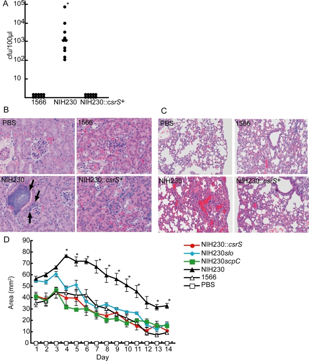
As shown in Figure 7A, the NIH230 strain caused bacteremia in mice 24 h after intraperitoneal injection whereas the bacteremia was barely detected in mice infected with NIH230::csrS+as well as the 1566 strain (p = 0.005 compared with non-invasive isolates, and p = 0.005 compared with invasive isolates +CsrS). Histopathologically, in the mice injected with the NIH230 strain, bacteria formed clusters in interstitial tissues in the kidneys and the lungs, accompanied congestion and no inflammatory cells at infectious foci (Figure 7B, 7C and data not shown). Contrarily, no significant pathological alterations were observed in the mice injected with the 1566 and NIH230::csrS + strains (Figure 7B and 7C). Figure 7D shows that subcutaneous infection of NIH230 formed the infected lesions with area significantly larger than those of 1566 and NIH230::csrS +. These results suggest that the invasive GAS isolates are more virulent in vivo than non-invasive GAS, and impair PMN function in vivo, owing, at least in part, to the mutations in the csrS gene.
Figure 7. Mutations of the csrS, SLO and ScpC regulate in vivo virulence of GAS in a mouse model.
(A) Number of GAS organisms recovered from the blood (100 µL) of each Male ddY mice injected intravenously with 1×10⁷ CFU in 100 ml suspension of GAS in PBS. Blood was taken 24 h after injection and the bacterial count was determined after plating on agar. The (-) bar represents median values. *(p<0.05) estimated by ANOVA. Histopathological changes in the (B) kidney and (C) lungs of mice infected with GAS. Each tissue was extracted 24 h after injecting GAS (1×10⁷ CFU). The black arrows indicate clusters of bacteria. (D) The course of subcutaneous infection in hairless mice injected with 1×10⁷ CFU in 100 µL suspension of GAS in PBS. Lesion area and body weight were measured daily after infection. Values are mean±SEM (n = 5). *Area of skin lesion in NIH230 infected mice was significantly higher than all other groups (p<0.05) estimated by ANOVA.
scpC and slo are insufficient singly for the pathogenesis of invasive infections
Finally, we assessed the influence of enhanced expression of the scpC or the slo gene on the virulence in a mouse model. As shown in Table 1, NIH230scpC and NIH230slo exerted the LD50 value 3–10 fold lower than that of the non-invasive isolate 1566, but 10–30 fold higher than that of severe invasive isolates. Subcutaneous inoculation of NIH230scpC and NIH230slo yielded the local infected lesions with area comparable to those of 1566 and NIH230::csrS + during the course of infection (Figure 7D). These results suggest that enhanced expression of ScpC and SLO in invasive GAS plays an important role in vivo virulence of GAS infection.
Discussion
It have been demonstrated that CsrS/R is a member of the two-component regulatory systems for regulating the multipe virulence factors of GAS, by using genetically- manipulated GAS mutants [20]. The present study demonstrate that the loss-of-functional mutations in csrS gene which were accumulated in clinically isolated GAS from patients with severe invasive infections, but not with emm-matched non-invasive strains. The csrS mutations enhanced the expression of scpC and slo, associated with the evasion of PMN functions and in vivo virulence. Introduction of the intact csrS gene into the severe invasive GAS restored the susceptibility to the killing by PMN and abrogated the activity for inhibition of PMN recruitment and survival, thus, demonstrating an instructional role of the loss-of-functional mutations in csrS gene for the evasion of PMN functions, providing unique pathophysiology of invasive GAS infections. Previous studies using animal-passaged GAS have shown that mutations in both csrS and csrR gene are important for the invasive phenotype [5], [6], and mutation frequency of csrS and csrR seems to be the same [21]. However, the severe invasive isolates analyzed in this study accumulated mutations in the csrS gene but not in the csrR gene (Figure 5B and data not shown). Then we further examined whether severe invasive isolates of other than emm49 genotype have the mutation of the csrS and the csrR genes. The frequency of the mutation in the csrS gene is higher than that in csrR (csrS mutation∶csrR mutation = 59∶19) (manuscript in preparation), suggesting the csrS mutation is more important in comparison with that of csrR in the clinical isolates regardless of emm genotypes. Furthermore, the expression of some human invasive disease-associated genes [5] including slo was enhanced in the csrS mutant (Figure 5B), but not in the csrR mutant [20]. On the contrary of a dogma that CsrS/R is a definitive member of the two-component regulatory systems, which involve a coordinate pair of proteins known as the sensor kinase and the response regulator [20], CsrS may transmit a signal not only to CsrR but also to other regulators. This dominant role of CsrS is the first important observation in this study, and its mutation is possibly more important than that of csrR in terms of etiopathogenesis of human severe invasive diseases.
Numbers of studies has pointed out virulent factors to evade host defense using genetically-manipulated GAS and animal models [18], [22], [23], although the significance of each factor to invasive infection is diverse and sometimes controversial, perhaps due to lack of proper non-invasive counterpart. As examples, SpeB [22] and SLS [23] have been proposed as an invasive infection-associated factor by its cytotoxic effect, however, speB and sagA expression is not enhanced in any csrS-mutated severe invasive GAS isolate used in this study and others [5], [24]. Furthermore, SLS hemolytic activity of invasive GAS is significantly decreased as compared with non-invasive strains (data not shown) and SLS-deletion in invasive GAS did not affect PMN survival at all, excluding the possibility for the role of SLS in PMN necrosis seen in this study. Extracellular deoxyribonuclease (DNase) is a virulence factor that protects emm1 type GAS against neutrophil killing by degrading the DNA framework of neutrophil extracelluar traps (NETs) [25], [26]. However, we confirmed that addition of DNase in the culture did not alter the level of PI-positive PMN, meaning bright PI staining of PMN is not due to release of NETs from PMN (Figure S1A). DNase activity of the emm49 severe invasive GAS was lower than that of non-invasive GAS (Figure S1B), possibly due to the difference of emm type. The expression of DNase as well as the slo and the scpC genes in emm1-genotype strains was enhanced under the csrS mutation [5]. These suggest that DNase may be important but redundant for induction of invasive diseases. Therefore, the second important observation in the present study is that an essential requirement of csrS mutation for invasive infection is associated with increased expression of ScpC and SLO and in vitro evasion of PMN functions, though we do not exclude the possibility that other CsrS-regulating factors contribute to the escape of invasive GAS from host defense.
SLO and ScpC independently enable GAS to escape from PMN functions; Present data using clinical isolated GAS and a scpC-deletion mutant (Figure 4A and 4B) show that enhanced production of serine proteinase ScpC in virulent GAS is essential to impair PMN migration in vitro by degradation of IL-8, as others partially have demonstrated [17]–[19]. The present study also uncovers that increased activity of SLO from invasive GAS isolates induces rapid and extensive necrosis to human PMN. SLO is a cholesterol-binding pore-forming hemolysin as well as cytotoxic for other cells [15]. A study has demonstrated SLO from invasive GAS lyse PMN [27], however this effect is likely due to complement activation by SLO [28] or PMN activation [29] but not due to cytotoxity of SLO itself as judged by their flow cytometry profiles which are distinct from ours (Figure 2A). In the present study, we observed that SLO concentration in a short-time culture with severe invasive GAS did not reach the threshold level to kill PMN by formation of pores (data not shown) and that PMN did not undergo necrosis upon incubation with culture media of severe invasive GAS (Figure 3B), leading to the novel possibility that PMN are probably killed following encounter with invasive GAS in a contact-dependent manner. PMN-binding GAS may make a small interface containing a high concentration of SLO between bacteria and PMN, which resembles to killing mechanism of killer cells to target cells [30]. Collaboration of SLO with other toxins may be critical to induce PMN necrosis as similarly mechanism has been reported [31], although it remains to be examined whether there exist explore interaction-associated molecules on both host and bacterial membrane is needed.
In contrast to the previous view [18], we observed that both of ScpC and SLO together, but not each of them, mediated sufficient in vivo virulence (Table 1), thus compatible with the notion that plural virulence-associated factors under the regulation of csrS abrogate PMN bactericidal functions and induce invasive diseases in in vivo animal model. Consistently, the high mortality and histopathological findings which lacks PMN infiltration in mice tissues infected with csrS-mutated GAS (Figure 7) are similar to those seen in clinical invasive GAS infections [32]. Thus, these results suggest that the ability of incompetence for PMN functions by individual GAS strain may determine the induction and clinical outcome of invasive diseases. Several clinical reports seem to support this hypothesis; Leukocytopenia seen in patients with STSS is more severe than that with non-STSS [33], and invasive GAS-infected patients with leukocytopenia show worse prognosis than those without leukocytopenia [33], [34]. Furthermore, predisposing factors for severe invasive GAS infection [9], such as diabetes mellitus [35], liver cirrhosis [36], and congestive heart failure [37] are known to impair PMN function. These evidences suggest that the level of PMN function is one of the critical factors to determine the threshold for the onset of invasive GAS infection, which may be the reason for rare outbreaks of invasive GAS infections.
Thus, enhanced expression of virulence factors that could evade PMN function is a key issue at first step to cause invasive bacterial infections. A further study in which collates clinical with bacterial/immunological data may provide with novel clues for early diagnosis and therapeutics of invasive bacterial infections.
Methods
Bacterial strains and culture
The S. pyogenes strains used in this study are described in Table S1 [8], [38]. Escherichia coli DH5α was used as the host for plasmid construction and was grown in liquid Luria-Bertani medium with shaking or on agar plates at 37°C. S. pyogenes was cultured in Todd-Hewitt broth supplemented with 0.5% yeast extract (THY medium) without agitation or on tryptic soy agar supplemented with 5% sheep blood. Cultures were grown at 37°C in a 5% CO2 atmosphere. When required, antibiotics were added to the medium at the following final concentrations: erythromycin, 500 µg/mL for E. coli and 1 µg/mL for S. pyogenes; spectinomycin (Sp), 25 µg/mL for E. coli and S. pyogenes both. The growth of S. pyogenes was turbidimetrically monitored at 600 nm using MiniPhoto 518R (Taitec, Tokyo, Japan).
Animals
Male 5–6-week-old outbred ddY and hairless mice were purchased from SLC (Shizuoka, Japan) and were maintained in a specific pathogen-free (SPF) condition. All animal experiments were performed according to the guidelines of the Ethics Review Committee of Animal Experiments of the National Institute of Infectious Diseases, Japan.
Isolation of human PMN
PMN were taken from nine healthy volunteers which were composed of 25–52 years old, 7 males and 2 female, and were isolated from venous blood of them using in accordance with a protocol approved by the Institutional Review Board for Human Subjects, National Institute of Infectious Diseases.
DNA manipulation
DNA amplifications by PCR, DNA restriction-endonuclease digestions, ligations, plasmid preparations, and agarose gel electrophoresis were performed according to standard techniques [42]. PCR reactions were performed using TaKaRa Ex Taq (TaKaRa Bio, Tokyo, Japan). Nucleotide sequence was determined by using the automated sequencer ABI PRISM 3100 Genetic Analyzer (Applied Biosystems, Tokyo, Japan)
Transformation
Calcium chloride (CaCl2) competent E. coli cells were prepared and transformed according to a standard protocol [39]. Electrocompetent S. pyogenes cells were prepared as described [38].
Construction of deletion or deficient mutants
Construction of the csrS mutants. A 1002-bp DNA fragment containing the 5′ terminal of csrS and the adjacent upstream chromosomal DNA was amplified from the 1566 chromosomal DNA using the primers for csrSdel1 and csrSdel2 (Table S2), and a 1104-bp fragment containing the 3′ terminal of csrS and the adjacent downstream chromosomal DNA was amplified from the NIH230 chromosomal DNA using the primers for csrSdel3 and csrS4del4 (Table S2); these 2 PCR products were ligated by BamHI and EcoRI and by EcoRI and PstI, respectively. The digested fragments were cloned into the erythromycin-resistant and temperature-sensitive shuttle vector pJRS233 [40] in order to create the plasmid pJRSΔcsrS. This plasmid was then purified from E. coli and introduced into the strains NIH230 and 1566 by electroporation. The transformants were selected by observing the growth on erythromycin agar at 30°C. The cells in which pJRSΔcsrS had been integrated into the chromosome were selected by the growth of the transformants at 39°C with erythromycin selection. The plasmid-integrated strain was serially passaged in a liquid culture at 30°C without erythromycin selection in order to facilitate the excision of the plasmid and thus, leaving the desired mutation in the chromosome. The replacement of the native csrS gene by the csrS-deleted mutant allele was verified by PCR, and the resultant strains were named as NIH230ΔcsrS and 1566ΔcsrS, respectively.
Construction of the slo mutant. A 1061-bp DNA fragment containing the internal region of slo was amplified from the NIH230 chromosomal DNA using the primers for slo-del3 and slo-del4 (Table S2). The PCR products were ligated by BamHI and EcoRI. This fragment was then cloned into the integration shuttle vector pSF152 [41] to create the plasmid pSF152slo that was then used for the chromosomal inactivation of the slo gene, as described previously [40]. The inactivated mutant strain NIH230slo (slo::aad9 Spr) was then selected by using spectinomycin-containing agar plates. Deficiency of the native slo gene was verified by PCR. Loss of SL hemoltitic activity of these mutants was confirmed by the standard SLS hemolysis assay (Figure 3C).
Construction of the sagA mutants. A 635-bp DNA fragment containing the 5′ terminal of sagA and the adjacent upstream chromosomal DNA was amplified from the NIH230 chromosomal DNA using the primers for sagA0-Xb and sagA2-Bm (Table S2) and a 1037-bp fragment containing the 3′ terminal of sagA and the adjacent downstream chromosomal DNA was amplified from the NIH230 chromosomal DNA using the primers for sagA3-Bm and sagA4-Ps; these 2 PCR products were ligated by XbaI and BamHI and by BamHI and PstI, respectively. The digested fragments were then cloned into the temperature-sensitive shuttle vector pJRS233 to create the plasmid pJRSΔsagA that was then used to create NIH230ΔsagA, as described above. Loss of SLS hemoltitic activity of these mutants was confirmed by the standard SLS hemolysis assay.
Construction of the scpC mutant. A 1240-bp DNA fragment containing the internal region of scpC was amplified from the NIH230 chromosomal DNA using the primers for scpC-del5 and scpC-del6 (Table S2). The PCR products were ligated by BamHI and EcoRI. This fragment was then cloned into the integration shuttle vector pSF152 [41] to create the plasmid pSF152scpC that was then used to create NIH230scpC, as described above.
Construction of the strains integrating the intact csrS gene
The csrS gene replacement was performed by allelic recombination. Specifically, the chromosomal DNA derived from the GAS strain 1566 was purified and used as a template for the PCR amplification of the csrS gene. The primers used were 5′-GGGGATCCTGAGATTCCTCTCACTAAAC-3′ (sense) and 5′-GGGAATTCTCTAATACACTATTTTACC-3′ (antisense). The PCR fragment was ligated into the plasmid pSF152 [41], and the resultant plasmid pSFcsrS was used for chromosomal integration into the mutated csrS gene of isolates from patients of severe invasive infections, as described previously [41]. The integrated strains (Spr) were then selected by using spectinomycin (Sp)-containing agar plates. The integration of the csrS gene was confirmed by PCR.
RT-PCR
S. pyogenes was grown in THY media at 37°C without aeration, and total RNA was extracted at OD600 of 0.75 by using the RNeasy Mini extraction kit (Qiagen). RT-PCR was performed by using a One Step RNA PCR Kit (AMV) (TaKaRa Shuzo Co., Kyoto, Japan) according to the manufacturer's recommendation using the RT-PCR primer pairs shown in Table S3.
GAS infection in a mouse model
To determine LD50, We injected several dilutions of 0.5 mL of GAS isolate suspensions in phosphate-buffered saline (PBS) intraperitoneally into male 5–6-week-old ddY outbred mice (5 mice/dilution, 9 dilutions for each GAS isolate). The exact numbers of the colony-forming units of the injected bacteria were determined by incubating adequate dilutions of each GAS sample on sheep blood agar plates. The data were analyzed for significance according to the Probit method to determine the LD50 values for a 7-day period. For a subcutaneous infection model, male hairless mice Hos:Hr-1 were injected 1×10⁷ CFU in 100 µl suspension of GAS in PBS. Lesion area and body weight were measured daily, and analyzed
Histopathology
For histological analysis, the tissues from GAS-infected mice were fixed in 10% formalin/PBS. The paraffin-embedded sections were stained with hematoxylin and eosin (Sapporo General Pathology Laboratory Co. Ltd., Hokkaido, Japan).
Phagocytosis and killing assay
Phagocytosis and killing assay by PMN were performed as previously described with some modifications [14]. 5×10⁵ human PMN and 5×10⁶ bacteria opsonized with human plasma and labeled with alexa 488 (Invitrogen, Carlsbad, CA) in a well of 24 well plates. After incubation for 60 min, PMN were harvested and stained with alexa-594 labeled anti-alexa 488 polyclonal Ab (Invitrogen) in order to distinguish non-phagocytosed but attached bacteria to PMN. The proportion of phagocytosed PMN were analyzed by FACS Calibur (BD Biosciences, San Jose, CA), For killing assay, PMN and opsonized bacteria in the same MOI as phagocytosis assay were incubated for 2 hours at 37°C, adding antibiotics at 60 min to eliminate non-phagocytosed bacteria. Corrected PMN were lysed in 0.1% saponin / PBS for 20 min on ice. Bacteria were washed with PBS and cultured on soy beans agar plate overnight, for counting the number of colonies.
Migration assay
Chemotaxis assay were performed as previously described with modification [42]. Briefly, 5×10⁵ PMN in RPMI medium containing 25 mM HEPES and 1% FCS were in Transwell inserts (3 µm pore size, Coaster, Corning, NY) placed in 24-well plates containing 600 µl medium, or 100 nM IL-8 solution (Peprtec, London, UK), which were incubated with or without 5×10⁶ bacteria for 1 hour at 37°C in advance of the assay. After 1 hour incubation, cells in the lower wells were collected and 104 10 µm microsphere beads (Polysciences Inc., Warrington, MA) were added. Cells were stained with propidium iodine (Sigma, St Louis, MI) for flow cytometry to quantify viable PMN and were analyzed using FACSCalibur. In some experiments, cholesterol (Sigma), 25 µg/mL anti-SLO polyclonal Ab (American Research Product, Inc., Belmont, MA), or rabbit IgG was added in 24 well plates.
ELISA
The amount of IL-8 in supernatant after incubation with bacteria was determined by Ready-to-Go human IL-8 ELISA kit (eBioScience, San, Diego, CA) according to manufacturers' protocol.
SLO-hemolysis assay
The activity of SLO in supernatant is measured as previously described [43]. Briefly, overnight culture supernatants of various strains were subjected to centrifugation, and were filtrated through a 0.45 µm membrane. Dithiothreitol and trypan blue were then added to each sample to a final concentration of 4 mM and 13 µg/mL respectively, and the mixtures were incubated at room temperature for 10 min. A 0.2 ml aliquot of 5% (v/v) sheep erythrocyte in PBS was added to 0.4 ml of each treated sample. After 30 min incubation at 37°C, the mixtures were subjected to centrifugation, and absorbance of the supernatants fluids was measured at 540 nm. To confirm that hemolysis was due to SLO, control reaction including culture supernatants to which water-soluble cholesterol (Sigma), a specific SLO inhibitor, had been added to yield a final concentration of 250 µg/mL.
SLS-hemolytic assay
Overnight culture of various strains were frozen at −80°C, thawed, and centrifuged to obtain the supernatants. Serially diluted culture supernatants (0.1 ml) in PBS containing 250 µg/mL water-soluble cholesterol were incubated at room temperature for 10 min. A 0.1 ml aliquot of 5% (v/v) sheep erythrocyte was added and incubated for 1 h at 37°C. The mixture were subjected to brief centrifugation, and absorbance of the supernatants fluids was measured at 540 nm. To confirm that hemolysis was due to SLS, control reaction including culture supernatants to which trypan blue (Sigma), a specific SLS inhibitor, had been added to yield a final concentration of 13 µg/ml.
Supporting Information
DNase activity is not involved in the virulence of emm49 severe invasive GAS isolates. a) To investigate the role of DNase in PMN survival, the viability of PMN that migrated in lower wells of transwell system was estimated as PMNs were applied into the upper well (5×10⁵ cells) of a transwell system, and lower wells consisted of IL-8 in the presence or absence of DNase I (100 mg/ml at a final concentration), together with either non-invasive GAS (1566), or invasive GAS (NIH230). PMN migrated in lower wells were stained with propidium iodine and were analyzed using flow cytometry. b) Activity of DNase in emm49 GAS. 10 ng of Calf thymus DNA was incubated with or without culture supernatants from non-invasive, severe invasive, and CsrS-transduced severe invasive GAS for 15 min at 37°C. Activity to degrade calf thymus DNA was visualized by 1% agarose gel electrophoresis. Methods in vitro migration assay As shown 5×10⁵ PMN in RPMI medium containing 25 mM HEPES and 1% FCS were in Transwell inserts (3 µm pore size, Coaster) placed in 24-well plates containing 600 µl medium, 100 nM IL-8 solution (Peprtec), 100 µg/mL deoxyribonuclease I (Sigma, St Louis, MI) which were incubated with or without 5×10⁶ bacteria for 1 hour at 37°C in advance of the assay. After 1 hour incubation, cells in the lower wells were collected and 104 10 µm microsphere beads (Polysciences) were added. Cells were stained with propidium iodine (Sigma) for flow cytometry to quantify viable PMN and were analyzed using FACSCalibur (BD BioScience). DNase activity assays Supernatants were collected from overnight cultures of bacterial strains grown in THB. Calf thymus DNA (10 ng) was combined with bacterial supernatant in final volume of 50 ml buffer (300 mM Tris-HCl (pH 7.5), 3 mM CaCl2, 3 mM MgCl2) for 15 min at 37°C. To halt DNase activity, 10 ml of 0.5 M EDTA (pH 8.0) was added to the reaction. Visualization of DNA degrad tion was done in 1% agarose gel electrophoresis.
(0.60 MB TIF)
Strains and plasmids used in this study
(0.03 MB DOC)
Primers used for the construction of deletion mutants
(0.03 MB DOC)
Primers used in RT-PCR
(0.03 MB DOC)
Acknowledgments
We thank Drs L. Tao and J.R. Scott for providing the plasmids used in this study, Drs J.J. Ferretti, H. Malke, M. Ohnishi, K. Kobayashi for their helpful advices, Dr H. Hasegawa for comments on pathology, and Dr K. Yamamoto for animal experiments. We also thank Ms Y. Nakamura for excellent technical assistance.
Footnotes
Competing Interests: The authors have declared that no competing interests exist.
Funding: This work was supported by a grant (H19-Shinkou-Ippan-002, to H.W) from the Ministry of Health, Labour and Welfare of Japan.
References
- 1.Bisno AL, Brito MO, Collins CM. Molecular basis of group A streptococcal virulence. Lancet Infect Dis. 2003;3:191–200. doi: 10.1016/s1473-3099(03)00576-0. [DOI] [PubMed] [Google Scholar]
- 2.Cunningham MW. Pathogenesis of group A streptococcal infections. Clin Microbiol Rev. 2000;13:470–511. doi: 10.1128/cmr.13.3.470-511.2000. [DOI] [PMC free article] [PubMed] [Google Scholar]
- 3.Davies HD, McGeer A, Schwartz B, Green K, Cann D, et al. Invasive group A streptococcal infections in Ontario, Canada. Ontario Group A Streptococcal Study Group. N Engl J Med. 1996;335:547–554. doi: 10.1056/NEJM199608223350803. [DOI] [PubMed] [Google Scholar]
- 4.Mitchell TJ. The pathogenesis of streptococcal infections: from tooth decay to meningitis. Nat Rev Microbiol. 2003;1:219–230. doi: 10.1038/nrmicro771. [DOI] [PubMed] [Google Scholar]
- 5.Sumby P, Whitney AR, Graviss EA, DeLeo FR, Musser JM. Genome-wide analysis of group a streptococci reveals a mutation that modulates global phenotype and disease specificity. PLoS Pathog. 2006;2:e5. doi: 10.1371/journal.ppat.0020005. [DOI] [PMC free article] [PubMed] [Google Scholar]
- 6.Walker MJ, Hollands A, Sanderson-Smith ML, Cole JN, Kirk JK, et al. DNase Sda1 provides selection pressure for a switch to invasive group A streptococcal infection. Nat Med. 2007;13:981–985. doi: 10.1038/nm1612. [DOI] [PubMed] [Google Scholar]
- 7.Ikebe T, Murai N, Endo M, Okuno R, Murayama S, et al. Changing prevalent T serotypes and emm genotypes of Streptococcus pyogenes isolates from streptococcal toxic shock-like syndrome (TSLS) patients in Japan. Epidemiol Infect. 2003;130:569–572. [PMC free article] [PubMed] [Google Scholar]
- 8.Ikebe T, Endo M, Ueda Y, Okada K, Suzuki R, et al. The genetic properties of Streptococcus pyogenes emm49 genotype strains recently emerged among severe invasive infections in Japan. Jap J Infect Dis. 2004;57:187–188. [PubMed] [Google Scholar]
- 9.ith A, Lamagni TL, Oliver I, Efstratiou A, George RC, et al. Invasive group A streptococcal disease: should close contacts routinely receive antibiotic prophylaxis? Lancet Infect Dis. 2005;5:494–500. doi: 10.1016/S1473-3099(05)70190-0. [DOI] [PubMed] [Google Scholar]
- 10.Hidalgo-Grass C, Dan-Goor M, Maly A, Eran Y, Kwinn LA, et al. Effect of a bacterial pheromone peptide on host chemokine degradation in group A streptococcal necrotising soft-tissue infections. Lancet. 2004;363:696–703. doi: 10.1016/S0140-6736(04)15643-2. [DOI] [PubMed] [Google Scholar]
- 11.Nathan C. Neutrophils and immunity: challenges and opportunities. Nat Rev Immunol. 2006;6:173–182. doi: 10.1038/nri1785. [DOI] [PubMed] [Google Scholar]
- 12.Urban CF, Lourido S, Zychlinsky A. How do microbes evade neutrophil killing? Cell Microbiol. 2006;8:1687–1696. doi: 10.1111/j.1462-5822.2006.00792.x. [DOI] [PubMed] [Google Scholar]
- 13.Voyich JM, Musser JM, DeLeo FR. Streptococcus pyogenes and human neutrophils: a paradigm for evasion of innate host defense by bacterial pathogens. Microbes Infect. 2004;6:1117–1123. doi: 10.1016/j.micinf.2004.05.022. [DOI] [PubMed] [Google Scholar]
- 14.Kobayashi SD, Braughton KR, Whitney AR, Voyich JM, Schwan TG, et al. Bacterial pathogens modulate an apoptosis differentiation program in human neutrophils. Proc Natl Acad Sci U S A. 2003;100:10948–10953. doi: 10.1073/pnas.1833375100. [DOI] [PMC free article] [PubMed] [Google Scholar]
- 15.Bhakdi S, Bayley H, Valeva A, Walev I, Walker B, et al. Staphylococcal alpha-toxin, streptolysin-O, and Escherichia coli hemolysin: prototypes of pore-forming bacterial cytolysins. Arch Microbiol. 1996;165:73–79. doi: 10.1007/s002030050300. [DOI] [PubMed] [Google Scholar]
- 16.Hirsch JG, Bernheimer AW, Weissmann G. Motion picture study of the toxic action of streptolysins on leucocytes. J Exp Med. 1963;118:223–228. doi: 10.1084/jem.118.2.223. [DOI] [PMC free article] [PubMed] [Google Scholar]
- 17.Edwards RJ, Taylor GW, Ferguson M, Murray S, Rendell N, et al. Specific C-terminal cleavage and inactivation of interleukin-8 by invasive disease isolates of Streptococcus pyogenes. J Infect Dis. 2005;192:783–790. doi: 10.1086/432485. [DOI] [PubMed] [Google Scholar]
- 18.Hidalgo-Grass C, Mishalian I, Dan-Goor M, Belotserkovsky I, Eran Y, et al. A streptococcal protease that degrades CXC chemokines and impairs bacterial clearance from infected tissues. EMBO J. 2006;25:4628–4637. doi: 10.1038/sj.emboj.7601327. [DOI] [PMC free article] [PubMed] [Google Scholar]
- 19.Sumby P, Zhang S, Whitney AR, Falugi F, Grandi G, et al. A chemokine-degrading extracellular protease made by group A Streptococcus alters pathogenesis by enhancing evasion of the innate immune response. Infect Immun. 2008;76:978–985. doi: 10.1128/IAI.01354-07. [DOI] [PMC free article] [PubMed] [Google Scholar]
- 20.Federle MJ, McIver KS, Scott JR. A response regulator that represses transcription of several virulence operons in the group A streptococcus. J Bacteriol. 1999;181:3649–3657. doi: 10.1128/jb.181.12.3649-3657.1999. [DOI] [PMC free article] [PubMed] [Google Scholar]
- 21.Engleberg NC, Heath A, Miller A, Rivera C, DiRita VJ. Spontaneous mutations in the CsrRS two-component regulatory system of Streptococcus pyogenes result in enhanced virulence in a murine model of skin and soft tissue infection. J Infect Dis. 2001;183:1043–1054. doi: 10.1086/319291. [DOI] [PubMed] [Google Scholar]
- 22.Terao Y, Mori Y, Yamaguchi M, Shimizu Y, Ooe K, et al. Group A streptococcal cysteine protease degrades C3 (C3b) and contributes to evasion of innate immunity. J Biol Chem. 2008;283:6253–6260. doi: 10.1074/jbc.M704821200. [DOI] [PubMed] [Google Scholar]
- 23.Miyoshi-Akiyama T, Takamatsu D, Koyanagi M, Zhao J, Imanishi K, et al. Cytocidal effect of Streptococcus pyogenes on mouse neutrophils in vivo and the critical role of streptolysin S. J Infect Dis. 2005;192:107–116. doi: 10.1086/430617. [DOI] [PubMed] [Google Scholar]
- 24.Kansal RG, McGeer A, Low DE, Norrby-Teglund A, Kotb M. Inverse relation between disease severity and expression of the streptococcal cysteine protease, SpeB, among clonal M1T1 isolates recovered from invasive group A streptococcal infection cases. Infect Immun. 2000;68:6362–6369. doi: 10.1128/iai.68.11.6362-6369.2000. [DOI] [PMC free article] [PubMed] [Google Scholar]
- 25.Sumby P, Barbian KD, Gardner DJ, Whitney AR, Welty DM, et al. Extracellular deoxyribonuclease made by group A streptococcus assists pathogenesis by enhancing evasion of the innate immune response. Proc Natl Acad Sci U S A. 2005;102:1679–1684. doi: 10.1073/pnas.0406641102. [DOI] [PMC free article] [PubMed] [Google Scholar]
- 26.Buchanan JT, Simpson AJ, Aziz RK, Liu GY, Kristian SA, et al. DNase expression allows the pathogen group A streptococcus to escape killing in neutrophil extracellular traps. Curr Biol. 2006;16:396–400. doi: 10.1016/j.cub.2005.12.039. [DOI] [PubMed] [Google Scholar]
- 27.Sierig G, Cywes C, Wessels MR, Ashbaugh CD. Cytotoxic effects of streptolysin o and streptolysin s enhance the virulence of poorly encapsulated group a streptococci. Infect Immun. 2003;71:446–455. doi: 10.1128/IAI.71.1.446-455.2003. [DOI] [PMC free article] [PubMed] [Google Scholar]
- 28.Bhakdi S, Tranum-Jensen J. Complement activation and attack on autologous cell membranes induced by streptolysin-O. Infect Immun. 1985;48:713–719. doi: 10.1128/iai.48.3.713-719.1985. [DOI] [PMC free article] [PubMed] [Google Scholar]
- 29.Walev I, Hombach M, Bobkiewicz W, Fenske D, Bhakdi S, et al. Resealing of large transmembrane pores produced by streptolysin O in nucleated cells is accompanied by NF-kappaB activation and downstream events. FASEB J. 2002;16:237–239. doi: 10.1096/fj.01-0572fje. [DOI] [PubMed] [Google Scholar]
- 30.Pipkin ME, Lieberman J. Delivering the kiss of death: progress on understanding how perforin works. Curr Opin Immunol. 2007;19:301–308. doi: 10.1016/j.coi.2007.04.011. [DOI] [PMC free article] [PubMed] [Google Scholar]
- 31.Madden JC, Ruiz N, Caparon M. Cytolysin-mediated translocation (CMT): a functional equivalent of type III secretion in gram-positive bacteria. Cell. 2001;104:143–152. doi: 10.1016/s0092-8674(01)00198-2. [DOI] [PubMed] [Google Scholar]
- 32.Bakleh M, Wold LE, Mandrekar JN, Harmsen WS, Dimashkieh HH, et al. Correlation of histopathologic findings with clinical outcome in necrotizing fasciitis. Clin Infect Dis. 2005;40:410–414. doi: 10.1086/427286. [DOI] [PubMed] [Google Scholar]
- 33.Eriksson BK, Andersson J, Holm SE, Norgren M. Epidemiological and clinical aspects of invasive group A streptococcal infections and the streptococcal toxic shock syndrome. Clin Infect Dis. 1998;27:1428–1436. doi: 10.1086/515012. [DOI] [PubMed] [Google Scholar]
- 34.Hasegawa T, Hashikawa SN, Nakamura T, Torii K, Ohta M. Factors determining prognosis in streptococcal toxic shock-like syndrome: results of a nationwide investigation in Japan. Microbes Infect. 2004;6:1073–1077. doi: 10.1016/j.micinf.2004.06.001. [DOI] [PubMed] [Google Scholar]
- 35.Marhoffer W, Stein M, Maeser E, Federlin K. Impairment of polymorphonuclear leukocyte function and metabolic control of diabetes. Diabetes Care. 1992;15:256–260. doi: 10.2337/diacare.15.2.256. [DOI] [PubMed] [Google Scholar]
- 36.Propst-Graham KL, Preheim LC, Vander Top EA, Snitily MU, Gentry-Nielsen MJ. Cirrhosis-induced defects in innate pulmonary defenses against Streptococcus pneumoniae. BMC Microbiol. 2007;7:94. doi: 10.1186/1471-2180-7-94. [DOI] [PMC free article] [PubMed] [Google Scholar]
- 37.Iversen PO, Woldbaek PR, Tønnessen T, Christensen G. Decreased hematopoiesis in bone marrow of mice with congestive heart failure. Am J Physiol Regul Integr Comp Physiol. 2002;282:R166–172. doi: 10.1152/ajpregu.2002.282.1.R166. [DOI] [PubMed] [Google Scholar]
- 38.Ikebe T, Endoh M, Watanabe H. Increased expression of ska gene in emm49-genotyped Streptococcus pyogenes strains isolated from patients of severe invasive streptococcal infections. Jap J Infect Dis. 2005;58:272–275. [PubMed] [Google Scholar]
- 39.Sambrook J, Fritsch EF, Maniatis T. T. Molecular cloning: a laboratory manual, 2nd ed. Cold Spring Harbor, , NY: Cold Spring Harbor Laboratory; 1990. [Google Scholar]
- 40.Perez-Casal J, Price JA, Maguin E, Scott JR. An M protein with a single C repeat prevents phagocytosis of Streptococcus pyogenes: use of a temperature-sensitive shuttle vector to deliver homologous sequences to the chromosome of S. pyogenes. Mol Microbiol. 1993;8:809–819. doi: 10.1111/j.1365-2958.1993.tb01628.x. [DOI] [PubMed] [Google Scholar]
- 41.Tao L, LeBlanc DJ, Ferretti JJ. Novel streptococcal integration shuttle vectors for gene cloning and inactivation. Gene. 1992;120:105–110. doi: 10.1016/0378-1119(92)90016-i. [DOI] [PubMed] [Google Scholar]
- 42.Ato M, Stäger S, Engwerda CR, Kaye PM. Defective CCR7 expression on dendritic cells contributes to the development of visceral leishmaniasis. Nat Immunol. 2001;3:1185–1191. doi: 10.1038/ni861. [DOI] [PubMed] [Google Scholar]
- 43.Ruiz N, Wang B, Pentland A, Caparon M. Streptolysin O and adherence synergistically modulate proinflammatory responses of keratinocytes to group A streptococci. Mol Microbiol. 1998;27:337–346. doi: 10.1046/j.1365-2958.1998.00681.x. [DOI] [PubMed] [Google Scholar]
- 44.Working Group on Severe Streptococcal Infections. Defining the group A streptococcal toxic shock syndrome. JAMA. 1993;269:390–391. [PubMed] [Google Scholar]
Associated Data
This section collects any data citations, data availability statements, or supplementary materials included in this article.
Supplementary Materials
DNase activity is not involved in the virulence of emm49 severe invasive GAS isolates. a) To investigate the role of DNase in PMN survival, the viability of PMN that migrated in lower wells of transwell system was estimated as PMNs were applied into the upper well (5×10⁵ cells) of a transwell system, and lower wells consisted of IL-8 in the presence or absence of DNase I (100 mg/ml at a final concentration), together with either non-invasive GAS (1566), or invasive GAS (NIH230). PMN migrated in lower wells were stained with propidium iodine and were analyzed using flow cytometry. b) Activity of DNase in emm49 GAS. 10 ng of Calf thymus DNA was incubated with or without culture supernatants from non-invasive, severe invasive, and CsrS-transduced severe invasive GAS for 15 min at 37°C. Activity to degrade calf thymus DNA was visualized by 1% agarose gel electrophoresis. Methods in vitro migration assay As shown 5×10⁵ PMN in RPMI medium containing 25 mM HEPES and 1% FCS were in Transwell inserts (3 µm pore size, Coaster) placed in 24-well plates containing 600 µl medium, 100 nM IL-8 solution (Peprtec), 100 µg/mL deoxyribonuclease I (Sigma, St Louis, MI) which were incubated with or without 5×10⁶ bacteria for 1 hour at 37°C in advance of the assay. After 1 hour incubation, cells in the lower wells were collected and 104 10 µm microsphere beads (Polysciences) were added. Cells were stained with propidium iodine (Sigma) for flow cytometry to quantify viable PMN and were analyzed using FACSCalibur (BD BioScience). DNase activity assays Supernatants were collected from overnight cultures of bacterial strains grown in THB. Calf thymus DNA (10 ng) was combined with bacterial supernatant in final volume of 50 ml buffer (300 mM Tris-HCl (pH 7.5), 3 mM CaCl2, 3 mM MgCl2) for 15 min at 37°C. To halt DNase activity, 10 ml of 0.5 M EDTA (pH 8.0) was added to the reaction. Visualization of DNA degrad tion was done in 1% agarose gel electrophoresis.
(0.60 MB TIF)
Strains and plasmids used in this study
(0.03 MB DOC)
Primers used for the construction of deletion mutants
(0.03 MB DOC)
Primers used in RT-PCR
(0.03 MB DOC)


