Abstract
Previous studies using in vivo candidiasis models have demonstrated that the concentration-associated pharmacodynamic indices, the maximum concentration of a drug in serum/MIC and 24-h area under the curve (AUC)/MIC, are associated with echinocandin treatment efficacy. The current investigations used a neutropenic murine model of disseminated Candida albicans and C. glabrata infection to identify the 24-h AUC/MIC index target associated with a stasis and killing endpoint for the echinocandin, micafungin. The kinetics after intraperitoneal micafungin dosing were determined in neutropenic infected mice. Peak levels and AUC values were linear over the 16-fold dose range studied. The serum drug elimination half-life ranged from 7.5 to 16 h. Treatment studies were conducted with 4 C. albicans and 10 C. glabrata isolates with micafungin MICs varying from 0.008 to 0.25 μg/ml to determine whether similar 24-h AUC/MIC ratios were associated with efficacy. The free drug AUC/MICs associated with stasis and killing (1-log) endpoints were near 10 and 20, respectively. The micafungin exposures associated with efficacy were similar for the two Candida species. Furthermore, the free drug micafungin exposures required to produce stasis and killing endpoints were similar to those recently reported for another echinocandin, anidulafungin, against the identical Candida isolates in this model.
Antifungal pharmacodynamic studies have been helpful to optimize dosing regimen design and provide a rationale for the development of susceptibility breakpoints for several antifungal drugs including the triazoles, amphotericin B, and flucytosine. Recent studies have begun to apply these pharmacodynamic principles to understand the exposure and response relationship for drugs from the echinocandin class (10, 15, 46, 50). These compounds exhibit broad-spectrum activity against Candida and Aspergillus species, including emerging Candida species such as C. glabrata (13, 20, 41, 42). Previous investigations with other echinocandins, including micafungin (D. Andes, unpublished observations) have demonstrated that the concentration-associated pharmacodynamic indices, the maximum concentration of a drug in serum (Cmax)/MIC and the area under the curve (AUC)/MIC, drive treatment efficacy for this drug class in candidiasis models (2, 9, 21, 26). The present study was performed to determine whether the magnitude of the pharmacokinetic/pharmacodynamic index required for efficacy is similar for C. albicans and C. glabrata strains, including several previously characterized caspofungin-resistant organisms. These study observations were also compared to recently published data with the echinocandin anidulafungin in this model with the same strains (9). The results from these studies provide a pharmacodynamic rationale in support of the current clinical dosing regimens. Furthermore, the data provide information useful regarding the susceptibility breakpoints for this new compound.
MATERIALS AND METHODS
Organisms.
Fourteen clinical Candida isolates were used: four C. albicans isolates (K-1, 580, 98-17, and 98-210) and ten C. glabrata isolates (570, 513, 5592, 5376, 33609, 32930, 33616, 34341, 35315, and 37661). Six of the isolates have been previously described and were kindly provided by J. Knudsen (28). These six clinical isolates were collected after prolonged caspofungin exposure and characterized as caspofungin resistant. The organisms were maintained, grown, and quantified on Sabouraud dextrose agar (SDA) plates. At 24 h prior to the study, the organisms were subcultured at 35°C. The isolates were chosen to include those with varying echinocandin susceptibilities. We also attempted to choose isolates based upon a relatively similar degree of fitness in this animal model, as determined by the amount of growth in the kidneys of untreated animals over 48 h (9).
Antifungal agent.
Micafungin was obtained from Astellas and was prepared in 0.15 M NaCl.
In vitro susceptibility testing.
MICs were determined by using Clinical Laboratory Standards Institute (CLSI) method M27-A2 (12, 41). The MIC endpoints for micafungin included both partial (CLSI recommended and referred to as M1) and complete growth inhibition readings relative to that of the drug-free control well (referred to as M2). Determinations were performed in duplicate on four separate occasions. Final results are expressed as the mean of these results.
Animals.
Six-week-old ICR/Swiss specific-pathogen-free female mice weighing 23 to 27 g were used for all studies (Harlan Sprague-Dawley, Indianapolis, IN). Animals were housed in groups of five and allowed access to food and water ad libitum. Animals were maintained in accordance with American Association for Accreditation of Laboratory Care criteria. Animal studies were approved by the Animal Research Committee of the William S. Middleton Memorial VA Hospital and the University of Wisconsin.
Infection model.
A neutropenic, murine, disseminated candidiasis model was used for all studies (2, 4, 5, 6). Mice were rendered neutropenic (polymorphonuclear cells < 100/mm3) by injecting cyclophosphamide (Mead Johnson Pharmaceuticals, Evansville, IN) subcutaneously 4 days before (150 mg/kg of body weight), 1 day before (100 mg/kg) infection, and 2 days after infection (100 mg/kg). Prior studies have demonstrated this regimen produces neutropenia (absolute neutrophil counts remained at or below 100/mm3 throughout the 96-h study). Organisms were subcultured on SDA 24 h prior to infection. The inoculum was prepared by placing three to five colonies into 5 ml of sterile pyrogen-free 0.9% saline warmed to 35°C. The final inoculum was adjusted to a 0.6 transmittance at 530 nm. Fungal counts of the inoculum determined by viable counts on SDA were 6.1 ± 0.51 log10 CFU/ml.
Disseminated infection with the Candida organisms was achieved by injection of 0.1 ml of inoculum via lateral tail vein 2 h prior to start of drug therapy. At the end of the study period, animals were sacrificed by CO2 asphyxiation. After sacrifice, the kidneys of each mouse were immediately removed and placed in sterile 0.9% saline at 4°C. The homogenate was then serially diluted 1:10, and aliquots were plated on SDA for viable fungal colony counts after incubation for 24 h at 35°C. The lower limit of detection was 100 CFU/ml. The results were expressed as the mean CFU/kidneys for three mice.
Pharmacokinetic analyses.
The single-dose pharmacokinetics of micafungin were determined in infected neutropenic ICR/Swiss mice after intraperitoneal administration of 80, 20, and 5 mg/kg administered in 0.2-ml volumes. Blood from groups of three isofluorane-anesthetized mice (nine mice for each dose level) was collected at each of eight time points (1, 2, 4, 6, 8, 12, 24, and 48 h). Serum was collected by centrifugation, and samples were stored at −80°C until drug assay. Samples were analyzed by microbiologic assay using C. albicans K1 as the assay organism (30). The lower limit of detection of the assay was 0.12 μg/ml. The mean intraday variation was <7%.
A noncompartmental model was used in the kinetic analysis (3). Pharmacokinetic parameters, including elimination half-life and concentration at time zero (C0) were calculated via nonlinear least-squares techniques. The AUC was calculated by the trapezoidal rule. For treatment doses in which no kinetics were determined, pharmacokinetic indices were estimated by linear extrapolation from the highest and lowest dose levels used in the kinetic studies described above. Protein binding was considered based upon previous reports of binding of micafungin in rodents and humans (99.75%) (25, 51).
Pharmacodynamic index magnitude determinations.
Fourteen Candida strains, including of four C. albicans (K1, 98-17, 580, and 98-210) and ten C. glabrata (5376, 570, 513, 5592, 33609, 32930, 33616, 34341, 35315, and 37661) isolates, were used for in vivo experiments. Infection in neutropenic mice was produced with each strain as described above. Micafungin dosing studies were designed to vary the magnitude of the pharmacodynamic indices and to produce treatment effects that included no effect to maximal effect (based on results from pilot treatment studies not presented). Six total dose levels varied from 0.078 to 80 mg/kg/24 h. Doses were administered every 24 h for the 4-day study period. Groups of three mice were used for each dosing regimen. At the end of the study, mice were euthanized, and the kidneys were immediately processed for CFU determinations.
Data analysis.
A sigmoid dose-effect model was used to model the in vivo potency of micafungin. The model is derived from the Hill equation: E = (Emax × DN)/(ED50N + DN), where E is the observed effect (change in log10 CFU/kidney compared to untreated controls at the end of the treatment period), D is the total dose of micafungin over the entire treatment period, Emax is the maximum effect of micafungin compared to untreated controls, ED50 is the dose required to achieve 50% of the Emax, and N is the slope of the dose-effect relationship. The indices Emax, ED50, and N were calculated by using nonlinear least-squares regression. The correlation between efficacy and the 24-h AUC/MIC for the group of Candida isolates was determined by nonlinear least-squares regression analysis (Sigma Stat; Jandell Scientific Software, San Rafael, CA). The coefficient of determination (R2) was used to estimate the variance that could be due to regression with each the pharmacodynamic index. Calculations were performed using both total- and free-drug concentrations.
To allow a comparison of the potency of micafungin against the study organisms, we calculated the 24-h static dose and the doses required to achieve a 1-log reduction in colony counts using the above model. The magnitude of the pharmacokinetic/pharmacodynamic index associated with each endpoint dose was calculated from the following equation: log10 D = log10 [E/Emax − E]/N + log ED50, where E is the amount of control growth over the treatment period in untreated animals for the static dose calculation, E is the control growth as described above +1 log for the calculation of the dose (D) associated with a 1-log kill, and N is the slope of the dose-response curve.
RESULTS
In vitro susceptibility testing. The study organisms and the MICs against micafungin are listed in Table 1. The 24-h MICs for the 14 Candida organisms studied varied by >30-fold (range, 0.008 to 0.25 μg/ml). The MICs using the partial (50%) endpoint (M1) were either the same or twofold lower than those using a complete inhibition endpoint (M2).
TABLE 1.
In vitro and in vivo efficacies of micafungin against select C. albicans and C. glabrata in a disseminated candidiasis modela
| Strain | MIC1 (μg/ml) | MIC2 (μg/ml) | Static dose (mg/kg/24 h) | 24-h AUC/MIC1
|
1-Log kill (mg/kg/24 h) | 24-h AUC/MIC1
|
||
|---|---|---|---|---|---|---|---|---|
| Total drug | Free drug | Total drug | Free drug | |||||
| CA98-17 | 0.03 | 0.06 | 2.53 | 2,333 | 5.9 | 4.60 | 4,233 | 10.6 |
| CA98-210 | 0.016 | 0.03 | 2.84 | 5,200 | 13.0 | 10.4 | 18,400 | 46.0 |
| CA580 | 0.008 | 0.016 | 1.98 | 7,857 | 19.6 | 2.36 | 1,686 | 4.21 |
| CAK1 | 0.016 | 0.03 | 2.84 | 5,200 | 13.0 | 4.08 | 7,533 | 18.8 |
| CG35315 | 0.06 | 0.12 | 2.4 | 1,103 | 2.8 | 3.77 | 1,733 | 4.33 |
| CG37661 | 0.25 | 0.25 | 9.25 | 984 | 2.5 | 20.6 | 2,080 | 5.2 |
| CG34341 | 0.06 | 0.12 | 2.18 | 1,003 | 2.51 | 4.26 | 1,960 | 4.9 |
| CG32930 | 0.008 | 0.016 | 0.06 | 236 | 0.50 | 1.69 | 3,107 | 7.8 |
| CG33609 | 0.016 | 0.016 | 0.41 | 753 | 1.9 | 1.40 | 2,600 | 6.5 |
| CG513 | 0.016 | 0.03 | 1.34 | 2,467 | 6.2 | NA | NA | NA |
| CG5592 | 0.016 | 0.03 | 1.80 | 3,333 | 8.4 | 5.46 | 10,000 | 25.0 |
| CG5376 | 0.008 | 0.016 | 0.90 | 3,571 | 8.9 | 2.0 | 7,886 | 19.7 |
| CG570 | 0.016 | 0.03 | 3.76 | 6,933 | 17.3 | 6.70 | 12,066 | 30.2 |
| CG33616 | 0.25 | 0.50 | 3.88 | 428 | 1.1 | 7.89 | 848 | 2.20 |
| Mean ± SD | 2,957 ± 2,493 | 7.5 ± 6.2 | 5,702 ± 5,239 | 14.3 ± 13.1 | ||||
Strain prefixes: CA, C. albicans; CG, C. glabrata. The static dose is the dose required to produce an organism burden in kidneys the same as that at the start of therapy. The 1-log kill is the dose required to produce an organism burden in kidneys the 1 log10 lower than that at the start of therapy. NA, not available.
Pharmacokinetics.
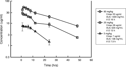
The serum time course of micafungin in infected neutropenic mice after intraperitoneal doses of 80, 20, and 5 mg/kg is shown in Fig. 1. Peak serum drug levels and the AUC increased in a linear fashion with dose escalation. Peak levels were achieved within the 2 h for each of the doses and ranged from 7.0 ± 0.80 to 53 ± 0.62 μg/ml. The serum elimination half-life ranged from 7.5 to 16 h. The AUC0-∞, as determined by the trapezoidal rule, ranged from 138 to 1,400 mg·h/liter with the lowest and highest doses, respectively. Free-drug calculations were based on previously determined protein binding in mice and humans using equilibrium dialysis (99.75% bound) (24).
FIG. 1.
Serum pharmacokinetics of micafungin following single, intraperitoneal doses of 80, 20, or 5 mg/kg. Concentrations were measured by using a microbiologic assay. Each symbol represents data from three mice. The error bars represent the standard deviation. Cmax represents the observed peak serum concentration. The AUC was calculated from 0 to infinity using the trapezoid rule.
Magnitude of the pharmacodynamic index associated with efficacy.
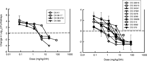
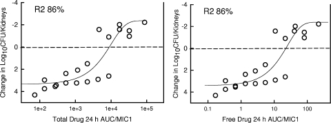
Both Cmax/MIC and AUC/MIC have been shown to be important predictors of the in vivo efficacy of the echinocandins. For comparison of the index magnitude among strains with various MICs, we utilized the 24-h AUC/MIC index. To determine whether the index magnitude was similar among Candida strains, we studied the activities of the 24-h dosing regimen of micafungin against four strains of C. albicans and ten strains of C. glabrata. The dose-response curves for micafungin against these various strains are shown in Fig. 2. At the start of therapy, the mice infected with C. albicans had 4.06 ± 0.23 log10 CFU/kidney of Candida (range, 3.57 to 4.28 log10 CFU/kidneys). Each of the four C. albicans isolates grew similarly in the kidneys of untreated animals. However, the growth of the C. glabrata strains was less than the other Candida strains studied. This difference is similar to that recently reported for these organisms in this model (9). The range of organism growth in control animals over the treatment period was 3.91 ± 0.56 log10 CFU/kidney for C. albicans to 1.95 ± 0.79 log10 CFU/kidney for C. glabrata. The C. glabrata isolates demonstrated relatively equivalent growth compared among the group of ten isolates. In general, the shapes of the dose-response curves were similar for all strains. The location of the dose-response curve was related to the MIC of the organism. The static dose, the doses associated with a 1-log killing, and the associated total and free-drug 24-h AUC/MIC are shown in Table 1. The extent of organism killing varied among the strains and was related to the micafungin exposure-MIC relationship. The majority of strains exhibited an extensive drop in the numbers of CFU/kidneys after micafungin therapy over the 4-day study compared to the numbers of CFU for the untreated controls (mean ± the standard deviation reduction in organism burden, 4.92 ± 1.3 log10 CFU/kidneys). For the micafungin regimens with 24-h dosing, the free-drug 24-h AUC/MICs associated with a static effect against the 14 Candida strains was 7.5 ± 6.2 using free-drug concentrations and the partial inhibition MIC endpoint (M1). The differences in the AUC/MIC associated with this endpoint were not statistically different among the organisms (P = 0.44). The relationship between the micafungin total- and free-drug AUC/MIC ratios and efficacy with the 14 strains are displayed in Fig. 3 for C. albicans and Fig. 4 for C. glabrata. The exposure response relationship among the treatment groups was strong (24-h AUC/MIC R2 = 86% for C. albicans and 24-h AUC/MIC R2 = 58% for C. glabrata). The in vivo micafungin drug exposures associated with a 1-log reduction in organism burden are also shown in Table 1. The mean micafungin drug exposures relative to the MIC necessary to produce a 1-log reduction in yeast burden were only twofold greater than that associated with fungal stasis in this model.
FIG. 2.
Relationship between micafungin dose and effect on burden of organisms in kidneys of mice after 96 h of therapy against four C. albicans (left) and ten C. glabrata (right) isolates. Six dose levels were used for each strain. Micafungin was administered intraperitoneally every 24 h for four doses. Each escalating dose varied fourfold. Each symbol represents the mean CFU/kidneys for three mice. Efficacy on the y axis is expressed as the change in CFU/kidneys compared to the burden of organisms compared to the start of therapy. The error bars represent the standard deviation. The horizontal dashed line represents the burden of organisms at the start of therapy.
FIG. 3.
Relationship between micafungin total (left) and free (right) drug 24-h AUC/MIC and efficacy against four C. albicans strains. Each symbol represents the mean CFU/kidneys from three mice. Efficacy on the y axis is expressed as the change in CFU/kidneys compared to the burden of organisms compared to the start of therapy. The dashed horizontal line represents the burden of organisms in thighs at the start of therapy. The sigmoid line represents the best-fit curve using the sigmoid Emax model. The R2 is the coefficient of determination.
FIG. 4.
Relationship between micafungin total (left)- and free (right)-drug 24-h AUC/MIC and efficacy against ten C. glabrata strains. Each symbol represents the mean CFU/kidneys from three mice. Efficacy on the y axis is expressed as the change in CFU/kidneys compared to the burden of organisms compared to the start of therapy. The dashed horizontal line represents the burden of organisms in thighs at the start of therapy. The sigmoid line represents the best-fit curve using the sigmoid Emax model. The R2 is the coefficient of determination.
DISCUSSION
The study of antimicrobial pharmacodynamics explores the relationships among drug exposure, in vitro susceptibility, and treatment outcome (1, 3, 14, 16). These investigations have been useful for optimizing treatment regimens, defining clinical resistance, and guiding the development of susceptibility breakpoints (1, 3, 7, 8, 14, 16, 43, 45). The majority of these studies are undertaken using either in vitro or animal infection models, and the results are used to predict outcome in humans. The ability to translate anti-infective pharmacodynamic results from experimental models to humans is because the antimicrobial drug target resides in the pathogen. Thus, the exposure relative to the organism is independent of the host model system. For example, studies have demonstrated that an organism responds similarly if the AUC/MIC exposure is accomplished in a mouse or in a human.
Studies have begun to explore these pharmacodynamic relationships for drugs from the most recently U.S. Food and Drug Administration-approved antifungal drug class, the echinocandins (2, 13, 17, 18, 19, 26, 39, 40, 47, 48, 49, 50). The results from these investigations have demonstrated concentration-dependent killing. Furthermore, time course studies have shown that these antifungal effects persist for prolonged periods after exposure (long postantifungal effects). The pharmacodynamic indices associated with this pattern killing and growth suppression include both Cmax/MIC and AUC/MIC. Not surprisingly, studies examining the impact of each of the pharmacodynamic indices have demonstrated that both of these indices are closely associated with treatment efficacy for each of the available echinocandin drugs (2, 9, 26, 50). Both pharmacodynamic relationships support a dosing strategy that includes administration of large, but infrequent doses of echinocandin. Recent retrospective analysis of the clinical use of micafungin in candidemia demonstrated a statistically significant impact of dose level on time to successful response (35). The clinical impact of the micafungin pharmacodynamic exposure has also been demonstrated in a trial of esophageal candidiasis (10). In this trial, the two micafungin dosing regimens were examined and included a regimen of 150 mg daily in comparison to a regimen of 300 mg every other day. The total drug exposure or AUC would be similar for the two regimens. Of note, the clinical and microbiologic efficacy was similar for both regimens, a finding consistent with results from the preclinical models and demonstrating the importance of the AUC/MIC index. It will be interesting to see whether additional lengthening of the dosing interval can be explored in clinical trials. In animal model studies, the dosing interval of echinocandins has been successfully lengthened to every 7 days while maintaining efficacy (2, 21).
The definition of an optimal dosing strategy leads logically to the next pharmacodynamic question: what is the optimal pharmacodynamic target or, for echinocandins, the Cmax/MIC or AUC/MIC exposure needed for efficacy? In other words, what drug dose is needed for a favorable treatment outcome? Drugs from the echinocandin class, including caspofungin, micafungin, and anidulafungin, exert potent activity against many fungal pathogens, including Candida species (20, 41, 42). Reproducible susceptibility testing methods have been adopted for these compounds. Recent suggestions have been made by the CLSI to define in vitro susceptibility breakpoints for the available echinocandins. The rationale for these decisions is similar to that used for the other available antifungal compounds and includes MIC distribution, clinical outcome relative to MIC, and pharmacodynamic analyses from experimental and clinical data sets. Unfortunately, there has been limited investigation of optimal pharmacodynamic target with the echinocandins.
The potency and MIC distribution of this class is similar (narrow range) against C. albicans and C. glabrata. The most common clinical Candida species for which echinocandin in vitro potency is reduced is Candida parapsilosis. Most C. parapsilosis isolates are roughly 50- to 100-fold less susceptible to echinocandins than are other common Candida species. Clinical trials have demonstrated the effectiveness of these compounds for management of both mucosal and systemic candidiasis against all Candida species (10, 11, 29, 32, 33, 38, 44, 46). In these large trials, the majority of organisms exhibited very low MICs, and there has been no discernible relationship between in vitro susceptibility and treatment efficacy. The only isolates with elevated MICs are the few C. parapsilosis infections, and patients infected with these isolates fared well in these trials. However, case reports describing treatment failure and elevated MICs for C. albicans and C. glabrata have begun to accumulate (23, 25, 28, 31, 33). The goal of the present study was to begin to determine the amount of drug relative to the MIC or the magnitude of the predictive pharmacodynamic index required for the treatment efficacy of micafungin. In addition, we wanted to determine whether the pharmacodynamic target was similar among Candida species (C. albicans and C. glabrata) and echinocandin drugs (micafungin and anidulafungin). In designing these experiments we attempted to utilize strains with various in vitro susceptibilities to micafungin. The less-susceptible strains used in these studies have been previously clinically characterized and studied in this model with another echinocandin, anidulafungin (9, 28). Similar to previous reports, response to the echinocandin therapy in the present studies was related to the organism MIC with both endpoints (9, 13).
In the present study we considered the 24-h AUC/MIC index in pharmacodynamic calculations. In addition, because previous pharmacodynamic studies with other antimicrobials have demonstrated the importance of considering protein binding, we accounted for both the total (protein bound) and free (unbound) drug concentrations in these analyses (7, 9, 36, 49). Furthermore, since it not known which echinocandin treatment endpoint in this in vivo model will correlate with the outcome in patients we considered both an inhibitory endpoint (static dose) and a killing endpoint (1-log reduction). In studies with all 14 organisms we observed an inhibitory effect relative to the organism burden at the start of therapy. We also observed a 1-log reduction in vivo with all but one of the isolates. The amount of drug required for these treatment endpoints appeared related to the organism MIC. These data suggest a strong relationship between exposure and effect and further demonstrate the relevance of the MIC.
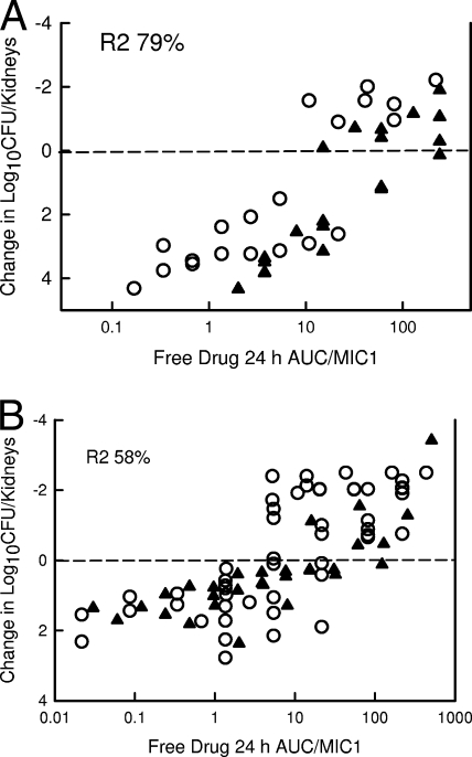
Recent pharmacodynamic studies with the echinocandin anidulafungin in this model with the same organisms demonstrated a similar pharmacodynamic relationship (9). The 24-h free-drug AUC/MIC values associated with a static effect and 1-log reduction were 18 ± 15 and 39 ± 45, respectively. However, if one only considers total drug concentrations, it would appear that the micafungin exposure needed for efficacy (AUC/MIC) was more than 1,000 AUC/MIC units greater than that for anidulafungin. However, with free-drug considerations, the values are similar (based on statistical comparison using the Mann-Whitney rank sum test [P = 0.53]) since the degree of protein binding is greater for micafungin than for anidulafungin. In fact, the entire dose-response relationship for these two echinocandins against this set of C. albicans and C. glabrata isolates is also very similar (Fig. 5).
FIG. 5.
(A) Relationship between micafungin (○) and anidulafungin (▴) free-drug 24-h AUC/MIC and efficacy against four C. albicans strains. Each symbol represents the mean CFU/kidneys from three mice. Efficacy on the y axis is expressed as the change in CFU/kidneys compared to the burden of organisms compared to the start of therapy. The dashed horizontal line represents the burden of organisms in thighs at the start of therapy. (B) Relationship between micafungin (○) and anidulafungin (▴) free-drug 24-h AUC/MIC and efficacy against ten C. glabrata strains. Each symbol represents the mean CFU/kidneys from three mice. Efficacy on the y axis is expressed as the change in CFU/kidneys compared to the burden of organisms compared to the start of therapy. The dashed horizontal line represents the burden of organisms in thighs at the start of therapy.
Human pharmacokinetics with micafungin demonstrate a long half-life and protein binding values essentially the same as those observed in mice (22, 24, 27, 34). In healthy volunteers, a dose of 100 mg/day produces a steady-state AUC of 126 mg·h/liter (free-drug value of 0.32 mg·h/liter). Recent population pharmacokinetic studies reported a similar exposure that was dependent upon the weight of the patient (range, 83 to 121 mg·h/liter) (22). A global survey of more than 2,500 target Candida isolates observed MICs varying from 0.007 to 2.0 μg/ml. The MIC50 and MIC90 in the present study were 0.015 and 0.03 μg/ml for C. albicans and 0.015 and 0.015 μg/ml for C. glabrata, respectively. If one considers the inhibitory pharmacodynamic targets identified in the current in vivo model, the 100-mg/day micafungin dosing regimen would exceed the free-drug 24-h AUC/MICs for both C. albicans and C. glabrata for more than 90% of organisms (9, 41, 42). Thus far, there is minimal clinical data to reliably discern the impact of echinocandin MICs on treatment outcome. The current in vivo pharmacodynamic studies would suggest that at least for C. albicans and C. glabrata the lack of correlation between MIC and outcome is related to the very low MIC distribution observed in these clinical trials (29, 32, 37, 44).
In summary, these studies identify the pharmacodynamic target needed to achieve a static and killing endpoint against C. albicans and C. glabrata in a neutropenic murine disseminated candidiasis model. Consideration of this target relative to human micafungin pharmacokinetics for the 100-mg/day dosing regimen would predict that this regimen achieves an inhibitory pharmacodynamic target against C. albicans and C. glabrata organisms with MICs up to 0.06 μg/ml. Future studies should attempt to examine the impact of higher MICs observed with the C. parapsilosis species and the less-susceptible C. albicans and C. glabrata.
Acknowledgments
This research was funded by a grant from Astellas.
Footnotes
Published ahead of print on 14 July 2008.
REFERENCES
- 1.Ambrose, P. G., S. M. Bhavnani, C. M. Rubino, A. Louie, T. Gumbo, A. Forrest, and G. L. Drusano. 2007. Pharmacokinetics-pharmacodynamics of antimicrobial therapy: it's not just for mice anymore. Clin. Infect. Dis. 44:79-86. [DOI] [PubMed] [Google Scholar]
- 2.Andes, D., K. Marchillo, J. Lowther, A. Bryskier, T. Stamstad, and R. Conklin. 2003. In vivo pharmacodynamics of HMR 3270, a glucan synthase inhibitor, in a murine candidiasis model. Antimicrob. Agents Chemother. 47:1187-1192. [DOI] [PMC free article] [PubMed] [Google Scholar]
- 3.Andes, D., and W. A. Craig. 2007. In vivo pharmacodynamic activity of the glycopeptide dalbavancin. Antimicrob. Agents Chemother. 51:1633-1642. [DOI] [PMC free article] [PubMed] [Google Scholar]
- 4.Andes, D., and M. I. van Ogtrop. 1999. Characterization and quantitation of the pharmacodynamics of fluconazole in a neutropenic murine model of disseminated candidiasis model. Antimicrob. Agents Chemother. 43:2116-2120. [DOI] [PMC free article] [PubMed] [Google Scholar]
- 5.Andes, D., and M. I. van Ogtrop. 2000. In vivo characterization of the pharmacodynamics of flucytosine in a neutropenic murine candidiasis model. Antimicrob. Agents Chemother. 44:938-942. [DOI] [PMC free article] [PubMed] [Google Scholar]
- 6.Andes, D. 2001. In vivo pharmacodynamics of amphotericin B against selected Candida species. Antimicrob. Agents Chemother. 45:922-926. [DOI] [PMC free article] [PubMed] [Google Scholar]
- 7.Andes, D. 2003. In vivo pharmacodynamics of antifungal drugs in treatment of candidiasis. Antimicrob. Agents Chemother. 47:1179-1186. [DOI] [PMC free article] [PubMed] [Google Scholar]
- 8.Andes, D., K. Marchillo, J. Nett, A. Pitula, and J. Smith. 2006. In vivo fluconazole pharmacodynamics and resistance development in a previously susceptible Candida albicans population examined by microbiologic and transcriptional profiling. Antimicrob. Agents Chemother. 50:2384-2394. [DOI] [PMC free article] [PubMed] [Google Scholar]
- 9.Andes, D., D. J. Diekema, M. A. Pfaller, R. A. Prince, K. Marchillo, J. Ashbeck, and J. Hou. 2008. In vivo pharmacodynamic characterization of anidulafungin in a neutropenic murine candidiasis model. Antimicrob. Agents Chemother. 52:539-550. [DOI] [PMC free article] [PubMed] [Google Scholar]
- 10.Buell, D., L. Kovanda, T. Drake, et al. 2007. Alternative day dosing of micafungin in the treatment of esophageal candidiasis, abstr. M719. 47th Intersci. Conf. Antimicrob. Agents Chemother. American Society for Microbiology, Washington, DC.
- 11.Chandrasekar, P. H., and J. D. Sobel. 2006. Micafungin: a new echinocandin. Clin. Infect. Dis. 42:1171-1178. [DOI] [PubMed] [Google Scholar]
- 12.Clinical Laboratory Standards Institute. 2002. Reference method for broth dilution antifungal susceptibility testing of yeasts; approved standard. Document M27-A2. National Committee for Clinical Laboratory Standards, Wayne, PA.
- 13.Cota, J., M. Carden, J. R. Graybill, L. K. Najvar, D. S. Burgess, and N. P. Wiederhold. 2006. In vitro pharmacodynamics of anidulafungin and caspofungin against Candida glabrata isolates, including strains with decreased caspofungin susceptibility. Antimicrob. Agents Chemother. 50:3926-3928. [DOI] [PMC free article] [PubMed] [Google Scholar]
- 14.Craig, W. A. 1998. Pharmacokinetic/pharmacodynamic parameters: rationale for antibacterial dosing of mice and men. Clin. Infect. Dis. 26:1-12. [DOI] [PubMed] [Google Scholar]
- 15.Denning, D. W. 2003. Echinocandin antifungal drugs. Lancet 362:1142-1151. [DOI] [PubMed] [Google Scholar]
- 16.Drusano, G. L. 2004. Antimicrobial pharmacodynamics: critical interactions of bug and drug. Nat. Rev. Microbiol. 2:289-300. [DOI] [PubMed] [Google Scholar]
- 17.Ernst, E. J., M. E. Klepser, and M. A. Pfaller. 2000. Postantifungal effects of echinocandin, azole, and polyene antifungal agents against Candida albicans and Cryptococcus neoformans. Antimicrob. Agents Chemother. 44:1108-1111. [DOI] [PMC free article] [PubMed] [Google Scholar]
- 18.Ernst, E. J., M. E. Klepser, M. E. Ernst, S. A. Messer, and M. A. Pfaller. 1999. In vitro pharmacodynamic properties of MK-0991 determined by timekill methods. Diagn. Microbiol. Infect. Dis. 33:75-80. [DOI] [PubMed] [Google Scholar]
- 19.Ernst, M. E., M. E. Klepser, E. J. Wolfe, and M. A. Pfaller. 1996. Antifungal dynamics of LY 303366, an investigational echinocandin B analog, against Candida subsp. Diagn. Microbiol. Infect. Dis. 26:125-131. [DOI] [PubMed] [Google Scholar]
- 20.Espinel-Ingroff, A. 2003. In vitro antifungal activities of anidulafungin and micafungin, licensed agents, and the investigational triazole posaconazole as determined by NCCLS methods for 12,052 fungal isolates: review of the literature. Rev. Iberoam. Micol. 20:121-136. [PubMed] [Google Scholar]
- 21.Gumbo, T., G. L. Drusano, W. Liu, R. W. Kulawy, C. Fregeau, V. Hsu, and A. Louie. 2007. Once-weekly micafungin therapy is as effective as daily therapy for disseminated candidiasis in mice with persistent neutropenia. Antimicrob. Agents Chemother. 51:968-974. [DOI] [PMC free article] [PubMed] [Google Scholar]
- 22.Gumbo, T., J. Hiemenz, L. Ma, J. J. Keirns, D. N. Buell, and G. L. Drusano. 2008. Population pharmacokinetics of micafungin in adult patients. Diagn. Microbiol. Infect. Dis. 60:329-331. [DOI] [PubMed] [Google Scholar]
- 23.Hakki, M., J. F. Staab, and K. A. Marr. 2006. Emergence of a Candida krusei isolate with reduced susceptibility to caspofungin during therapy. Antimicrob. Agents Chemother. 50:2522-2524. [DOI] [PMC free article] [PubMed] [Google Scholar]
- 24.Hebert, M. F., H. E. Smith, T. C. Marbury, K. S. Swan, W. B. Smith, W. S. Townsend, D. Buell, J. Keirns, and I. Bekersky. 2005. Pharmacokinetics of micafungin in healthy volunteers, volunteers with moderate liver disease, and volunteers with renal dysfunction. J. Clin. Pharmacol. 45:1145. [DOI] [PubMed] [Google Scholar]
- 25.Laverdiere, M., R. G. Lalonde, J. G. Baril, D. C. Sheppard, S. Park, and D. S. Perlin. 2006. Progressive loss of echinocandin activity following prolonged use for treatment of Candida albicans oesophagitis. J. Antimicrob. Chemother. 57:705-708. [DOI] [PubMed] [Google Scholar]
- 26.Louie, A., M. Deziel, W. Liu, M. F. Drusano, T. Gumbo, and G. L. Drusano. 2005. Pharmacodynamics of caspofungin in a murine model of systemic candidiasis: importance of persistence of caspofungin in tissues to understanding drug activity. Antimicrob. Agents Chemother. 49:5058-5068. [DOI] [PMC free article] [PubMed] [Google Scholar]
- 27.Keirns, J., T. Sawamoto, M. Holum, D. Buell, W. Wisemandle, and A. Alak. 2007. Steady-state pharmacokinetics of micafungin and voriconazole after separate and concomitant dosing in healthy adults. Antimicrob. Agents Chemother. 51:787-790. [DOI] [PMC free article] [PubMed] [Google Scholar]
- 28.Krogh-Madsen, M., M. C. Arendrup, L. Heslet, and J. Dahl Knudsen. 2006. Amphotericin B and caspofungin resistance in Candida glabrata isolates recovered from a critically ill patient. Clin. Infect. Dis. 42:938-944. [DOI] [PubMed] [Google Scholar]
- 29.Kuse, E. R., P. Chetchotisakd, C. A. da Cunha, M. Ruhnke, C. Barrios, D. Raghunadharao, J. S. Sekhon, A. Freire, V. Ramasubramanian, I. Demeyer, M. Nucci, A. Leelarasamee, F. Jacobs, J. Decruyenaere, D. Pittet, A. J. Ullmann, L. Ostrosky-Zeichner, O. Lortholary, S. Koblinger, H. Diekmann-Berndt, and O. A. Cornely. 2007. Micafungin versus liposomal amphotericin B for candidaemia and invasive candidosis: a phase III randomized double-blind trial. Lancet 369:1519-1527. [DOI] [PubMed] [Google Scholar]
- 30.Martinez, M., and D. A. Stevens. 2006. Bioassay for azoles in the presence of caspofungin and visa versa, abstr. A2076. 46th Intersci. Conf. Antimicrob. Agents Chemother. American Society for Microbiology, Washington, DC.
- 31.Miller, C. D., B. W. Lomaestro, S. Park, and D. S. Perlin. 2006. Progressive esophagitis caused by Candida albicans with reduced susceptibility to caspofungin. Pharmacotherapy 26:877-880. [DOI] [PubMed] [Google Scholar]
- 32.Mora-Durate, J. M., R. Betts, C. Rotstein, A. Pescolombo, L. Thompson-Moya, J. S. Meitana, R. Lupinacci, C. Sable, N. Kartsonis, and J. Perfect. 2002. Comparison of caspofungin and amphotericin B for invasive candidiasis. N. Engl. J. Med. 347:2020-2029. [DOI] [PubMed] [Google Scholar]
- 33.Moudgal, V., T. Little, D. Boikov, and J. A. Vazquez. 2005. Multiechinocandin- and multiazole-resistant Candida parapsilosis isolates serially obtained during therapy for prosthetic valve endocarditis. Antimicrob. Agents Chemother. 49:767-769. [DOI] [PMC free article] [PubMed] [Google Scholar]
- 34.Nakagawa, Y., Y. Ichii, Y. Saeki, M. Kodaka, K. Suzuki, and S. Kishino. 2007. Plasma concentration of micafungin in patients with hematologic malignancies. J. Infect. Chemother. 13:39-45. [DOI] [PubMed] [Google Scholar]
- 35.Ota, Y., K. Tatsuno, S. Okugawa, S. Yanagimoto, T. Kitazawa, A. Fukushima, K. Tsukada, and K. Koike. 2007. Relationship between the initial dose of micafungin and its efficacy in patients with candidemia. J. Infect. Chemother. 13:208-212. [DOI] [PubMed] [Google Scholar]
- 36.Paderu, P., G. Garcia-Effron, S. Balashov, G. Delmas, S. Park, and D. S. Perlin. 2007. Serum differentially alters the antifungal properties of echinocandin drugs. Antimicrob. Agents Chemother. 51:2253-2256. [DOI] [PMC free article] [PubMed] [Google Scholar]
- 37.Pappas, P. G., J. H. Rex, J. Lee, R. J. Hamill, R. A. Larsen, W. Powderly, C. A. Kauffman, N. Hyslop, J. E. Mangino, S. Chapman, H. W. Horowitz, J. E. Edwards, W. E. Dismukes, et al. 2003. A prospective observational study of candidemia: epidemiology, therapy, and influences on mortality in hospitalized adult and pediatric patients. Clin. Infect. Dis. 37:634-643. [DOI] [PubMed] [Google Scholar]
- 38.Pappas, P. G., C. M. Rotstein, R. F. Betts, M. Nucci, D. Talwar, J. J. De Waele, J. A. Vazquez, B. F. Dupont, D. L. Horn, L. Ostrosky-Zeichner, A. C. Reboli, B. Suh, R. Digumarti, C. Wu, L. L. Kovanda, L. J. Arnold, and D. N. Buell. 2007. Micafungin versus caspofungin for treatment of candidemia and other forms of invasive candidiasis. Clin. Infect. Dis. 45:883-893. [DOI] [PubMed] [Google Scholar]
- 39.Petraitiene, R., V. Petraitis, A. H. Groll, T. Sein, R. L. Schaufele, A. Francesconi, J. Bacher, N. A. Avila, and T. J. Walsh. 2002. Antifungal efficacy of caspofungin (MK-0991) in experimental pulmonary aspergillosis in persistently neutropenic rabbits: pharmacokinetics, drug disposition, and relationship to galactomannan antigenemia. Antimicrob. Agents Chemother. 46:12-23. [DOI] [PMC free article] [PubMed] [Google Scholar]
- 40.Petraitis, V., R. Petraitiene, A. H. Groll, K. Roussillon, M. Hemmings, C. A. Lyman, T. Sein, C. Bacher, I. Bekersky, and T. J. Walsh. 2002. Comparative antifungal activities and plasma pharmacokinetics of micafungin (FK463) against disseminated candidiasis and invasive pulmonary aspergillosis in persistently neutropenic rabbits. Antimicrob. Agents Chemother. 46:1857-1869. [DOI] [PMC free article] [PubMed] [Google Scholar]
- 41.Pfaller, M. A., L. Boyken, R. J. Hollis, S. A. Messer, S. Tendolkar, and D. J. Diekema. 2006. Global surveillance of in vitro activity of micafungin against Candida: a comparison with caspofungin by CLSI-recommended methods. J. Clin. Microbiol. 44:3533-3538. [DOI] [PMC free article] [PubMed] [Google Scholar]
- 42.Pfaller, M. A., and D. J. Diekema. 2007. Epidemiology of invasive candidiasis: a persistent public health problem. Clin. Microbiol. Rev. 20:133-163. [DOI] [PMC free article] [PubMed] [Google Scholar]
- 43.Pfaller, M. A., D. J. Diekema, and D. J. Sheehan. 2006. Interpretive breakpoints for fluconazole and Candida revisited: a blueprint for the future of antifungal susceptibility testing. Clin. Microbiol. Rev. 19:435-447. [DOI] [PMC free article] [PubMed] [Google Scholar]
- 44.Reboli, A. C., C. Rotstein, P. G. Pappas, S. W. Chapman, D. H. Kett, D. Kumar, R. Betts, M. Wible, B. P. Goldstein, J. Schranz, D. S. Krause, T. J. Walsh, et al. 2007. Anidulafungin versus fluconazole for invasive candidiasis. N. Engl. J. Med. 356:2472-2482. [DOI] [PubMed] [Google Scholar]
- 45.Rex, J. H., M. A. Pfaller, J. N. Galgiani, M. S. Bartlett, A. Espinel-Ingroff, M. A. Ghannoum, M. Lancaster, F. C. Odds, M. G. Rinadli, T. J. Walsh, A. L. Barry, et al. 1997. Development of interpretive breakpoints for antifungal susceptibility testing: conceptual framework and analysis of in vitro and in vivo correlation data for fluconazole, itraconazole, and Candida infections. Clin. Infect. Dis. 24:235-247. [DOI] [PubMed] [Google Scholar]
- 46.Vazquez, J. A., and J. D. Sobel. 2006. Anidulafungin: a novel echinocandin. Clin. Infect. Dis. 43:215-222. [DOI] [PubMed] [Google Scholar]
- 47.Walsh, T. J., J. Lee, P. Kelly, J. Bacher, J. Leeciones, V. Thomas, C. L. Lyman, D. Coleman, R. Gordee, and P. A. Pizzo. 1991. Antifungal effects of the nonlinear pharmacokinetics of cilofungin, a 1,3-3-glucan synthetase inhibitor, during continuous and intermittent intravenous infusions in treatment of experimental disseminated candidiasis. Antimicrob. Agents Chemother. 35:1321-1328. [DOI] [PMC free article] [PubMed] [Google Scholar]
- 48.Warn, P. A., A. Sharp, G. Morrissey, and D. W. Denning. 2002. In vivo activity of micafungin in a persistently neutropenic murine model of disseminated infection caused by Candida tropicalis. J. Antimicrob. Chemother. 50:1071-1074. [DOI] [PubMed] [Google Scholar]
- 49.Wiederhold, N. P., L. K. Najvar, R. Bocanegra, D. Molina, M. Olivo, and J. R. Graybill. 2007. In vivo efficacy of anidulafungin and caspofungin against Candida glabrata and association with in vitro potency in the presence of sera. Antimicrob. Agents Chemother. 51:1616-1620. [DOI] [PMC free article] [PubMed] [Google Scholar]
- 50.Wiederhold, N. P., and R. E. Lewis. 2003. The echinocandin antifungals: an overview of the pharmacology, spectrum, and clinical efficacy. Expert Opin. Investig. Drugs 12:1313-1333. [DOI] [PubMed] [Google Scholar]
- 51.Yamato, Y., H. Kanedo, T. Hashimoto, M. Katashima, K. Ishibashi, A. Kawamura, M. Terakawa, and A. Kagayama. 2002. Pharmacokinetics of the antifungal drug micafungin in mice, rats, and dogs and in its in vitro protein binding and distribution to blood cells. Jpn. J. Chemother. 50:74-79. [Google Scholar]


