Abstract
Although extensively studied biochemically, members of the Protein 4.1 superfamily have not been as well characterized genetically. Studies of coracle, a Drosophila Protein 4.1 homologue, provide an opportunity to examine the genetic functions of this gene family. coracle was originally identified as a dominant suppressor of EgfrElp, a hypermorphic form of the Drosophila Epidermal growth factor receptor gene. In this article, we present a phenotypic analysis of coracle, one of the first for a member of the Protein 4.1 superfamily. Screens for new coracle alleles confirm the null coracle phenotype of embryonic lethality and failure in dorsal closure, and they identify additional defects in the embryonic epidermis and salivary glands. Hypomorphic coracle alleles reveal functions in many imaginal tissues. Analysis of coracle mutant cells indicates that Coracle is a necessary structural component of the septate junction required for the maintenance of the transepithelial barrier but is not necessary for apical–basal polarity, epithelial integrity, or cytoskeletal integrity. In addition, coracle phenotypes suggest a specific role in cell signaling events. Finally, complementation analysis provides information regarding the functional organization of Coracle and possibly other Protein 4.1 superfamily members. These studies provide insights into a range of in vivo functions for coracle in developing embryos and adults.
INTRODUCTION
The Protein 4.1 gene superfamily consists of a functionally diverse group of proteins that nonetheless share highly conserved structural features. Members of this family include Protein 4.1, Drosophila Coracle, Merlin (the protein product of the Neurofibromatosis 2 [NF2] gene), the Ezrin, Radixin, and Moesin (ERM) proteins, talin, Drosophila Expanded, several protein tyrosine phosphatases, and others (Rees et al., 1990; McCartney and Fehon, 1997). All contain a functional domain of 200–300 aa that is typically found in the N-terminal half of the protein and is thought to interact with the cytoplasmic domain of particular transmembrane proteins, thereby localizing the protein to the cytoplasmic face of the plasma membrane (Rees et al., 1990). In addition, Protein 4.1 contains within this region sites for interaction with p55 (Alloisio et al., 1993) and hDLG (Lue et al., 1994), members of the membrane-associated guanylate kinase homologues family of proteins, which also interact with the cytoplasmic tail of transmembrane proteins. Some members of the Protein 4.1 and membrane-associated guanlylate kinase homologues superfamilies are found at intercellular junctions, the primary sites of cell–cell contact and cellular communication (Fehon et al., 1997; Ponting et al., 1997). Recent work, in particular the identification of the NF 2 tumor suppressor gene as a member of this family (Rouleau et al., 1993; Trofatter et al., 1993), has generated considerable interest in possible functions of these proteins in mediating cellular interactions that occur within the junctional complex.
Although most functional data regarding Protein 4.1 family members are based on biochemical studies of the proteins they encode, limited genetic data on some of these genes are also available. In humans, loss of Merlin function results in the bilateral schwannomas and other benign tumors that characterize the NF2 disease (Martuza and Eldridge, 1988). Homozygous mutant mice in which this gene has been “knocked out” fail to survive to the point of embryonic gastrulation and have defects in the extra-embryonic tissues (McClatchey et al., 1997). Protein 4.1 mutations in humans have been associated with hemolytic anemias, but although this protein is widely expressed, defects in other tissues have not been reported (Conboy et al., 1993). Injection of Protein 4.1 antisense oligonucleotides into Xenopus embryos results in various defects, including retinal degeneration and abnormal body size (Giebelhaus et al., 1988). In addition, studies of Drosophila expanded, a divergent member of this superfamily, indicate that like the NF2 gene, expanded seems to be necessary to restrict cellular proliferation (Boedigheimer and Laughon, 1993; Boedigheimer et al., 1993). However, with the exception of expanded, none of these genes has been studied in a system that is amenable to mutagenesis and phenotypic characterization.
To better understand the in vivo functions of these proteins, we have initiated a molecular–genetic analysis of coracle, a Drosophila homologue of Protein 4.1. Severe coracle mutations result in a failure of dorsal closure and lethality late in the process of embryonic development (Fehon et al., 1994). Confocal and immunoelectron microscopy using specific antibodies have shown that Coracle localizes to septate junctions, a primary site of cell–cell contact and growth regulation in Drosophila epithelial cells. Other proteins known to be associated with this junctional region include the products of the discs large (dlg) (Woods and Bryant, 1989) and Neurexin (Nrx) (Baumgartner et al., 1996) genes. Mutations in dlg result in disrupted apical–basal cellular polarity and overgrowth in the imaginal epithelia (Abbott and Natzle, 1992; Woods et al., 1997), whereas mutations in Nrx show dorsal closure defects and disruption of the blood–brain barrier (Baumgartner et al., 1996). In addition, coracle mutations dominantly suppress eye phenotypes associated with the EgfrElp mutation, a gain of function mutation in Egfr (Baker and Rubin, 1992; Fehon et al., 1994). Mutations in Egfr have been shown to affect cell proliferation in imaginal discs (Diaz-Benjumea and Garcia-Bellido, 1990; Xu and Rubin, 1993). Together, these results implicate the septate junction in mediating cellular interactions necessary for normal growth control in Drosophila epithelia.
We present here the results of screens for new coracle alleles, and their embryonic and adult phenotypes. One advantage of such an approach is that is does not require previous knowledge of the functions of a particular gene, nor do such notions bias it if they do exist. Our results indicate that coracle provides essential functions throughout development in various epithelia, including the embryonic epidermis and salivary glands, and adult structures such as the eyes, wings, ocelli, and other tissues. We provide evidence that coracle function is required for the maintenance of the transepithelial barrier function of the septate junction and suggest that this role of the septate junction is crucial for the establishment of a unique apical environment. Interestingly, despite its structural similarity to a vertebrate cytoskeletal protein and its localization to a major component of the apical junctional complex, Coracle does not appear to be required for epithelial integrity, apical–basal polarity, or organization of the actin cytoskeleton.
MATERIALS AND METHODS
Isolation of coracle Alleles
The coracle alleles used in this study were generated in three independent genetic screens. cor1 and cor2 were isolated and characterized previously (Fehon et al., 1994). Further alleles were isolated in an F2 lethal screen. cn; ry506 males were mutagenized with methane–sulfonic acid ethyl ester and mated to y w; Sco/CyO females according to standard procedures (Grigliatti, 1986). F1 males were individually mated to y w; cor2 px sp/CyO females, and the resulting F2 progeny were screened for lethality over the cor2 px sp chromosome. Fertile F1 crosses (10,046) were screened, and 12 independent alleles, cor3–cor14, were recovered. All chromosomes were “cleaned” by recombination of flanking markers. Except where noted in Table 1, all lines are rescuable in homozygous condition by a Ubiquitin-promoter–driven cor+ cDNA transgene, P{w+mc, Up-cor+=cor+}, which we abbreviate as the P{Up-cor+} transgene (Fehon et al., 1994).
Table 1.
Summary of coracle alleles
| Allele | Lethal period | Embryonic phenotypea | Adult escapers | P{Up-cor+} rescue? | Phenotypic categoryb | EgfrElp suppression |
|---|---|---|---|---|---|---|
| cor1 | E | DS/FC | N | Y | I | +++ |
| cor2 | E | DH/FC | N | Y | S | +++ |
| cor3 | E | DH/FC | N | Y | S | + |
| cor4 | E | DH/FC | N | Y | S | ++ |
| cor5 | E | DH/FC | N | Y | S | ++ |
| cor6 | E | AO/DH/FC | N | P | S | ++ |
| cor7 | E/L | FC | Y | Y | I | +/++ |
| cor8 | E/L | FC | Y | P | W | + |
| cor9 | E/L | FC | N | N | W | N |
| cor10 | E/L | FC | Y | Y | W | + |
| cor11 | E/L | FC | N | N | W | N |
| cor12 | E | DS/FC | N | Y | I | ++++ |
| cor13 | E | DS/FC | N | Y | I | +++ |
| cor14 | L | N | Y | Y | W | ++ |
| cor15 | N | N | Y | Y | W | ++++ |
Abbreviations: DS, dorsal scab; DH, dorsal hole; AO, anterior open; FC, faint cuticle; E, embryo; L, larva; N, none; Y, yes; P, partial; I, intermediate; S, strong; W, weak; ++++, eye appears wild-type; +++, eye is wild-type in size with some roughening; ++, eye is smaller than wild-type with some roughening; +, eye is less rough than an EgfrElp eye in a wild-type background.
See text for explanation of categories.
cor15 was isolated in a separate F1 visible screen. cn; ry506 males were mutagenized with methane–sulfonic acid ethyl ester and mated to cor2 px sp/CyO females according to standard procedures (Grigliatti, 1986). The resulting F1 progeny were screened for males in which a visible phenotype in the eye or other cuticular structures were evident over cor2 px sp. F1 males (24,590) were screened, and 214 individuals were selected. Individuals that bred true and segregated with the second chromosome were retained for further analysis. In this screen one allele, cor15, was isolated.
Complementation Analysis
We performed pairwise crosses between all coracle alleles plus two deficiencies that uncover the coracle locus. Crosses were performed in duplicate at 18 and 25°. Embryos were collected on apple juice agar plates as described (Wieschaus et al., 1984). A total of 250 embryos per cross were followed, and the level of embryonic lethality was determined. Larvae that crawled away were transferred to vials, and the number of larvae that pupariated was determined. Finally, the number of adults that eclosed was counted. Sufficient numbers of offspring were examined so that at least 200 coracle mutant offspring would be expected if the allelic combination was viable. Mutant viability was calculated by dividing the number of mutant flies by the number of balancer class siblings that eclosed. Surviving coracle hypomorphs were examined for phenotypes. Scanning electron microscopy was performed on a Philips model 501 microscope (FEI Company, Hillsboro, OR) as described previously (Rebay et al., 1993). Fertility of the coracle hypomorphs was examined by crossing one to five corx/cory males and females to w1118 females and males, respectively, and examining vials for viable offspring.
Cuticle Preparations
Cuticle preparations were performed as described (Szüts et al., 1997) with the following modifications. The cuticles were expanded in 1× PBS, 0.1% Triton X-100 for 20 min at 65°C, and then allowed to settle overnight at room temperature through diluted Hoyer’s solution (2:1:1, Hoyer’s:lactic acid:ddH2O). Cuticles were then mounted in diluted Hoyer’s solution and allowed to clear for several days.
Transmission Electron Microscopy
Fixation of embryos for transmission electron microscopy was performed as described (Tepass and Hartenstein, 1994) with the following modifications. After the devitellinization step, the late stage 17 embryos were bisected midway between the anterior and posterior ends. Additionally, the electron microscopy fixation and osmication steps were doubled in length to 4 and 2 h, respectively. A subset of embryos was devitellinized with methanol to investigate the cuticular phenotype, which was more apparent under these conditions. Sections (30–80 nm) were cut on an AO/Reichert Ultracut ultramicrotome (American Optical Scientific Instruments, Buffalo, NY) and were analyzed on a Zeiss EM 10A electron microscope (Carl Zeiss, Oberkochen, West Germany).
Dye Permeability Experiments
Progeny from a cross of cor5/CyO, P{w+; hsMerlinGFP} were collected for 1 h, aged for 10–12 h, and then heat-shocked for 1 h at 38°C to induce the expression of the Merlin-GFP transgene in all heterozygous and homozygous balancer class embryos. The embryos were subsequently aged 3–4 h and then dechorionated in 50% commercial bleach (12% sodium hypochlorite). The embryos were arrayed on apple juice plates, transferred to double-stick tape on glass coverslips, and desiccated in a closed container containing Drierite (W. A. Hammond Drierite Company, Xenia, OH) for 20 min. The embryos were then covered with halocarbon oil (Halocarbon, North Augusta, SC). Rhodamine-labeled dextran (Mr, 10,000; Molecular Probes, Eugene, OR) in injection buffer (Rubin and Spradling, 1982) was then injected into the hemocoel using a micromanipulator under a microscope, and the embryos were examined on a Zeiss LSM 410 laser scanning confocal microscope with a krypton/argon laser (Carl Zeiss, Thornwood, NY).
Genetic Interactions with Egfr
Ten corx/CyO males were mated to 20 EgfrElp/CyO virgin females, and the corx/EgfrElp offspring were examined for suppression of the EgfrElp/+ dominant rough eye phenotype. Crosses were performed at 25°C. Specimens were prepared for scanning electron microscopy on a Philips model 501 microscope (FEI Company, Hillsboro, OR) as described previously (Rebay et al., 1993).
Clonal Analysis
The cor4 allele was crossed onto an FRT43D chromosome (Xu and Rubin, 1993). w1118, hsFLP; FRT43D P{y+} virgin females were mated to y w; FRT43D cor4/CyO males. Mitotic clones were induced 76 h after egg laying by two 1-h heat shocks at 38°C separated by a 1-h 25°C recovery and examined in late third instar imaginal discs.
Immunofluorescence
Embryos aged 0–14 h were collected from y w; FRT43D cor5/CyO adults and fixed and stained as described previously (Fehon et al., 1991). Third instar wing imaginal discs were dissected, fixed, and stained as described previously (Fehon et al., 1994). Primary antibodies were used at the following dilutions: guinea pig anti-Coracle, 1:10,000 (embryos) or 1:5000 (discs); mouse anti-Neurexin, 1:1000; mouse anti-Notch (C17.9C6), 1:3000; rabbit anti-Armadillo (gift from M. Peifer, University of North Carolina, Chapel Hill, NC), 1:500; mouse anti-Crumbs (gift from E. Knust, Universität Düsseldorf, Düsseldorf, Germany), 1:100; rabbit anti–βH-Spectrin (gift from G. Thomas, Pennsylvania State University, University Park, PA), 1:2000; rabbit anti–α-Spectrin (gift from D. Kiehart, Duke University, Durham, NC) 1:1000; and mouse anti-Moesin, 1:10,000. All secondary antibodies were from Jackson ImmunoResearch (West Grove, PA). Rhodamine-conjugated phalloidin (Molecular Probes) was added with the primary antibody at a dilution of 1:1000.
RESULTS
Isolation of coracle Point Mutations and Deficiencies
The coracle gene was simultaneously identified as a Drosophila Protein 4.1 homologue and in a screen for dominant suppressors of the rough eye phenotype caused by EgfrElp (Fehon et al., 1994). Initial phenotypic analysis of two alleles, cor1 and cor2, indicated that coracle has an essential role during embryonic dorsal closure. However, the limited number of alleles and the lack of an existing deficiency with which to examine the null phenotype limited our ability to thoroughly examine coracle phenotypes in embryonic and adult development. To better understand the functions of coracle, we have conducted genetic screens for new coracle alleles, resulting in the identification of 13 new coracle mutations (Table 1).
To aid in the characterization of these alleles, we have also identified two small deficiencies that uncover coracle. One was originally identified as a gamma ray-induced allele of enabled, Df(2R)enbGC8, which had been mapped to the 56A-F interval (Konsolaki and Schupbach, 1998). The second deficiency, Df(2R)corP97, was induced by imprecise excision of P{white-un3}AA48 in region 56B (B. McCartney and R. Fehon, unpublished data). Both of these deficiencies fail to complement the cor1 and cor2 point mutations (as well as enb point mutations), and quantitative hybridization analysis using a coracle cDNA probe shows that both deficiencies remove all coracle coding sequences (our unpublished results). Thus, these deficiencies are useful for determining the null coracle phenotype and the genetic function of coracle point mutations.
Classification of coracle Alleles
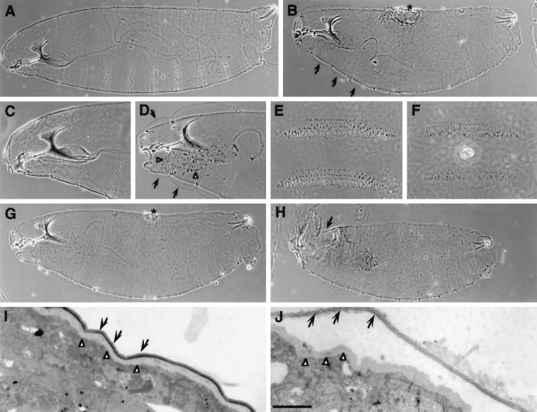
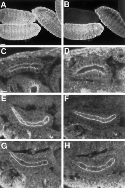
To assess its severity, each of the coracle alleles was examined in homozygous condition and in trans with Df(2R)corP97. We then ranked these alleles into three classes, strong, intermediate, and weak, on the basis of the severity of embryonic phenotype when in trans with Df(2R)corP97 (Table 1). Strong alleles are characterized by moderately to highly penetrant dorsal defects (dorsal hole or scab) (Figure 1, B and G), whereas intermediate alleles are embryonic lethal but only rarely display dorsal defects. Weak alleles are either embryonic or larval lethal and show no dorsal defects. Two alleles, cor14 and cor15, show no embryonic lethality when homozygous or in trans with the deficiency and are classified as weak alleles. Most strong and all intermediate alleles show a greater penetrance of the dorsal closure phenotype when in trans with Df(2R)corP97 than when homozygous, indicating that the dorsal closure defect represents the null phenotype for the coracle locus. Only one allele, cor5, displays a consistently strong dorsal closure phenotype in either condition. cor5 mutant embryos also fail to express any detectable Coracle protein (Figure 2B), whereas all other alleles express detectable amounts of Coracle (our unpublished results). We therefore conclude that cor5 is a null allele.
Figure 1.
Cuticular phenotypes of coracle mutant embryos. Phase-contrast photomicrographs of wild-type (A, C, and E) and coracle mutant embryos (B, D, and F). (A) Wild-type embryonic cuticular pattern, lateral view. (B) A similarly oriented cor5/Df(2)corP97 embryo demonstrating the characteristic dorsal hole (*) and detached cuticle (arrows) phenotypes. (C and D) The anterior ends of wild-type and cor2/Df(2)corP97 mutant embryos, respectively. Delaminated cuticle (arrows) in the coracle mutant embryo is obvious in the naked area of each segment. In addition, the necrotic remains of the salivary glands characteristic of coracle mutant embryos are apparent (arrowhead). (E and F) High-magnification views of the ventral denticle belts from the third and fourth abdominal segments in a wild-type (E) and a cor5/Df(2)corP97 mutant embryo (F). Note that there are fewer denticles and that they are less distinctly formed in coracle mutants, although the overall pattern appears normal. (G and H) Differences between the phenotypes of cor6/Df(2)corP97 (G) and cor6/cor6 (H). Although cor6/Df(2)corP97 embryos show a typical severe coracle embryonic phenotype with a dorsal hole (*), cor6/cor6 embryos display a clear anterior–open phenotype (arrow) that is not observed in any of the other coracle alleles. (I and J) Transmission electron micrographs of the epidermis and cuticle of late stage 17 wild-type (I) and cor5 (J) mutant embryos. Note that in the cor5 mutant tissue the epicuticle (black arrows) has separated from the procuticle (white arrowheads). Bar (I and J), 1 μm.
Figure 2.
Apical–basal cellular polarity is normal in coracle null mutant embryos. (A and B) Confocal projections of a field of embryos double-labeled with anti–α-Spectrin (A) as a labeling control and anti-Coracle (B) to demonstrate that cor5 mutant embryos do not express detectable amounts of Coracle. (C–H) Wild-type (C, E, and G) and cor5 mutant (D, F, and H) stage 14 embryonic salivary glands stained with anti-Armadillo (C and D), anti-Notch (E and F), and anti-Crumbs (G and H) antisera. The subcellular localization of all three proteins is normal in cor5 mutant cells. Bars, 50 μm (A and B), 10 μm (C–H).
One allele, cor6, is unusual in that it displays a highly penetrant anterior open phenotype in homozygous condition (Figure 1H), whereas when placed in trans over a deficiency it displays a characteristic dorsal closure phenotype (Figure 1G). This result indicates that either the cor6 chromosome carries a second site mutation that is epistatic to coracle or that the cor6 mutation conveys a neomorphic or possibly antimorphic function on the coracle gene. In support of the latter notion, we find that cor6 is rescuable in a dose-dependent manner by the P{Up-cor+} transgene (Fehon et al., 1994), although this rescue is incomplete (Table 1). One dose of P{Up-cor+} rescues 11% of the expected progeny, whereas two doses of the transgene rescue 29% of the expected progeny. By contrast, one dose of the P{Up-cor+} transgene rescues 96% of the expected cor5 homozygotes. In addition, the viability of cor6/+ heterozygotes is enhanced by P{Up-cor+} (our unpublished results), indicating that cor6 is partially dominant. On the basis of these results, we conclude that the cor6 allele has antimorphic functions.
Defects in coracle Mutant Embryonic and Imaginal Epithelial Cells
Several studies have suggested that coracle and some of the other genes required for dorsal closure may play a role in epithelial morphogenesis (reviewed in Noselli, 1998). To better assess the functions of coracle in the embryonic epidermis, we have examined phenotypes of null and strongly hypomorphic mutant embryos. Cuticle preparations of coracle mutant embryos display characteristic epidermal phenotypes in addition to dorsal closure defects. In coracle mutant embryos the cuticle appears to be thinner than normal and often appears to split into two layers in cuticle preparations (Figure 1, B, D, I, and J). The cuticular thinning is most obvious on the ventral surface where the denticle belts appear faint in cuticle preparations. Although the overall segmental pattern is normal, denticle belts contain fewer than normal denticles (Figure 1, E and F), and there are correspondingly fewer hairs on the dorsal side. Ultrastructural examination of the epidermis and cuticle reveals that the apparent delamination of the cuticle results from a failure of the epicuticle to adhere to the procuticle (Figure 1, I and J). All of the embryonic lethal alleles also display salivary gland defects, which are apparent as necrotic material that remains in cuticle preparations (Figure 1D). This salivary gland defect was observed in embryos that had been aged beyond the stage at which wild-type embryos hatch as larvae, suggesting that this necrosis is a very late effect.
The salivary glands have been shown previously to express coracle at high levels (Fehon et al., 1994; Ward et al., 1998). To study the effects of loss of coracle function at the cellular level in epithelial cells, we examined the morphology of salivary gland epithelia in cor5 mutant embryos using several molecular markers for the plasma membrane. These experiments were performed in midembryogenesis (stage 14), before the salivary gland necrosis described earlier is apparent. Markers for the adherens junction (Armadillo, Figure 2, C and D, and Notch, Figure 2, E and F) and the apical membrane (Crumbs, Figure 2, G and H) all displayed normal subcellular localizations in cor5 mutant cells. It is important to note that these embryos had no detectable Coracle protein and that coracle is not expressed maternally (Fehon et al., 1994). These results indicate that the apical–basal polarity of epithelial cells is not grossly affected by loss of Coracle function.
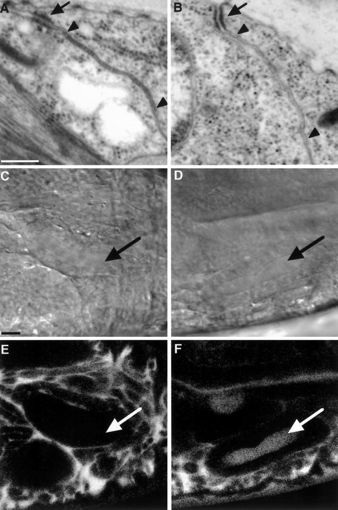
Baumgartner et al. (1996) reported that strongly hypomorphic alleles of Neurexin lack pleated septate junctions in ectodermal epithelia and display transepithelial barrier defects (Baumgartner et al., 1996). We reported recently (Ward et al., 1998) that in cor5 mutant embryos, Neurexin is mislocalized, raising the possibility that the integrity of the septate junction is compromised in coracle mutant embryos. To investigate this idea, we conducted an ultrastructural examination of the epidermis and internal epithelia of late stage 17 mutant embryos. Similar to the observations regarding loss of Neurexin, we found that the septate junction in cor5 mutant epithelia was disrupted. Although the adherens junction appeared unaffected, the individual septae that characterize the pleated septate junction were always absent in cor5 mutant tissue (Figure 3, compare A with B). We occasionally observed electron-dense material in the intermembranal space, but this material was not organized into an array of discrete septae.
Figure 3.
The ultrastructure and transepithelial barrier function of the septate junction is disrupted in the embryonic epithelia of coracle mutant embryos. (A and B) Transmission electron micrographs of the apical junctional complex in the epidermis of late stage 17 wild-type (A) and cor5 mutant (B) embryos. Although the adherens junction appears normal in the cor5 mutant tissue (black arrows; compare B with A), the region of the septate junction lacks the individual septae that characterize the pleated septate junction in the wild-type tissue (region between the arrowheads in A). (C–F) The transepithelial barrier function of the septate junction is disrupted in cor5 mutant embryos. (C–F) Differential interference contrast micrographs (C and D) and confocal optical sections (E and F) of stage 16 wild-type (C and E) and cor5 mutant (D and F) embryos showing the diffusion of a 10-kDa rhodamine–dextran injected into the hemocoel. Note that in the wild-type embryo the dextran fails to cross the salivary gland epithelium (arrows), whereas in the mutant embryo the dextran crosses the salivary gland epithelium as well as the epidermis. Bars, 250 nm (A and B), 10 μm (C–F).
To examine the functional consequences of this lack of an organized septate junction, we tested the transepithelial barrier function of coracle mutant epithelia using a 10-kDa rhodamine-labeled dextran. We injected this dye into the hemocoel of stage 16 embryos. Although in wild-type embryos the dextran was confined to the hemocoel even 1 h after injection, in cor5 mutant embryos the dextran rapidly crossed the salivary gland epithelium and filled the luminal space (Figure 3F). The epidermis, hindgut, and trachea were similarly compromised. Taken together, these results demonstrate the essential requirement for coracle in the integrity of the transepithelial barrier function of the septate junction.
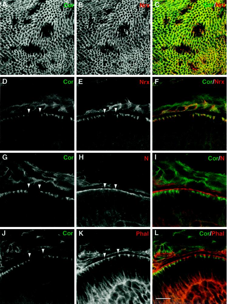
To examine further the effects of loss of coracle function in epithelial cells, we used somatic mosaic analysis to generate cor− cells in the imaginal epithelium. cor4 somatic clones were generated ∼76 h after egg laying using the FLP recombinase/FRT target system (Xu and Rubin, 1993). coracle mutant clones were observed in late third instar wing imaginal discs using anti-Coracle antibodies to identify mutant cells (Figure 4) (at least 10 clones were observed for each antiserum used). The cor4 allele was chosen for this analysis because it is a strong allele that disrupts the ability of Coracle to associate with the plasma membrane (Ward et al., 1998). Cells within the mutant clones were contiguous with the rest of the imaginal epithelium and appeared normally shaped. As previously described for embryonic tissues, we observed that loss of coracle function in imaginal epithelial cells resulted in disruption of Neurexin localization (Figure 4, A–F). In addition, Neurexin protein was not readily detectable in the center of cor− clones, suggesting that in imaginal epithelia Neurexin is not stable in the absence of coracle function. To assess apical–basal polarity and cytoskeletal organization in cor− cells, we stained imaginal discs with antibodies for Notch (Figure 4, G–I), βH-Spectrin, and Moesin (our unpublished results). As in the embryonic epidermis, these markers for the apical junctional region were localized normally in mutant cells. In addition, the distribution of filamentous actin was examined using rhodamine phalloidin and was also found to be normal (Figure 4, J–L). These results indicate that coracle function is not necessary for overall apical–basal polarity or cytoskeletal organization in imaginal tissues; however, when adult flies were examined for the presence of cor− tissue, no mutant clones could be observed either in the eye (using a w+ marker) or in the thorax (using a y+ marker for bristles), indicating that cor− cells do not persist to the adult stage (our unpublished results).
Figure 4.
Neurexin is affected but apical–basal polarity is normal in imaginal cells lacking coracle function. (A–L) Confocal micrographs of clones of cor4 cells that were induced at ∼76 h of development (early third instar) using the FLP/FRT system. A, D, G, and J display Coracle staining, B and E display Nrx staining, H displays Notch staining, and K displays filamentous actin stained with rhodamine–phalloidin. C, F, I, and L show merged images. (A–C) Projections of tangential optical sections through the apical portion of a wing imaginal disk that has been stained with anti-Coracle (A) and anti-Neurexin (B) antibodies. Note in B that in the absence of Coracle, Neurexin staining is also diminished. (D–L) Optical cross sections of the wing imaginal epithelium stained for Nrx and other apical membrane markers. Loss of Nrx staining in cor4 cells is also apparent in cross section (E; arrowheads mark borders of the clone), whereas staining for Notch (H), an apically localized transmembrane protein, and filamentous actin (K), which is concentrated in the apical junctional region, appear to be unaffected. Bar, 10 μm.
coracle Postembryonic Phenotypes
As shown above, the majority of coracle alleles are embryonic lethal; however, some weak alleles and several hypomorphic allelic combinations did produce adult escapers (Figure 6 and Table 1). For example, cor7 was mostly embryonic lethal, but it produced 7% of expected viable, fertile adults and can be maintained as a homozygous stock at 25°C. cor14 was almost exclusively larval lethal, but 0.3% of the expected homozygous class survived to viable adults. cor15 was completely viable, with 100% of the expected homozygotes surviving to adulthood. All of the coracle alleles that produced adult escapers displayed a similar range of adult defects (see below), although the severity and penetrance of the phenotypes varied with genotype. All of these alleles were also cold sensitive (our unpublished results).
Figure 6.
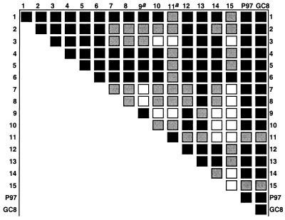
Complementation analysis of coracle alleles. Animals heterozygous for coracle alleles were crossed, and the percentage of expected cory/corx animals was calculated. Homozygous mutant phenotypes are indicated along the diagonal. Black indicates that the allelic combination was lethal, gray indicates that <75% of the expected progeny survive, and white indicates that 75% or more of the expected animals survive to adults. Note: a, not rescuable with P{Up-cor+}; P97, Df(2R)corP97; GC8, Df(2R)enbGC8.
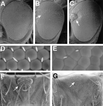
All coracle escapers display some degree of a rough eye phenotype. These eye defects ranged from a slight roughening of the eye, especially across the equator (Figure 5B), to eyes in which part of the eye tissue in the anterior equatorial region was replaced by head cuticle (Figure 5C). The penetrance of severe phenotypes increased with the strength of the coracle allele, to include up to 60% of the mutant adults. Histological sections show that the roughened appearance of coracle mutant eyes was caused by abnormally shaped, fused, improperly spaced, and occasionally misoriented ommatidia, rather than abnormal photoreceptor cell differentiation (our unpublished results). Essentially all of the ommatidia in the mutant eyes displayed normal numbers and positions of photoreceptors. Interommatidial bristles were also frequently lost in the roughened area (Figure 5E). Another very penetrant phenotype seen in coracle mutant animals was partial or total loss of the ocelli and associated bristles (Figure 5, compare G with F).
Figure 5.
Eye and head defects associated with coracle hypomorphs. Scanning electron micrographs of adult eyes (A–E; anterior is to the left) and dorsal surface of heads (F and G). (A) cor5/CyO (wild-type) eye showing normal morphology. (B) A cor2/cor15 eye displays roughening across the equator (arrow). (C) In a cor8/cor14 eye, an area of the eye has been replaced by head cuticle with associated bristles (arrow). (D) Higher-magnification view of a cor5/CyO (wild-type) eye near the equator. Notice the regular hexagonal shape of the ommatidia and the precise spacing of the bristles. (E) Higher-magnification view of a cor8/cor14 eye near the equator. Notice the irregularity in size and shape of the ommatidia and scarcity and misplacement of the bristles. (F) A dorsal view of a cor5/CyO (wild-type) head showing the normal arrangement of three ocelli and associated bristles. (G) A dorsal view of a cor4/cor15 head missing the ocelli and all associated bristles.
In addition to the eye defects, coracle hypomorphic escapers displayed a range of other pleiotropic defects. These included wing vein phenotypes (interrupted cross veins and truncated fifth veins), rotational defects in the male genital apparatus, kinked or curved bristles, and leg abnormalities. Also both male and female escapers displayed partial or complete sterility (our unpublished results).
Complementation Analysis
To better examine the genetic function of the coracle alleles, and to determine whether coracle has genetically separable functional domains, we performed pairwise complementation analysis between all of the coracle alleles and two deficiencies that uncover the locus. This analysis revealed some cases of partial complementation in combinations of lethal with viable alleles (Figure 6). In addition, some alleles displayed antimorphic properties.
The behavior of alleles in the complementation analysis did not always correspond to allelic strength as determined by embryonic phenotype. Although strong coracle alleles displayed similar phenotypes when in trans with the deficiencies (Table 1), they did not behave similarly when in trans with weaker alleles (Figure 6). For example, although cor4 and cor5 were lethal when in trans with weak alleles, cor3, another allele with a strong embryonic phenotype, almost fully complemented cor10 and cor11. cor3 also partially complemented several other alleles (cor7–cor9; Figure 6). In addition, cor2 partially complemented some weaker alleles (cor7–cor10, cor14; Figure 6), although it displayed a strong embryonic phenotype (Table 1). These results imply that there may be more than one functional domain within Coracle, and that mutations that disturb one functional domain can complement mutations that disrupt another.
Two other alleles, cor1 and cor6, displayed characteristics that were not expected from the embryonic phenotypic analysis. cor1, an allele that displayed an intermediate embryonic phenotype (Table 1), produced phenotypes similar to those of the strong alleles cor4 and cor5 when in trans with weaker coracle alleles (Figure 6). cor6, an allele that displayed a strong embryonic phenotype in combination with deficiencies (Table 1) and an anterior open phenotype when homozygous (Figure 1H), was unusual in that it failed to even partially complement cor15 (Figure 6). cor15, a weak allele (Table 1), was semiviable even in combination with deficiencies of the coracle locus and only displayed completely penetrant lethality in combination with cor6. These results suggest that cor1 and cor6 encode mutant proteins that have antimorphic functions and therefore interfere with residual coracle function encoded by weaker alleles such as cor15.
cor7, an intermediate allele based on homozygous embryonic phenotype (Table 1), was partially viable and complemented most weak alleles, although other intermediate alleles (for example, cor12) did not (Figure 6). In addition, two alleles, cor9 and cor11, were homozygous lethal with no adult escapers, but they were viable or semiviable with several weak alleles in the complementation analysis (Figure 6); however, neither of these alleles could be rescued by P{Up-cor+} (Table 1). This suggested either that these two mutations were not allelic to coracle, or that second-site lethals were associated with them. To determine which of these possibilities was true, the two alleles, along with the cor2 allele as a control, were mapped by recombination with a P-element, P{white-un3}AA48, that has been cytologically mapped to 56B, near the cytological position of coracle at 56C. All three alleles mapped within 1 cm of P{white-un3}AA48, indicating that cor9 and cor11 are allelic to coracle.
coracle Genetic Interactions with EgfrElp
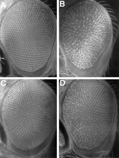
Two coracle alleles have been shown previously to effectively suppress the hypermorphic EgfrElp mutation in a dominant manner (Fehon et al., 1994). EgfrElp causes a roughening and a reduction in size of the Drosophila eye due to increased entry into S-phase and subsequent cell death (Baker and Rubin, 1992). To determine whether EgfrElp suppression by coracle is allele specific or is instead directly proportional to the level of coracle function, we examined the ability of the new coracle alleles to suppress the eye phenotype of EgfrElp (Table 1). Interestingly, the ability to suppress EgfrElp does not correlate strictly with phenotypic strength of coracle alleles. The two deficiencies that uncover the coracle locus (our unpublished results) and the null allele cor5 (Figure 7D and Table 1) only partially suppress the EgfrElp eye phenotype, less efficiently than does either cor1 or cor2 (Fehon et al., 1994). In addition, an intermediate and a weak coracle allele, cor12 and cor15, respectively, are the strongest suppressors of EgfrElp (Figure 7C and Table 1). These results indicate that the cor/EgfrElp interaction is not strictly dose sensitive, and instead suggest that some mutant Coracle proteins may interact with the EGFR pathway in an allele-specific manner.
Figure 7.
Genetic interactions between coracle and Egfr. Scanning electron micrographs of adult eyes. Anterior is to the left. (A) A cor5/CyO (wild-type) eye and a EgfrElp/CyO eye (B) that displays the dominant rough eye phenotype. (C) A cor12/EgfrElp eye showing full suppression of the EgfrElp rough eye phenotype. (D) A cor5/EgfrElp eye that displays moderate roughness.
DISCUSSION
Previous analyses have shown that coracle is required during the process of dorsal closure and that it interacts genetically with hypermorphic mutations at the Egfr locus (Fehon et al., 1994), but they have not examined the role of this conserved junctional protein in epithelial structure, morphogenesis, or apical–basal polarity. Here we have characterized the null phenotype of coracle mutations in embryos and in somatic mosaic clones of mutant cells. We show that although cells lacking coracle function display septate junction defects at the ultrastructural level and disruption of the transepithelial barrier, coracle mutations do not appear to affect apical–basal polarity or structural integrity of epithelial cells. Nonetheless, embryonic epithelia display a range of defects in coracle mutant animals that may result from disruption of an apical–basal barrier function that is dependent on coracle function. Data from somatic mosaic and genetic complementation analyses indicate that coracle functions throughout development of the imaginal epithelia and is necessary for differentiation of adult structures. Furthermore, complementation analysis suggests that the conserved amino terminal region (CNTR) of Coracle constitutes a functional domain and that sequences C-terminal to this domain may also be functionally significant.
Functional Domains within Coracle
In vitro studies of Protein 4.1, Coracle, and related family members indicate that the conserved N-terminal ∼300 aa region forms a discrete functional domain that interacts with the cytoplasmic tail of a transmembrane partner (Rees et al., 1990; Ward et al., 1998). In most family members, there is also at least one other functional domain within the protein (Rees et al., 1990; McCartney and Fehon, 1997). In the case of erythrocyte Protein 4.1, the second domain is known to bind to spectrin and actin, thereby linking the membrane to the cytoskeleton (Correas et al., 1986).
The complementation analysis described here provides a powerful tool for examining the functional organization of coracle. For example, nonsense or missense mutations that inactivate one functional domain should behave in an antimorphic or “dominant negative” manner because they interfere with wild-type protein by competing for binding to an interacting partner in a nonproductive manner. Of particular interest in this regard are the cor1 and cor2 mutations, because we have shown previously that both alleles result from nonsense mutations just 3′ to the CNTR (Fehon et al., 1994). Consistent with the notion that the CNTR encodes a functional domain (Ward et al., 1998), we find that cor1 displays an antimorphic phenotype in combination with hypomorphic coracle alleles (Figure 6). Furthermore, the fact that cor1 is expressed at greater levels than cor2 (Fehon et al., 1994) suggests that although cor2 should have a more severe embryonic phenotype, cor1 would have more severe antimorphic phenotypes, as we have observed. Additionally, we find that cor6, a missense mutation that disrupts the function of the CNTR (Ward et al., 1998), has antimorphic properties. These results also imply that Coracle sequences C-terminal to the CNTR constitute a separate functional domain.
If these alleles have antimorphic functions, then one might expect them to have dominant phenotypes in heterozygotes. Indeed, we have observed partially dominant lethality in flies heterozygous for the cor6 mutation that can be suppressed by adding an additional dose of cor+. However, given the high endogenous level of coracle expression in wild-type individuals and the relatively low levels of expression of cor1 and cor6 protein that have been observed (Fehon et al., 1994; Ward et al., 1998), it is not surprising that dominant antimorphic phenotypes are not readily observed in heterozygous flies. An antimorphic effect is more readily observed, however, when cor1 or cor6 is in trans with a hypomorphic allele that produces less functional coracle product (Figure 6). Thus, the hypomorphic coracle alleles we have isolated can be used as a “sensitized” system to detect antimorphic coracle products or potentially to identify other genes that function together with coracle at the septate junction.
Although we did not observe instances of two strong coracle alleles complementing one another to produce viable adults, there were several instances of partial interallelic complementation between strong and weak alleles. For example, cor3, which has a strong embryonic phenotype, appears to partially complement several of the weak alleles, whereas the other strong alleles do not (Figure 6). Interallelic complementation can be characteristic of mutations in proteins that interact with one another to form complexes (Clifford and Schüpbach, 1994). Although there are currently no biochemical data to indicate such an interaction for Coracle or Protein 4.1, there is evidence that other family members form dimeric complexes that are essential for normal function (Gary and Bretscher, 1993; Berryman et al., 1995). The genetic results presented here suggest that self interactions are also possible in Coracle and that additional biochemical experiments to test for possible interactions should be performed.
coracle Cellular Functions
Previous studies have suggested that septate junctions perform various functions within epithelial cells, including mediating cell–cell adhesion, promoting intercellular interactions (especially those related to cell proliferation), maintaining a diffusional barrier between the apical and basolateral surfaces of an epithelial sheet (the so called “gate” function), and maintaining a barrier within the plane of the plasma membrane to prevent diffusion of lipids and membrane-bound proteins between the apical and basolateral membrane domains (the “fence” function) (Noirot-Timothée and Noirot, 1980; Wood, 1990; Mandel et al., 1993; Woods and Bryant, 1993). Because Coracle is tightly associated with the septate junction and interacts directly with Neurexin, another component of the septate junction (Ward et al., 1998), we examined the effect of loss of coracle function on septate junction morphology, epithelial integrity, and apical–basal polarity within the membrane. Ultrastructural analysis shows that the normally pleated appearance of septate junctions in the embryonic epidermis is lost in embryos that lack coracle function (Figure 3B). This result is similar to that previously reported for Nrx mutations and is consistent with our recent finding that coracle function is required for the maintenance of Neurexin localization (Ward et al., 1998). Thus coracle function is necessary at the cytoplasmic face of the septate junction to localize Neurexin and possibly other proteins in the region of this intercellular junction that together function to form the pleated arrays that are characteristic of this structure.
Given this clear disruption of septate junction morphology, one might expect that many of the functions of this intercellular junction would also be perturbed by coracle mutations. Previous studies of the dlg gene, which encodes a PDZ (PSD-90, DLG, ZO-1)-repeat- containing protein that localizes to the septate junction in epithelial cells (Woods and Bryant, 1989), have demonstrated that dlg mutations display disruptions in apical–basal polarity and loss of epithelial organization (Abbott and Natzle, 1992; Woods et al., 1997); however, examination of markers for apical–basal polarity in embryonic and imaginal epithelia that lack coracle function shows no general effect on epithelial polarity (Figures 2 and 4). Thus coracle and indeed the pleated structure of the septate junction do not appear necessary for the fence function of septate junctions that separates the apical and basolateral membrane domains. In addition, although the characteristic pleated structure that appears to link adjacent cells is missing in these mutations, the integrity of embryonic and imaginal epithelia is unaffected (Figures 2 and 4), indicating that these structures also do not play an essential role in maintaining epithelial cell adhesion. Thus, by these morphological and molecular criteria coracle does not appear necessary for overall epithelial structure, either within an individual cell or within a sheet of cells.
Despite the absence of gross morphological defects, we do consistently see defects in epithelial differentiation of coracle mutant cells. In the embryo, we have found that there is a morphological defect in the cuticle secreted by the apical ends of epithelial cells that is manifested by a delamination between the epicuticle and procuticle layers (Figure 1J). At the light level, we also have observed that the cuticle produced in coracle mutant embryos appears thinner than in wild-type embryos, suggesting that the overall ability to synthesize or deposit cuticle is affected in these embryos (Figure 1). In addition, we have observed necrosis of the salivary gland epithelium (Figure 1D) (Ward et al., 1998). To our knowledge similar phenotypes have not been reported previously for other genes, although we have seen similar defects at the light level in Nrx mutant embryos (our unpublished results). Our experiments testing diffusion of fluorescent dextran molecules across embryonic epithelia indicate that Coracle is required for the gate function of the septate junction (Figure 3F), and a similar phenotype has been noted for Nrx mutations (Baumgartner et al., 1996). It is possible that the cuticular defects observed in both mutations are due to disruption of the ability of the embryonic epithelia to produce an effective barrier to diffusion between the apical and basolateral cell surfaces. This barrier may be important to maintain a particular microenvironment at the apical end of cells that is necessary for proper cuticular deposition.
Another phenotype that is shared by coracle and Nrx mutations is the disruption of the process of dorsal closure, a coordinated series of cell shape changes and rearrangements that occurs midway through embryogenesis (Young et al., 1993). Given the absence of gross epithelial defects in coracle mutant embryos or in mutant clones of cells in imaginal epithelia, it seems unlikely that the failure in dorsal closure is due to disruption of epithelial integrity, although it could represent a defect in the ability to modulate junctional contacts. Alternatively, it is possible that the transepithelial barrier that becomes established by the formation of septate junctions in the embryonic epithelium at the onset of dorsal closure is itself required for the process of dorsal closure to proceed properly. For example, signaling events during dorsal closure that are mediated by the product of the decapentaplegic gene, a secreted TGF-β–like peptide (Noselli, 1998), may require a unique apical environment to function effectively.
In imaginal tissues, coracle mutant cells are morphologically normal but fail to produce adult cuticular structures. This result could indicate that coracle mutant cells are incapable of differentiating adult cuticular structures, but this seems unlikely given that embryonic epithelial cells do differentiate in the absence of coracle function and that we find no evidence of cuticular scars from mutant clones in adults. Alternatively, this failure could represent an earlier loss of coracle mutant cells from the imaginal epithelium. One potential mechanism for this loss is cell competition, a well documented phenomenon in which cells that are at a proliferation disadvantage are lost from the imaginal epithelia (Simpson, 1979, 1981). This possibility is made more likely by the previous observation that coracle mutations suppress hypermorphic mutations in Egfr (see RESULTS) (Fehon et al., 1994), suggesting that Coracle may function together with this receptor to promote cell proliferation in imaginal tissues.
Implications for the 4.1 Superfamily
The experiments presented here provide insights into the in vivo functions of coracle, a Drosophila member of the Protein 4.1 superfamily. Although erythrocyte Protein 4.1 and other family members generally have been considered to play a structural role in linking transmembrane proteins to proteins in the cytoplasm (Rees et al., 1990), recently there has been increasing evidence that these proteins function in mediating intercellular signals. In particular, studies of the ERM proteins indicate that they play essential roles in mediating Rho-dependent signaling mechanisms that may function in the regulation of cell shape or establishment of cell–cell contacts (Hirao et al., 1996). In addition, the product of the NF2 gene, Merlin, is involved in regulating cell proliferation, although the mechanism by which this occurs is not yet understood (Trofatter et al., 1993; Lutchman and Rouleau, 1996). Other family members, such as the protein tyrosine phosphatases, also are likely to function in cell–cell signaling, although their precise functions are also unknown (Banville et al., 1994).
Genetic studies of coracle and other Protein 4.1 family members in Drosophila provide a powerful method for examining the in vivo functions of these proteins. Our data indicate that coracle is not necessary for overall maintenance of cell structure and apical–basal polarity or integrity of the actin cytoskeleton, as might have been assumed from previous studies of Protein 4.1 function in erythrocytes. Instead, the data presented here indicate that coracle is required for establishment of a transepithelial barrier provided by septate junctions. Although this function is likely to be important to all epithelial cells, it is currently unclear whether this cellular defect alone can explain all of the phenotypes that we observe in flies that carry coracle mutations. Further genetic and molecular–genetic studies of coracle and related family members in Drosophila, such as Merlin and Moesin (McCartney and Fehon, 1996), should continue to provide new insights into the functions of this family of membrane-associated proteins.
ACKNOWLEDGMENTS
We thank R. Murray and C. Campbell for technical assistance, L. Eibest for help with the scanning electron microscopy, S. Ward for assistance with the transmission electron microscopy, the Rubin lab and the Indiana stock center for fly stocks, M. Peifer, D. Kiehart, E. Knust, and G. Thomas for providing antibodies, and our colleagues in the Fehon lab for valuable suggestions and discussions. R.S.L. was supported by a National Institutes of Health training grant in cell and molecular biology (GM07184). R.E.W. was supported by a National Institutes of Health training grant in genetics (5T32 GM07754). This research was supported by grants from the National Science Foundation (IBN-92066555) and the American Cancer Society (DB-84846) to R.G.F.
REFERENCES
- Abbott LA, Natzle JE. Epithelial polarity and cell separation in the neoplastic l(1)dlg-1 mutant of Drosophila. Mech Dev. 1992;37:43–56. doi: 10.1016/0925-4773(92)90014-b. [DOI] [PubMed] [Google Scholar]
- Alloisio N, Dalla Venezia N, Rana A, Andrabi K, Texier P, Gilsanz F, Cartron J-P, Delaunay J, Chishti AH. Evidence that red blood cell protein p55 may participate in the skeleton-membrane linkage that involves protein 4.1 and glycophorin C. Blood. 1993;82:1323–1327. [PubMed] [Google Scholar]
- Baker NE, Rubin GM. Ellipse mutations in the Drosophila homologue of the EGF receptor affect pattern formation, cell division, and cell death in eye imaginal discs. Dev Biol. 1992;150:381–396. doi: 10.1016/0012-1606(92)90250-k. [DOI] [PubMed] [Google Scholar]
- Banville D, Ahmad S, Stocco R, Shen SH. A novel protein-tyrosine phosphatase with homology to both the cytoskeletal proteins of the band 4.1 family and junction-associated guanylate kinases. J Biol Chem. 1994;269:22320–22327. [PubMed] [Google Scholar]
- Baumgartner S, Littleton JT, Broadie K, Bhat MA, Harbecke R, Lengyel JA, Chiquet-Ehrismann R, Prokop A, Bellen HJ. A Drosophila neurexin is required for septate junction and blood-nerve barrier formation and function. Cell. 1996;87:1059–1068. doi: 10.1016/s0092-8674(00)81800-0. [DOI] [PubMed] [Google Scholar]
- Berryman M, Gary R, Bretscher A. Ezrin oligomers are major cytoskeletal components of placental microvilli: a proposal for their involvement in cortical morphogenesis. J Cell Biol. 1995;131:1231–1242. doi: 10.1083/jcb.131.5.1231. [DOI] [PMC free article] [PubMed] [Google Scholar]
- Boedigheimer M, Bryant P, Laughon A. Expanded, a negative regulator of cell proliferation in Drosophila, shows homology to the NF2 tumor suppressor. Mech Dev. 1993;44:83–84. doi: 10.1016/0925-4773(93)90058-6. [DOI] [PubMed] [Google Scholar]
- Boedigheimer M, Laughon A. expanded: a gene involved in the control of cell proliferation in imaginal discs. Development. 1993;118:1291–1301. doi: 10.1242/dev.118.4.1291. [DOI] [PubMed] [Google Scholar]
- Clifford R, Schüpbach T. Molecular analysis of the Drosophila EGF receptor homolog reveals that several genetically defined classes of alleles cluster in subdomains of the receptor protein. Genetics. 1994;137:531–550. doi: 10.1093/genetics/137.2.531. [DOI] [PMC free article] [PubMed] [Google Scholar]
- Conboy J, Chasis J, Winardi R, Tchernia G, Kan Y, Mohandas N. An isoform-specific mutation in the protein 4.1 gene results in hereditary elliptocytosis and complete deficiency of protein 4.1 in erythrocytes but not in nonerythroid cells. J Clin Invest. 1993;91:77–82. doi: 10.1172/JCI116203. [DOI] [PMC free article] [PubMed] [Google Scholar]
- Correas I, Speicher DW, Marchesi VT. Structure of the spectrin-actin binding site of erythrocyte protein 4.1. J Biol Chem. 1986;261:13362–13366. [PubMed] [Google Scholar]
- Diaz-Benjumea FJ, Garcia-Bellido A. Behaviour of cells mutant for an EGF receptor homologue of Drosophila in genetic mosaics. Proc R Soc Lond B Biol Sci. 1990;242:36–44. doi: 10.1098/rspb.1990.0100. [DOI] [PubMed] [Google Scholar]
- Fehon RG, Dawson IA, Artavanis-Tsakonas S. A Drosophila homologue of membrane-skeleton protein 4.1 is associated with septate junctions and is encoded by the coracle gene. Development. 1994;120:545–557. doi: 10.1242/dev.120.3.545. [DOI] [PubMed] [Google Scholar]
- Fehon RG, Johansen K, Rebay I, Artavanis-Tsakonas S. Complex cellular and subcellular regulation of Notch expression during embryonic and imaginal development of Drosophila: implications for Notch function. J Cell Biol. 1991;113:657–669. doi: 10.1083/jcb.113.3.657. [DOI] [PMC free article] [PubMed] [Google Scholar]
- Fehon RG, LaJeunesse D, Lamb R, McCartney BM, Schweizer L, Ward RE. Functional studies of the Protein 4.1 family of junctional proteins in Drosophila. Soc Gen Physiol Ser. 1997;52:149–159. [PubMed] [Google Scholar]
- Gary R, Bretscher A. Heterotypic and homotypic associations between ezrin and moesin, two putative membrane-cytoskeletal linking proteins. Proc Natl Acad Sci USA. 1993;90:10846–10850. doi: 10.1073/pnas.90.22.10846. [DOI] [PMC free article] [PubMed] [Google Scholar]
- Giebelhaus DH, Eib DW, Moon RT. Antisense RNA inhibits expression of membrane skeleton protein 4.1 during embryonic development of Xenopus. Cell. 1988;53:601–615. doi: 10.1016/0092-8674(88)90576-4. [DOI] [PubMed] [Google Scholar]
- Grigliatti T. Mutagenesis. In: Roberts D, editor. Drosophila: a practical approach. Oxford: IRL Press; 1986. pp. 39–58. [Google Scholar]
- Hirao M, Sato N, Kondo T, Yonemura S, Monden M, Sasaki T, Takai Y, Tsukita S, Tsukita S. Regulation mechanism of ERM (ezrin/radixin/moesin) protein/plasma membrane association: possible involvement of phosphatidylinositol turnover and Rho-dependent signaling pathway. J Cell Biol. 1996;135:37–51. doi: 10.1083/jcb.135.1.37. [DOI] [PMC free article] [PubMed] [Google Scholar]
- Konsolaki M, Schupbach T. windbeutel, a gene required for dorsoventral patterning in Drosophila, encodes a protein that has homologies to vertebrate proteins of the endoplasmic reticulum. Genes Dev. 1998;12:120–131. doi: 10.1101/gad.12.1.120. [DOI] [PMC free article] [PubMed] [Google Scholar]
- Lue RA, Marfatia SM, Branton D, Chishti AH. Cloning and characterization of hdlg: the human homologue of the Drosophila discs large tumor suppressor binds to protein 4.1. Proc Natl Acad Sci USA. 1994;91:9818–9822. doi: 10.1073/pnas.91.21.9818. [DOI] [PMC free article] [PubMed] [Google Scholar]
- Lutchman M, Rouleau GA. Neurofibromatosis type 2: a new mechanism of tumor suppression. Trends Neurosci. 1996;19:373–377. doi: 10.1016/S0166-2236(96)10044-8. [DOI] [PubMed] [Google Scholar]
- Mandel LJ, Bacallao R, Zampighi G. Uncoupling of the molecular “fence” and paracellular “gate” functions in epithelial tight junctions. Nature. 1993;361:552–555. doi: 10.1038/361552a0. [DOI] [PubMed] [Google Scholar]
- Martuza RL, Eldridge R. Neurofibromatosis 2 (bilateral acoustic neurofibromatosis) N Engl J Med. 1988;318:684–688. doi: 10.1056/NEJM198803173181106. [DOI] [PubMed] [Google Scholar]
- McCartney BM, Fehon RG. Distinct cellular and subcellular patterns of expression imply distinct functions for the Drosophila homologues of moesin and the neurofibromatosis 2 tumor suppressor, merlin. J Cell Biol. 1996;133:843–852. doi: 10.1083/jcb.133.4.843. [DOI] [PMC free article] [PubMed] [Google Scholar]
- McCartney BM, Fehon RG. The ERM family of proteins and their roles in cell-cell interactions. In: Cowin P, Klymkowsky MW, editors. Cytoskeletal-membrane interactions and signal transduction. Austin, TX: R.G. Landes Biocience; 1997. pp. 200–210. [Google Scholar]
- McClatchey AI, Saotome I, Ramesh V, Gusella JF, Jacks T. The Nf2 tumor suppressor gene product is essential for extraembryonic development immediately prior to gastrulation. Genes Dev. 1997;11:1253–1265. doi: 10.1101/gad.11.10.1253. [DOI] [PubMed] [Google Scholar]
- Noirot-Timothée C, Noirot C. Septate and scalariform junctions in arthropods. Int Rev Cytol. 1980;63:97–140. doi: 10.1016/s0074-7696(08)61758-1. [DOI] [PubMed] [Google Scholar]
- Noselli S. JNK signaling and morphogenesis in Drosophila. Trends Genet. 1998;14:33–38. doi: 10.1016/S0168-9525(97)01320-6. [DOI] [PubMed] [Google Scholar]
- Ponting CP, Phillips C, Davies KE, Blake DJ. PDZ domains: targeting signalling molecules to sub-membranous sites. Bioessays. 1997;19:469–479. doi: 10.1002/bies.950190606. [DOI] [PubMed] [Google Scholar]
- Rebay I, Fehon RG, Artavanis-Tsakonas S. Specific truncations of Drosophila Notch define dominant activated and dominant negative forms of the receptor. Cell. 1993;74:319–329. doi: 10.1016/0092-8674(93)90423-n. [DOI] [PubMed] [Google Scholar]
- Rees DJG, Ades SE, Singer SJ, Hynes RO. Sequence and domain structure of talin. Nature. 1990;347:685–689. doi: 10.1038/347685a0. [DOI] [PubMed] [Google Scholar]
- Rouleau GA, et al. Alteration in a new gene encoding a putative membrane-organizing protein causes neurofibromatosis type 2. Nature. 1993;363:515–521. doi: 10.1038/363515a0. [DOI] [PubMed] [Google Scholar]
- Rubin GM, Spradling AC. Genetic transformation of Drosophila with transposable element vectors. Science. 1982;218:348–353. doi: 10.1126/science.6289436. [DOI] [PubMed] [Google Scholar]
- Simpson P. Parameters of cell competition in the compartments of the wing disc of Drosophila. Dev Biol. 1979;69:182–193. doi: 10.1016/0012-1606(79)90284-7. [DOI] [PubMed] [Google Scholar]
- Simpson, P. (1981). Growth and cell competition in Drosophila. J. Embryol. Exp. Morph. 65(suppl), 77–88.
- Szüts D, Freeman M, Bienz M. Antagonism between EGFR and Wingless signalling in the larval cuticle of Drosophila. Development. 1997;124:3209–3219. doi: 10.1242/dev.124.16.3209. [DOI] [PubMed] [Google Scholar]
- Tepass U, Hartenstein V. The development of cellular junctions in the Drosophila embryo. Dev Biol. 1994;161:563–596. doi: 10.1006/dbio.1994.1054. [DOI] [PubMed] [Google Scholar]
- Trofatter JA, et al. A novel moesin-, ezrin-, radixin-like gene is a candidate for the neurofibromatosis 2 tumor suppressor. Cell. 1993;72:791–800. doi: 10.1016/0092-8674(93)90406-g. [DOI] [PubMed] [Google Scholar]
- Ward REI, Lamb RS, Fehon RG. A conserved functional domain of Drosophila Coracle is required for localization at the septate junction and has membrane-organizing activity. J Cell Biol. 1998;140:1463–1473. doi: 10.1083/jcb.140.6.1463. [DOI] [PMC free article] [PubMed] [Google Scholar]
- Wieschaus E, Nüsslein-Volhard C, Jürgens G. Mutations affecting the pattern of the larval cuticle in Drosophila melanogaster. III. Zygotic loci on the X-chromosome and fourth chromosome. Roux’s Arch Dev Biol. 1984;193:296–307. doi: 10.1007/BF00848158. [DOI] [PubMed] [Google Scholar]
- Wood RL. The septate junction limits mobility of lipophilic markers in plasma membranes of Hydra vulgaris (attenuata) Cell Tissue Res. 1990;259:61–66. [Google Scholar]
- Woods DF, Bryant PJ. Molecular cloning of the lethal (1) discs large-1 oncogene of Drosophila. Dev Biol. 1989;134:222–235. doi: 10.1016/0012-1606(89)90092-4. [DOI] [PubMed] [Google Scholar]
- Woods, D.F., and Bryant, P.J. (1993). Apical junctions and cell signalling in epithelia. J Cell Sci 17(suppl), 171–181. [DOI] [PubMed]
- Woods DF, Wu JW, Bryant PJ. Localization of proteins to the apico-lateral junctions of Drosophila epithelia. Dev Genet. 1997;20:111–118. doi: 10.1002/(SICI)1520-6408(1997)20:2<111::AID-DVG4>3.0.CO;2-A. [DOI] [PubMed] [Google Scholar]
- Xu T, Rubin G. Analysis of genetic mosaics in developing and adult Drosophila tissues. Development. 1993;117:1223–1237. doi: 10.1242/dev.117.4.1223. [DOI] [PubMed] [Google Scholar]
- Young PI, Richman AM, Ketchum AS, Kiehart DP. Morphogenesis in Drosophila requires nonmuscle myosin heavy chain function. Genes Dev. 1993;7:29–41. doi: 10.1101/gad.7.1.29. [DOI] [PubMed] [Google Scholar]


