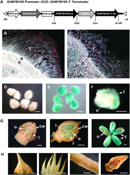
Figure 1.—
Histochemical localization of GUS activity in the transgenic cotton with the GhMYB109∷GUS fusion gene. (A) A schematic of the GhMYB109 Promoter∷GUS fusion construct used for cotton transformation. (B and C) Dark-field micrographs of 8-μm-thick longitudinal (B) and cross (C) sections of 3-DPA ovules. A high level of GUS activity represented by pink dots was found only in the fiber cells. f, fiber; e, epidermis; o, outer integument of ovule; i, inner integument of ovule. (D–H) Bright field of micrographs and photographs of ovules and other tissues in the transgenic and nontransformed plants.(D–F) GUS staining in ovules at 3 DPA. No GUS staining was detected in the ovules of the nontransformed cotton (D). Strong GUS activity was observed in the fibers of the transgenic plants (E and F). (F) A longitudinal section of a transgenic ovule. (G) GUS staining in each stage of transgenic cotton bolls: 1 DPA, 3 DPA, and 5 DPA (from left to right). The first two panels are longitudinal sections of cotton bolls. cw, carpel wall; l, loculus. (H) GUS staining in other tissues of the transgenic cotton. No GUS activity was detected in leaf, sepal, stem, and flower bud before anthesis (from left to right). Bars, 100 μm in A and B; 1 mm in D–F; 2 mm in G; 1 cm in H.

