Abstract
Research and surveillance activities involving airborne pathogens rely on the capture and enumeration of pathogens suspended in aerosols. The objective of this study was to estimate the analytical sensitivity (detection threshold) of each of 4 air samplers for Porcine reproductive and respiratory syndrome virus (PRRSV) and swine influenza virus (SIV). In a 5-min sampling period under controlled conditions, the analytical sensitivity of the AGI-30 (Ace Glass, Vineland, New Jersey, USA), AGI-4 (Ace Glass), SKC BioSampler (SKC, Eighty Four, Pennsylvania, USA), and Midwest Micro-Tek sampler (Midwest Micro-Tek, Brookings, South Dakota, USA) was calculated at 1 × 101.1, 1 × 101.3, 1 × 101.1, and 1 × 101.2 median tissue culture infectious dose (TCID50) equivalents for PRRSV and 1 × 101.4, 1 × 101.1, 1 × 101.6, and 1 × 101.2 TCID50 equivalents for SIV [per 60 L (5-min sampling period)]. Despite marked differences in sampler design, no statistically significant difference in analytical sensitivity was detected between the samplers for collection of artificially produced aerosols containing cell-culture-propagated PRRSV or SIV.
Résumé
Les activités de recherche et de surveillance impliquant les agents pathogènes transmis par voie aérienne se fient sur la capture et l’énumération de ces agents en suspension dans les aérosols. L’objectif de la présente étude était d’estimer la sensibilité analytique (seuil de détection) de chacun de 4 échantillonneurs d’air pour le virus du syndrome reproducteur et respiratoire porcin (PRRSV) et le virus de l’influenza porcin (SIV). Durant une période d’échantillonnage de 5 minutes en conditions contrôlées, la sensibilité analytique des échantillonneurs AGI-30 (Ace Glass, Vineland, New Jersey, USA), AGI-4 (Ace Glass), SKC BioSampler (SKC, Eighty Four, Pennsylvania, USA) et Midwest Micro-Tek (Midwest Micro-Tek, Brookings, South Dakota, USA), exprimée en équivalent de dose infectante médiane de culture de tissu (TCID50), a été calculée comme étant 1 × 101,1, 1 × 101,3, 1 × 101,1, et 1 × 101,2 pour le PRRSV et 1 × 101,4, 1 × 101,1, 1 × 101,6, et 1 × 101,2 pour le SIV [par volume de 60 L (période d’échantillonnage de 5 minutes)]. Malgré des différences majeures dans le design des échantillonneurs, aucune différence statistiquement significative dans la sensibilité analytique n’a été détectée entre les échantillonneurs pour la collecte d’aérosols artificiellement produits contenant du PRRSV ou du SIV propagés en culture cellulaire.
(Traduit par Docteur Serge Messier)
Airborne transmission of viral pathogens is a significant risk to human and animal health and a challenge to disease control programs. Viruses of concern include high-risk agents such as Severe acute respiratory syndrome coronavirus (1), influenza viruses (2), and foot-and-mouth disease virus (3). Airborne transmission of viruses requires release of infectious virus from a host or reservoir, transport in air, deposition in a susceptible host, entry into permissive cells, and productive infection.
Conceptually simple, airborne transmission is actually complex and dynamic. The quantity of infectious virus released from a host is affected by stage of infection, virulence of pathogen, and various host factors (4). Retention of infectivity during transport is highly variable among viruses and reflects virus-specific resistance to inactivation by environmental factors (temperature, relative humidity, solar ultraviolet radiation, etc.) (5). Dispersion of airborne particles is affected by particle shape, particle size, and atmospheric conditions (wind speed and direction, topography, etc.) (6). If infectious virus reaches a susceptible host, infection is not a certainty; its probability depends on dose, virus strain, and host-associated factors, such as gender and age (7).
Given the complexity of the process, it is understandable that aerobiology research historically has been qualitative and descriptive. However, the complex analyses needed to develop accurate models of airborne transmission for pathogens of concern require quantitative data. By definition, such an approach is based on enumeration of airborne virus (infectious and noninfectious) at various stages in the transmission process. The first step in this process is collection and enumeration of virus in air samples.
Air samplers take in environmental air and collect airborne pathogens by filtration, bubbling, or impaction. Impingement — impaction into a liquid medium — is considered the most effective approach for the recovery of viruses (8). All impingers function by directing a jet of air into a liquid collection medium and trapping viral particles therein. Aside from this common feature, impingers vary in design parameters, including the number, angle, and distance of the nozzle(s) relative to the liquid collection medium, flow rate, and quantity of medium in the collection reservoir. Given the variety of sampler designs, differences in sampler performance are to be expected. Therefore, the objective of this research was to compare the performance of several impingers on the basis of their analytical sensitivity; that is, the lowest detectable quantity of airborne virus.
Four impingers were tested: AGI-30 (model 7540-10, Ace Glass, Vineland, New Jersey, USA); AGI-4, 6 L (model 7541-10, Ace Glass); SKC BioSampler (model 225-9595; SKC, Eighty Four, Pennsylvania, USA); and Midwest Micro-Tek sampler (Midwest Micro-Tek, Brookings, South Dakota, USA). The impingers were compared on the basis of the quantity of virus captured from aerosol particles generated by a 6-jet Collison nebulizer (model CN60; BGI, Waltham, Massachsetts, USA) in 60 L of air in a 5-min sampling period. Factors that potentially affected impinger performance, such as collection medium, sampling time, target pathogen, and the diagnostic assays used for quantification of the target viruses, were held constant (9).
The impingers differed in design characteristics. The AGI-30 samples 12.3 to 12.6 L of air per minute with a single nozzle placed at 90° and 30 mm from the bottom of the flask. The AGI-4 samples 6 L/min with a single nozzle placed at 90° and 4 mm from the flask bottom. The SKC BioSampler operates at 12.5 L/min with 3 nozzles, each moving air at approximately 4.2 L/min. The nozzles are placed at an angle to the sides and bottom of the flask, thereby causing the collection liquid to swirl on the sides and bottom during operation. The Midwest Micro-Tek sampler has a self-contained internal pump operating at a sampling rate of 400 L/min and a fan (rotating vanes) that swirls the collection fluid in the reservoir bowl during operation.
Two viruses were used in the study: a North American isolate of Porcine reproductive and respiratory syndrome virus (PRRSV) designated ATCC VR-2332 (American Type Culture Collection, Manassas, Virginia, USA) and an isolate of swine influenza virus (SIV) designated A/Swine/Iowa/73 (H1N1) (National Veterinary Service Laboratories, Ames, Iowa, USA). The viruses were propagated and harvested as previously described (9).
Virus concentration in the impinged samples was determined by means of quantitative real-time reverse transcriptase polymerase chain reaction (qRT-PCR) assays; the protocols are described elsewhere (10). For each assay, a standard curve was generated with the use of a series of virus standards containing PRRSV or SIV at a concentration of 101 to 106 median tissue culture infectious dose (TCID50) per milliliter. Sample results were reported as TCID50 equivalents.
Six virus suspensions representing 6 concentrations of PRRSV and SIV were prepared. The initial suspension was prepared by adding 10 mL of PRRSV (1 × 106.3 TCID50) and 10 mL of SIV (1 × 104.6 TCID50) to 80 mL of sterile phosphate-buffered saline (PBS), pH 7.4 (Invitrogen, Carlsbad, California, USA), and 0.01% (v/v) Antifoam A Emulsion (Sigma Chemical Company, St. Louis, Missouri, USA). Five serial 10-fold dilutions (10−1 to 10−5) were made by adding 10 mL of each subsequent dilution to 90 mL of PBS plus antifoam.
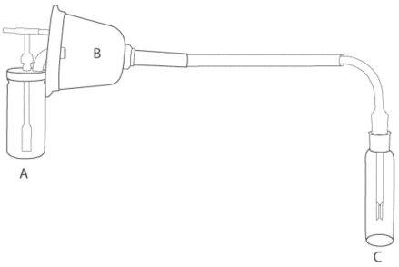
For the experiment, 1 of the 6 virus dilutions was aerosolized into a canine anesthesia mask (model 32393B1; SurgiVet, Waukesha, Wisconsin, USA) connected to an impinger by tubing (internal diameter 0.952 cm, wall thickness 0.317 cm) (Fisher Scientific, Hampton, New Hampshire, USA) (Figure 1). The aerosol was generated with a 6-jet Collison nebulizer operated at 20 lb/in2 of compressed air (model 00916734000; Sears Roebuck, Hoffman Estates, Illinois, USA). According to data generated by May (11), these conditions will produce 12 L/min of free air and aerosolize 9 mL of liquid per hour with a particle size of 2.0 μm. Four replicates were performed for each of the 4 impingers at each of the 6 virus dilutions. The air samplers were loaded with the manufacturers’ recommended quantity of collection fluid (PBS) and were operated for 5 min. Suspension fluid from the nebulizer and collection fluid from the impinger were collected immediately afterwards and stored at −80°C until tested. All procedures were carried out within a class II, type A2 biologic safety cabinet (Labgard 440; NuAire, Plymouth, Minnesota, USA). Upon completion of all replicates, samples were randomized and submitted to the Iowa State University Veterinary Diagnostic Laboratory for qRT-PCR assays.
Figure 1.
Experimental apparatus for estimating the analytical sensitivity of air samplers. A — collison nebulizer (BGI, Waltham, Massachusetts, USA); B — canine surgical mask; C — impinger (AGI-30; Ace Glass, Vineland, New Jersey, USA).
The experiment was designed to establish the relationship between the collection efficiency (quantity of virus aerosolized ÷ quantity virus captured) of each sampler across a series of dilutions. From this relationship, it was possible to estimate the analytical sensitivity of each sampler for the conditions under which the experiment was conducted. The total quantity of virus aerosolized was calculated as the (number of milliliters of suspension fluid aerosolized by nebulizer) × (the TCID50 equivalents/mL). The total quantity of virus captured was calculated as the (TCID50 equivalents/mL of impinger collection fluid) × (the number of milliliters of collection fluid). The collection efficiency of each sampler for PRRSV or SIV was analyzed by linear regression. The y-intercept derived from this analysis was an estimate of the minimum quantity of aerosolized virus necessary to result in detection by the impingers, that is, the analytical sensitivity of the impingers. These results were compared by analysis of variance (JMP; SAS Institute, Cary, North Carolina, USA) of the y-intercept from each replicate and reported as least square means. The null hypothesis stated that the means of the impingers were equal. A significance level of < 0.05 was used as the minimum acceptable P-value.
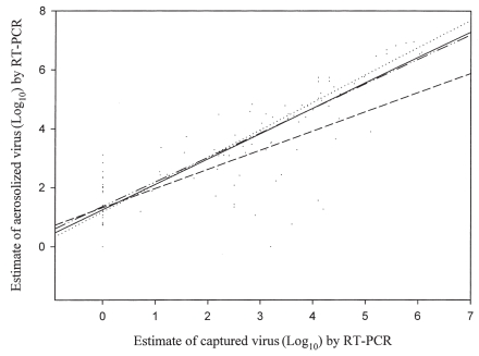
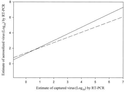
The calculated analytical sensitivity of the AGI-30, AGI-4, SKC BioSampler, and Midwest Micro-Tek samplers for PRRSV and SIV under the experimental sampling conditions are presented in Table I. No statistically significant difference in analytical sensitivity was detected among the impingers for collection of PRRSV (P = 0.88) or SIV (P = 0.97) or between the viruses for the AGI-30 (P = 0.27), AGI-4 (P= 0.99), SKC BioSampler (P= 0.54), and, Midwest Micro-Tek (P= 0.83) samplers. No statistically significant difference in analytical sensitivity was detected among the impingers (Figure 2) (P= 0.23) or between the viruses (Figure 3) (P = 0.97) for the collapsed data.
Table I.
Analytical sensitivity of air samplers for the detection of airborne Porcine reproductive and respiratory syndrome virus (PRRSV) and swine influenza virus (SIV)a
| Sampler and virus | Flow rate (L/min) | Analytical sensitivity (Log10) b |
|---|---|---|
| AGI-30c — PRRSV | 12.5 | 1 × 101.1 |
| — SIV | 12.5 | 1 × 101.4 |
| AGI-4 (6-L)c — PRRSV | 6.0 | 1 × 101.3 |
| — SIV | 6.0 | 1 × 101.1 |
| SKC BioSamplerd — PRRSV | 12.5 | 1 × 101.1 |
| — SIV | 12.5 | 1 × 101.6 |
| Midwest MicroTeke — PRRSV | 400 | 1 × 101.2 |
| — SIV | 400 | 1 × 101.2 |
The PRRSV was a North American isolate designated ATCC VR-2332 (American Type Culture Collection, Manassas, Virginia, USA); The SIV was A/Swine/Iowa/73 (H1N1) (National Veterinary Service Laboratories, Ames, Iowa, USA).
Values are mean estimates per 60 L of air in a 5-min sampling period from quantitative reverse transcriptase polymerase chain reaction assays based on median tissue culture infectious dose standards.
Ace Glass, Vineland, New Jersey, USA.
SCK, Eighty Four, Pennsylvania, USA.
Midwest Micro-Tek, Brookings, South Dakota, USA.
Figure 2.
Determination of analytical sensitivity across viruses for AGI-30 (– ·· –), AGI-4 (—), SKC BioSampler (····), and Midwest Micro-Tek (- - -) using linear regression analysis. RT-PCR — reverse transcriptase polymerase chain reaction.
Figure 3.
Determination of analytical sensitivity across impingers for Porcine reproductive and respiratory syndrome virus (—) and swine influenza virus (– –) by linear regression analysis.
Impingers can be characterized and compared on the basis of total collection, collection efficiency, or analytical sensitivity, or a combination. Comparisons made on the basis of total collection are relatively simple to perform and analyze. For example, Cage et al (12) reported that the AGI-30 collected more pollen and an equivalent number of spores per cubic meter when compared with Spin-Con, a high-volume cyclonic liquid impinger (Sceptor Industries, Kansas City, Missouri, USA) when operated on the roof of a hospital building.
Comparisons made on the basis of total collection do not address important issues in sampler performance, such as efficiency. Estimates of impinger efficiency provide much more information than do estimates of total collection but present technical challenges in the design and execution of the experiment. Sampler efficiency is based on the number of target particles recovered relative to the number available per volume of air. In this study, impinger efficiency was estimated for 4 samplers at each of six 10-fold dilutions of PRRSV and SIV. From these results it was possible to calculate the concentration below which aerosolized virus was undetectable for each impinger — that is, the sampler’s analytical sensitivity. Within the constraints of the experiment, 2 important conclusions can be made for the samplers and targets tested: marked differences in sampler design and function did not result in significant differences in collection efficiency, and airborne virus (infectious and noninfectious) will not be detected if present at concentrations below the impinger’s analytical sensitivity. This may provide an alternative explanation for negative results from air sampling for pathogens aerosolized in the field and under experimental conditions (13–18). The biological significance of undetectable levels of airborne virus will depend on whether the virus is infectious and whether exposure to such a level of virus will result in productive infection in the host.
Since impinger efficiency is known to vary by the mass and size of the target, the results of this study cannot be directly extrapolated to all airborne microorganisms, but they are relevant to other viruses. Additionally, the aerosols in this study were artificially produced and may yield different results than naturally produced aerosols — those generated or dispersed by animals.
Acknowledgments
This project was funded in part by Check-Off Dollars through the National Pork Board and the PRRS Coordinated Agricultural Project [United States Department of Agriculture: Cooperative State Research, Education, and Extension Service (CSREES) National Research Initiative, Competitive Grants Program]. The authors thank Dr. Kyoung-Jin Yoon, Dr. Karen Harmon, and the virology staff at the Veterinary Diagnostic Laboratory, College of Veterinary Medicine, Iowa State University, Ames, Iowa, USA for excellent technical assistance.
References
- 1.Wong SS, Yuen KY. Nasopharyngeal detection of severe acute respiratory syndrome-associated coronavirus RNA in health-care workers. Chest. 2006;129:12–13. doi: 10.1378/chest.129.1.12. [DOI] [PMC free article] [PubMed] [Google Scholar]
- 2.Tellier R. Review of aerosol transmission of influenza A virus. Emerg Infect Dis. 2006;12:1657–1662. doi: 10.3201/eid1211.060426. [DOI] [PMC free article] [PubMed] [Google Scholar]
- 3.Sellers RF, Parker J. Airborne excretion of foot-and-mouth disease virus. J Hyg (Lond) 1969;67:671–677. doi: 10.1017/s0022172400042121. [DOI] [PMC free article] [PubMed] [Google Scholar]
- 4.Alexandersen S, Quan M, Murphy C, Knight J, Zhang Z. Studies of quantitative parameters of virus excretion and transmission in pigs and cattle experimentally infected with foot-and-mouth disease virus. J Comp Pathol. 2003;129:268–282. doi: 10.1016/s0021-9975(03)00045-8. [DOI] [PubMed] [Google Scholar]
- 5.Ijaz MK, Karim YG, Sattar SA, Johnson-Lussenburg CM. Development of methods to study the survival of airborne viruses. J Virol Methods. 1987;18:87–106. doi: 10.1016/0166-0934(87)90114-5. [DOI] [PMC free article] [PubMed] [Google Scholar]
- 6.Morawska L. Droplet fate in indoor environments, or can we prevent the spread of infection? Indoor Air. 2006;16:335–347. doi: 10.1111/j.1600-0668.2006.00432.x. [DOI] [PubMed] [Google Scholar]
- 7.Stärk KD. The role of infectious aerosols in disease transmission in pigs. Vet J. 1999;158:164–181. doi: 10.1053/tvjl.1998.0346. [DOI] [PubMed] [Google Scholar]
- 8.Grinshpun SA, Willeke K, Ulevicius V, Donnelly J, Lin X, Mainelis G. Collection of airborne microorganisms: Advantages and disadvantages of different methods. J Aerosol Sci. 1996;27:S247–S248. [Google Scholar]
- 9.Hermann JR, Hoff SJ, Yoon KJ, Burkhardt AC, Evans RB, Zimmerman JJ. Optimization of a sampling system for recovery and detection of airborne porcine reproductive and respiratory syndrome virus and swine influenza virus. Appl Environ Microbiol. 2006;72:4811–4818. doi: 10.1128/AEM.00472-06. [DOI] [PMC free article] [PubMed] [Google Scholar]
- 10.Hermann J, Hoff S, Muñoz-Zanzi C, et al. Effect of temperature and relative humidity on the stability of infectious porcine reproductive and respiratory syndrome virus in aerosols. Vet Res. 2007;38:81–93. doi: 10.1051/vetres:2006044. Epub 2006 Dec 8. [DOI] [PubMed] [Google Scholar]
- 11.May KR. The Collison nebulizer: Description, performance and application. J Aerosol Sci. 1973;4:235–243. [Google Scholar]
- 12.Cage BR, Schreiber K, Barnes C, Portnoy J. Evaluation of four bioaerosol samplers in the outdoor environment. Ann Allergy Asthma Immunol. 1996;77:401–406. doi: 10.1016/S1081-1206(10)63339-X. [DOI] [PubMed] [Google Scholar]
- 13.Bourgueil E, Hutet E, Cariolet R, Vannier P. Air sampling procedure for evaluation of viral excretion level by vaccinated pigs infected with Aujeszky’s disease (pseudorabies) virus. Res Vet Sci. 1992;52:182–186. doi: 10.1016/0034-5288(92)90008-p. [DOI] [PubMed] [Google Scholar]
- 14.Donaldson AI, Wardley RC, Martin S, Ferris NP. Experimental Aujeszky’s disease in pigs: excretion, survival and transmission of the virus. Vet Rec. 1983;113:490–494. doi: 10.1136/vr.113.21.490. [DOI] [PubMed] [Google Scholar]
- 15.Wilkinson PJ, Donaldson AI, Greig A, Bruce W. Transmission studies with African swine fever virus. Infection of pigs by airborne virus. J Comp Pathol. 1977;87:487–495. doi: 10.1016/0021-9975(77)90037-8. [DOI] [PubMed] [Google Scholar]
- 16.Gillespie RR, Hill MA, Kanitz CL. Infection of pigs by aerosols of Aujeszky’s disease virus and their shedding of the virus. Res Vet Sci. 1996;60:228–233. doi: 10.1016/s0034-5288(96)90044-2. [DOI] [PubMed] [Google Scholar]
- 17.Trincado C, Dee S, Jacobson L, Otake S, Rossow K, Pijoan C. Attempts to transmit porcine reproductive and respiratory syndrome virus by aerosols under controlled field conditions. Vet Rec. 2004;154:294–297. doi: 10.1136/vr.154.10.294. [DOI] [PubMed] [Google Scholar]
- 18.Fano E, Pijoan C, Dee S. Evaluation of the aerosol transmission of a mixed infection of Mycoplasma hyopneumoniae and porcine reproductive and respiratory syndrome virus. Vet Rec. 2005;157:105–108. doi: 10.1136/vr.157.4.105. [DOI] [PubMed] [Google Scholar]


