Abstract
The hantaviruses are emerging infectious viruses that in humans can cause a cardiopulmonary syndrome or a hemorrhagic fever with renal syndrome. The nucleocapsid (N) is the most abundant viral protein, and during viral assembly, the N protein forms trimers and packages the viral RNA genome. Here, we report the NMR structure of the N-terminal domain (residues 1–74, called N1–74) of the Andes hantavirus N protein. N1–74 forms two long helices (α1 and α2) that intertwine into a coiled coil domain. The conserved hydrophobic residues at the helix α1-α2 interface stabilize the coiled coil; however, there are many conserved surface residues whose function is not known. Site-directed mutagenesis, CD spectroscopy, and immunocytochemistry reveal that a point mutation in the conserved basic surface formed by Arg22 or Lys26 lead to antibody recognition based on the subcellular localization of the N protein. Thus, Arg22 and Lys26 are likely involved in a conformational change or molecular recognition when the N protein is trafficked from the cytoplasm to the Golgi, the site of viral assembly and maturation.
Hantaviruses can cause two emerging infectious diseases known as the hantavirus cardiopulmonary syndrome (HCPS)3 and the hantavirus hemorrhagic fever with renal syndrome (1). Annually, there are over 150,000 cases of hantaviral infections reported world wide (2). Rodents are the primary reservoir of hantaviruses, and humans are normally infected by inhalation of aerosol contaminated with the excreta of infected rodents. The first reported cases of HCPS in North America (3) was caused by a novel hantaviral species (4, 5), the Sin Nombre virus, and had an initial mortality rate of 78%. HCPS has since been reported throughout the United States with a current mortality rate of 35% when correctly diagnosed (6). The major cause of HCPS in South America is the Andes virus, and person-to-person transmission of the Andes virus was reported in Argentina and Chile (7). Hantaviruses are known to invade and replicate primarily in endothelial cells, including the endothelium of vascular tissues lining the heart (8–10).
The genome of hantaviruses consists of three negative-stranded RNAs, which encode the nucleocapsid (N) protein, two integral membrane glycoproteins (G1 and G2), and an RNA-dependent RNA polymerase (L protein). The N protein is highly immunogenic (11, 12) and elicits a strong immune response, which confers protection in mice (13–15). It is highly conserved and is the most abundant viral protein, and it plays important roles in viral encapsidation, RNA packaging, and host-pathogen interaction (16). The N protein binds to viral proteins (16), host proteins (17–23), and viral RNA (24–28). The self-association of the N protein into trimers was shown by gradient fractionation and chemical cross-linking (29). Deletion mapping identified that regions at the N and C termini are important in N-N interaction (29–31), and a model of trimerization was proposed based on the head-to-head and tail-to-tail association of the N-terminal and C-terminal domains, respectively (30, 32).
The N-terminal region in the Sin Nombre virus N protein (residues 3–73) (33) and the Tula virus (residues 1–77) (34) were predicted to form coiled coil domains. Recently, the structure of the N-terminal coiled coil domain (residues 1–75 and 1–93) of the Sin Nombre virus was determined by crystallography (35). The highly conserved hydrophobic residues stabilize the structure of the coiled coil; however, there are highly conserved polar residues that appear to have no function in stabilizing the coiled coil domain. Here, we report the solution structure of the N-terminal 1–74 residues of the Andes virus N protein, which also forms a coiled coil domain. Further, we identified that the coiled coil contains distinct regions of positively and negatively charged surfaces involving conserved polar residues. We hypothesize that these regions are also important in N protein function. We used site-directed mutagenesis to alter the surface of the N protein and assayed for the subcellular localization of the N protein by immunocytochemistry. We used CD spectroscopy to confirm that mutations did not alter the coiled coil structure of the N1–74 (residues 1–74 of the N protein) domain. However, immunocytochemistry showed that despite the N protein being present throughout the cytoplasm, a monoclonal antibody only recognized the Arg22 and Lys26 mutants when nucleocapsids are associated with the Golgi, the site of viral assembly and maturation. We propose that the conserved surface residues Arg22 and Lys26 are important in the proper conformation or molecular recognition of the N protein.
EXPERIMENTAL PROCEDURES
Protein Expression and Purification of N1–74—The N1–74 domain of the Andes virus (strain 23) nucleocapsid protein was subcloned into pET151 (Invitrogen), which appends a 33-residue His6 tag and a TEV (tobacco etch virus) protease cleavage site at the N terminus. Isotopically (15N,13C) labeled protein was overexpressed in Escherichia coli BL21(DE3) (DNAY) grown in 1 liter of M9 minimal medium with [15N]ammonium chloride and [13C]glucose. The cells were grown at 37 °C to A600 0.8, induced with 1 mm isopropyl-β-d-thiogalactopyranoside, and incubated overnight (16 h) in a 15 °C shaker. The cells were harvested by centrifugation, resuspended in 30 ml of binding buffer (20 mm Tris-HCl, pH 8.0, 500 mm NaCl, 5 mm imidazole), and lysed by sonication. The cells were centrifuged at 22,500 × g for 15 min, and the supernatant was loaded on a Ni2+ affinity column (Sigma), washed with 35 ml of binding buffer, and eluted with elution buffer (500 mm NaCl, 20 mm Tris-HCl, pH 8.0, 250 mm imidazole). The purified His-tagged N1–74 was dialyzed into buffer (10 mm sodium phosphate, pH 6.9, 10 mm NaCl) and used for NMR structure determination. Typical NMR samples contained 1–1.4 mm N1–74. For CD spectroscopy, the His tag was cleaved by adding 0.08 mm TEV protease into purified His-tagged N1–74 and dialyzing the mixture in an 8000 molecular mass cut-off dialysis tubing in buffer (50 mm Tris-HCl, pH 8.0, 0.5 mm EDTA, 1 mm dithiothreitol) for 16 h at room temperature.
Mutagenesis of N1–74—Site-specific mutations in the N1–74 domain were introduced by PCR using the Stratagene QuikChange kit in two plasmids: (i) pET151-N1–74, used to overexpress recombinant His-tagged N1–74 in E. coli, and (ii) pcDNA3.1-AND-N, used to express full-length Andes virus N protein in a mammalian cell line for immunocytochemistry (see below). The mutations were confirmed by DNA sequencing.
NMR Spectroscopy—NMR data were acquired at 25 °C using a Bruker Avance 800 MHz spectrometer equipped with a cryoprobe, processed with NMRPipe (36), and analyzed with NMR-View (37). Backbone assignments were obtained from two-dimensional 1H-15N HSQC (38) and three-dimensional HNCA (39), CBCA(CO)NH (39), HNCACB (40), and HNCO (41). Secondary structures were identified from the Cα, Cβ, C′, and Hα chemical shifts (42). Side chain assignments were obtained from two-dimensional 1H-13C HMQC (43), three-dimensional HBHA(CO)NH (44), and three-dimensional 13C-edited HMQC-NOESY (tmix = 120 ms) (45). Nuclear Overhauser effect (NOE) cross-peaks were identified from three-dimensional 15N-edited NOESY-HSQC (tmix = 120 ms) (46) and three-dimensional 13C-edited HMQC-NOESY (tmix = 120 ms) (45). Hydrogen-deuterium exchange was performed by lyophilizing a 600-μl 15N-labeled NMR sample and resuspending in 600 μl of 50% D2O, 50% H2O, followed by acquisition of six consecutive 20-min two-dimensional 1H-15N HSQC spectra. Peak volumes were analyzed to identify residues with slower hydrogen-deuterium exchange rates.
Structure Calculation—NOE distance restraints were classified into upper bounds of 2.7, 3.5, 4.5, and 5.5 Å and lower bound of 1.8 Å based on peak volumes. Backbone dihedral angles in the α-helical regions were restrained to ϕ (-60 ± 20°) and Ψ (-40 ± 20°). Hydrogen bonding distance restraints were used for α-helical residues that showed slow hydrogen-deuterium exchange rates. Initial structures were generated by torsion angle dynamics in CYANA (47), followed by molecular dynamics and simulated annealing in AMBER7 (48), first in vacuo and then with the generalized Born potential to account for the effect of solvent during structure calculation. CYANA and AMBER structure calculation protocols have been described elsewhere (49). Iterative cycles of AMBER calculations followed by refinement of NMR-derived restraints were performed until the structures converged with low restraint violations and good statistics in the Ramachandran plot. A family of 20 lowest energy structures was analyzed using PRO-CHECK (50), and graphics were generated using Pymol. The surface electrostatic potentials were calculated using APBS (51) and visualized in Pymol.
CD Spectroscopy—N1–74 samples for CD spectroscopy contained 5–10 μm protein in buffer (25 μm Tris-HCl, pH 8, 3 μm EDTA, and 5 μm dithiothreitol). CD spectra were collected on a Jasco J-815 spectropolarimeter in triplicate. Wavelength scans were collected at 25 °C at a scanning rate of 50 nm/min. Thermal denaturation scans at 222 nm were acquired with a temperature ramp rate of 1 °C/min to a final temperature of 80 °C, followed by cooling at 1 °C/min to 25 °C. The melting temperature (Tm) was determined from calculating the first derivative of thermal denaturation plots using the Jasco CD software.
Immunocytochemistry—Immunocytochemistry was performed as reported (52). Briefly, Cos-7 cells (ATCC; no. CRL-1651) were grown overnight in 24-well plates with coverslips at 37 °C and 5% CO2 in Dulbecco's modified Eagle's medium containing 10% fetal bovine serum. The cells at 80% confluence were transfected using Lipofectamine 2000 (Invitrogen) with 0.8 μg of pcDNA3.1-AND-N plasmid, which expresses full-length wild type or mutated N protein. At 48 h after transfection, the cells were washed with ice-cold phosphate-buffered saline and fixed at room temperature with methanol:acetone (3:1) for 10 min. The cells were incubated in 10 mm glycine for 30 min and permeabilized in phosphate-buffered saline with 0.1% Triton X-100 for 30 min. Permeabilized cells were incubated with antibodies for 60 min at room temperature and washed for 5 min three times with 0.3% Tween in phosphate-buffered saline after each incubation. Goat serum (10%) was used as a blocking agent. Primary antibodies were of two sets: (i) rabbit polyclonal anti-hantavirus nucleocapsid (1:1000) (Immunology Consultants Laboratory; no. RSNV-55) and mouse monoclonal anti-hantavirus-nucleocapsid (1:1000) (Abcam; no. AB34757) or (ii) rabbit anti-Golgi matrix protein GM130 (1:200) (Calbiochem; no. CB1008) and mouse-anti-hantavirus nucleocapsid (1:1000). Secondary antibodies used were Alexa-Fluor-488 (1:1000) (Invitrogen; no. A11008) and Alexa-Fluor-594 (1:1000) (Invitrogen; no. A11005). Lastly, the cells were stained with 4′,6-diamidino-2-phenylindole (Bio-Genex; no. CS2010-06), mounted on slides, and visualized at 60× on an Olympus FV1000 confocal microscope. The images were cropped and adjusted using Adobe Photoshop CS2.
RESULTS
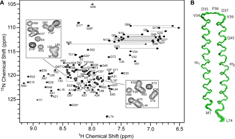
NMR Structure Determination of N1–74—The His-tagged N1–74 expressed well in soluble form in E. coli and yielded an excellent two-dimensional 1H-15N HSQC spectrum that showed distinct and well dispersed peaks (Fig. 1A). Nearly complete backbone assignments were obtained from three-dimensional HNCA, CBCA(CO)NH, HNCACB, and 15N-edited NOESY-HSQC. The histidine residues of the His tag were overlapped and could not be assigned unambiguously. The Cα, Hα, Cβ, and C′ secondary chemical shifts (supplemental Fig. S2) showed that the first 33 residues, which were part of the His tag, were in random coil orientation, and the native N1–74 sequence contained two α-helices (42). Side chain assignments were completed using two-dimensional 1H-13C HMQC, three-dimensional HBHA(CO)NH, and three-dimensional 13C-edited HMQC-NOESY. Manual analysis of three-dimensional 15N- and 13C-edited NOESY spectra identified 1432 unambiguous interproton distance restraints. The NOE restraints together with 73 ϕ and 62 Ψ dihedral angle restraints and 38 hydrogen bond restraints (supplemental Table S1) were used in structure calculation and refinement in CYANA and AMBER. The 20 low energy NMR structures of N1–74 converged into a family of structures (Fig. 1B) with low restraint violations and good Ramachandran plot statistics (supplemental Table S1).
FIGURE 1.
A, assigned 1H-15N HSQC spectrum of Andes virus N1–74 domain. The 33 N-terminal residues (shown with asterisks) are part of the His tag introduced by pET151. The boxes show expansions of the crowded regions. B, superposition of 20 low energy NMR structures of the Andes virus N1–74 coiled coil domain. N1–74 forms two α-helices, Met1–Val34 and Val39–Leu74. The anti-nucleocapsid monoclonal antibody used in immunocytochemistry below recognizes an epitope somewhere between residues 1 and 45.
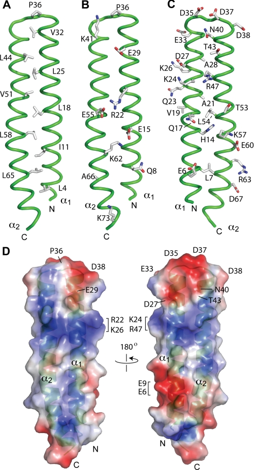
The N1–74 Coiled Coil Domain—N1–74 forms two well defined α-helices (α1, Met1–Val34; α2, Val39–Leu74) that are connected by an ordered acidic loop (Asp35-Pro36-Asp37-Asp38) (Fig. 1B). The two helices are intertwined into a coiled coil, and the helix α1-α2 interface is lined with hydrophobic amino acids positioned in every seventh residue on helix α1 (Leu4, Ile11, Leu18, Leu25, and Val32) and helix α2 (Leu44, Val51, Leu58, and Leu65) (Fig. 2A). This heptad repeat of hydrophobic residues is a hallmark of coiled coils and is highly conserved among hantaviruses (34). Together with Pro36, the heptad repeats of leucines, isoleucines, and valines form the hydrophobic core that stabilize the structure of the coiled coil (Fig. 3A). On the same face of the hydrophobic heptad, there is another seven-residue repeat, in this case, composed of polar residues on helix α1 (Gln8, Glu15, Arg22, and Glu29) (Fig. 2B), which are invariant among the hantaviruses (supplemental Fig. S1). Helix α2 also contains a polar heptad, however, with more residue variability at positions 41 (Lys), 48 (Arg/Gln/Glu), 55 (Glu/Gln), and 62 (Lys/Arg). These polar residues form two conserved salt bridges between helix α1-α2 (Glu15–Lys62 and Arg22–Glu55) (Fig. 2B). Gln8 and Lys41 are surface-exposed and do not form any salt bridges; however, they are invariant among the hantaviruses, suggesting some unknown function.
FIGURE 2.
A, heptad repeats of conserved hydrophobic residues form the interface of the helix α1 and α2 that stabilize the coiled coil domain. B, there is also a heptad repeat of polar residues, some of which (Arg22–Glu55 and Glu15–Lys62) form salt bridges that contribute in stabilizing the coiled coil. C, there are many highly conserved residues that point away from the coiled coil and thus are not involved in stabilizing the coiled coil. D, electrostatic surface potential map of N1–74. The orientation of the left panel is identical to that in A and is rotated 180° from the right panel, which is identical in the orientation of C. Conserved surface residues forming the acidic (red) and basic (blue) surfaces are indicated. Point mutations of Arg22 and Lys26 had a dramatic effect on the antibody recognition of the N protein in vivo.
FIGURE 3.
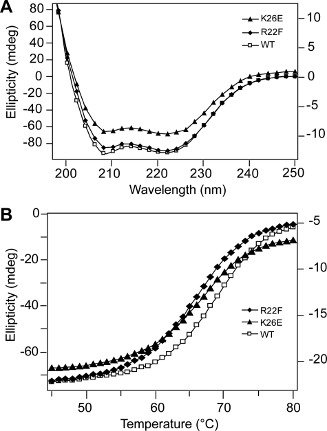
A, CD spectra of N1–74 wild type (WT) and point mutants (K26E and R22F) showing the characteristic α-helical dips at 208 and 222 nm. All other N1–74 point mutants (listed in Table 1) showed similar α-helical CD spectra. B, CD thermal denaturation curves, monitored at 222 nm, of wild type N1–74 and two point mutants, K26E and R22F. The rest of the point mutants showed similar thermal denaturation curves. The ellipticity scales on the y axes are shown on left (wild type and R22F) and right (K26E).
In addition to the conserved heptad repeats mentioned above, there are other highly conserved residues whose side chains are pointed toward the helix α1-α2 interface. These residues are nonpolar (Leu7 and Leu54), aromatic (His14), polar (Gln17, Asn40, and Thr43), or charged (Glu6, Glu15, Glu29, Lys41, Arg47, and Lys57), and their side chains are pointed toward the helix α1-α2 interface (Fig. 2C). The polar and charged residues in this group do not participate in any salt bridge or hydrogen bonding contacts; however, their polar moieties are pointed toward the surface of the coiled coil, whereas the aliphatic portion of their side chains are involved in hydrophobic interaction that contribute to the stabilization of the hydrophobic core. The methyl groups of two invariant alanines, Ala21 and Ala28, in helix α1 (Fig. 2C) are oriented toward the helix α1-α2 interface but do not contact any other residues on helix α2, indicating that small side chains are required in those positions.
Conserved Surface Residues—A striking feature of the N1–74 coiled coil is the presence of large numbers of highly conserved residues whose side chains are pointed away from the coiled coil. These residues are nonpolar (Ala66 and Val19), polar (Gln8 and Gln23), basic (Lys24, Lys26, Arg63, and Lys73), and acidic (Asp27, Glu33, Asp35, Asp37, Asp38, Glu60, and Asp67) (Fig. 2, B and C). These polar residues are identical (Gln8, Gln23, Lys24, Glu33, Asp35, Asp37, Arg63, and Lys73) or semi-identical (basic residues in position 26 and acidic residues in positions 27, 38, and 60) among hantaviruses (supplemental Fig. S1). Further, many residues in this group are clustered together on the surface. The first cluster (Gln23, Lys24, and Lys26 together with Arg47 and Arg22 discussed in the preceding paragraph) forms a basic surface (Fig. 2D), and the second cluster (Asp27, Glu33, Asp35, Asp37, and Asp38) forms an acidic surface (Fig. 2D). We mutagenized many residues in this group (see below) to test the hypothesis that these residues are important in molecular recognition rather than in stabilizing the coiled coil structure.
Electrostatic Surface of N1–74—The N1–74 domain is acidic (theoretical pI of 5.8), and the surface electrostatic potential map of N1–74 shows distinct regions of negatively charged (red) and positively charged (blue) surfaces (Fig. 2D). The tip of the coiled coil, where the loop connecting the two helices are located, is negatively charged (Fig. 2D) because of clustering of conserved acidic residues (Asp27, Glu29, Glu33, Asp35, Asp37, and Asp38) and polar residues (Asn40 and Thr43). Although the N1–74 domain is acidic, there are conserved basic residues (Arg22, Lys24, Lys26 and Arg47) that form a positively charged surface just below the negatively charged tip (Fig. 2D). Point mutations in this positively charged surface have a dramatic effect on the antibody recognition of the N protein in vivo (see below).
In addition, there is a smaller negatively charged surface formed by Glu9 and Glu6 (Fig. 2D). Residue 9 could be acidic (Glu or Asp) or basic (Arg or Lys). Residue 9 is acidic among American hantaviruses (which cause the cardiopulmonary syndrome) and Old World hantaviruses that are nonpathogenic or cause a milder form of hemorrhagic fever with renal syndrome. Residue 9 is basic among Old World hantaviruses that causes the severe form of hemorrhagic fever with renal syndrome.
Circular Dichroism Spectroscopy of N1–74—Point mutations were introduced in the basic (Arg22, Lys24, Lys26, and Arg47) and acidic (Glu33 and Asp38) surfaces. In addition, we mutated Gln23, which is near the basic region, and Pro36, which is near the acidic region. These residues are surface-exposed (Fig. 2D) and are nearly invariant among hantaviruses (supplemental Fig. S1). CD spectroscopy was used to assess the folding and stability of N1–74 mutants. Wild type and point mutants showed nearly identical CD spectra (Fig. 3A), indicating that the α-helical structure of N1–74 was preserved. In addition, the ratio of ellipticity at 222 and 208 nm can be used to characterize α-helices. A θ222/θ208 ratio of ∼1.0 indicates α-helices with extensive interhelical contacts as in coiled coils and helical bundles, whereas a θ222/θ208 ratio of ∼0.8 indicates extended α-helices with little interhelical contacts (53–55). All N1–74 constructs have a θ222/θ208 ratio higher than 0.9 (Table 1), suggesting that all mutants have the intact coiled coil structure. Further insight was provided by acquiring the CD melting temperatures (Fig. 3B and Table 1). Compared with wild type N1–74, the majority of mutants showed lower Tm, with D38R having the lowest value, whereas two mutants (K24A and R47A) showed higher Tm (Table 1). Nevertheless, all mutations were within ±5°Cof wild type Tm (Table 1), indicating that the mutations did not drastically alter the thermal stability of N1–74. Thus, the point mutations maintained the structural integrity of the N1–74 coiled coil.
TABLE 1.
Melting temperatures (Tm) and ellipticity (θ) ratio at 222 and 208 nm of N1–74
| N1–74 | Tm | θ222/θ208 | Change in surface property |
|---|---|---|---|
| °C | |||
| D38R | 64.3 ± 0.01 | 1.06 | Acidic to basic |
| E33K | 64.4 ± 0.02 | 1.05 | Acidic to basic |
| P36G | 66.0 ± 0.1 | 0.99 | Increased loop flexibility |
| R47E | 66.0 ± 0.03 | 1.07 | Basic to acidic |
| D38L | 66.2 ± 0.01 | 1.03 | Acidic to nonpolar |
| R22F | 66.4 ± 0.1 | 1.07 | Basic to bulky nonpolar |
| K26E | 67.0 ± 0.02 | 1.04 | Basic to acidic |
| E33L | 67.4 ± 0.01 | 1.07 | Acidic to nonpolar |
| Q23L | 68.5 ± 0.03 | 1.01 | Polar to nonpolar |
| R22M | 69.3 ± 1.2 | 1.06 | Basic to nonpolar |
| WT | 69.4 ± 0.1 | 0.99 | No change |
| K24A | 70.6 ± 0.01 | 1.04 | Basic to small nonpolar |
| R47A | 74.0 ± 0.02 | 1.02 | Basic to small nonpolar |
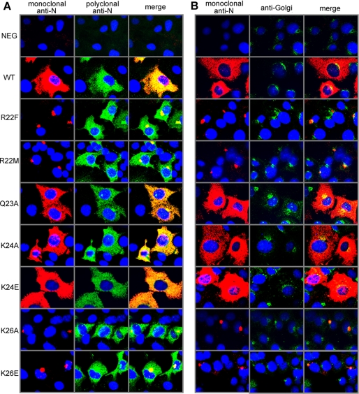
Immunocytochemistry of N Protein—Hantaviruses are believed to mature intracellularly; specifically, in the Golgi complex (56). During infection, the N protein was shown to localize cytoplasmically in the endoplasmic reticulum-Golgi intermediate compartment, presumably as they traffic from the endoplasmic reticulum to the Golgi (22). In addition, immunofluorescence of Cos-7 cells transfected with the N protein alone showed a granular pattern of staining in the perinuclear region (32, 34), suggesting colocalization with the Golgi. To test our hypothesis that the conserved surface residues of N1–74 are important in molecular interaction, we introduced point mutations designed to keep the N1–74 coiled coil domain intact while altering only specific surface residues and transfected full-length N protein in mammalian cells to observe the subcellular localization of the N protein. We used two types of anti-nucleocapsid antibodies, rabbit polyclonal and mouse monoclonal antibodies. The polyclonal antibody detected that wild type N and mutants (Arg22, Gln23, Lys24, and Lys26) were located throughout the cytoplasm (Fig. 4A). The monoclonal antibody also detected wild type N and the Gln23 and Lys24 mutants throughout the cytoplasm in a similar pattern of staining as the polyclonal antibody (Fig. 4A). However, the monoclonal antibody showed a dramatic difference between the recognition of wild type N and the Arg22 and Lys26 mutants (Fig. 4A). Using the monoclonal antibody, Arg22 and Lys26 mutants were observed in a compact location lateral to the nucleus (Fig. 4A). To further define the subcellular localization of these N mutants, a Golgi-specific antibody (targeting the Golgi matrix protein GM130) was used (Fig. 4B). The Arg22 and Lys26 mutants were only detected by the monoclonal antibody when the N protein colocalized with the Golgi (Fig. 4B); however, these mutants were also present throughout the cytoplasm as shown by the polyclonal antibody (Fig. 4A). Thus, for the Arg22 and Lys26 mutants, the monoclonal antibody was able to distinguish between two populations of the N protein based on its subcellular localization in the cytoplasm or in the Golgi, the site of viral assembly and maturation (56). Other mutants (Glu33, Asp35, Pro36, Asp37, Asp38, and Arg47) did not show this localization-dependent antibody recognition (supplemental Fig. S3).
FIGURE 4.
Immunocytochemistry of full-length N protein with point mutations in the N1–74 coiled coil domain. Cos-7 cells were transfected with a plasmid expressing Andes virus N protein. Two days after transfection, the cells were fixed for immunofluorescence microscopy and double labeled with monoclonal (red) and polyclonal (green) anti-nucleocapsid antibodies (A) and monoclonal anti-nucleocapsid antibody (red) and anti-Golgi antibody (green) (B). The cell nuclei were stained blue using 4′,6-diamidino-2-phenylindole. Point mutations in Arg22 and Lys26 showed a dramatic difference in the monoclonal antibody recognition of Golgi-associated N protein, suggesting that the conformation or molecular interaction (or both) of the N protein is different when it is in the cytoplasm or when it is associated with the Golgi. WT, wild type.
DISCUSSION
The NMR structure of the Andes virus N1–74 domain is similar to the recent crystal structure of the Sin Nombre virus nucleocapsid protein N-terminal coiled coil (N1–75) (35). The Cα root mean square deviation between the two structures is 1.3 Å. The crystal structure determination of the N protein addressed the issue of the trimerization of the N protein (35) because earlier models suggested the trimerization of the nucleocapsid N-terminal domain (29, 30, 32, 33). A proposed model of N protein trimerization involves, first, the association of three N-terminal domains, followed by the association of three C-terminal domain (34). However, crystallography revealed that the Sin Nombre nucleocapsid N-terminal domain was monomeric and formed a coiled coil structure, and conserved hydrophobic residues participate in helix-helix interaction that stabilize the coiled coil (35). Our NMR structure of the Andes virus N1–74 supports the crystallographic results; even at 1.4 mm, N1–74 remained monomeric in solution. Our results, however, do not preclude the trimerization of full-length N protein in vivo by another mechanism.
A feature of the N1–74 domain that had not been addressed in the literature is the role of many conserved polar residues whose side chains are pointed away from the coiled coil. Furthermore, the majority of these surface-exposed residues are not involved in polar interactions (Fig. 2D). Point mutations of these polar residues maintained the structural integrity and high thermal stability of the coiled coil (Fig. 3 and Table 1). For example, Arg22, which forms a salt bridge with a conserved residue Glu55, can be mutated (R22F or R22M) without disrupting the coiled coil structure of N1–74 (Table 1). R22F, which replaced arginine with a bulkier aromatic side chain, decreased the overall melting temperature by ∼3 °C (Table 1). This change is likely attributed to increased steric clash between phenylalanine and Glu55. However, the observation that R22M melts at a temperature comparable with that of wild type suggests that the salt bridge between Arg22 and Glu55 does not play a significant role in helix-helix interaction and that hydrophobic interaction is the major force stabilizing the coiled coil. A mutation in a nonpolar residue, Pro36, which is at the turn connecting the two α-helices of the coiled coil, had a Tm approximately four degrees lower than wild type, which is consistent with a mutation that increases the number of conformations available at the Pro36 turn and destabilizes the overall protein structure by uncoupling the helix-helix interaction. Nevertheless, all of the point mutations of the conserved surface residues maintained the coiled coil structure of N1–74 (Fig. 3 and Table 1).
Thus, there is no compelling structural reason for the high sequence conservation of surface residues. Furthermore, these polar residues are clustered together on the surface of the N1–74 domain and form distinct positively and negatively charged regions (Fig. 2D). We hypothesize that the reason for the clustering of conserved polar residues on the surface of N1–74 is that they are sites of molecular recognition involved in the proper function of the N protein. Our mutagenesis and immunocytochemistry data suggest that point mutations in this group had a dramatic effect on the antibody recognition of the N protein with respect to its subcellular localization (Fig. 4).
During infection, nucleocapsids are trafficked to the cytoplasm (22) to assemble into mature virions (56). Mammalian cells transfected with the N protein alone show a granular pattern of immunofluorescence (32, 34). This localization pattern is thought to be necessary for the nucleocapsid to perform its many functions in the establishment of an effective infection (22). We questioned whether the conserved polar surface residues in the coiled coil domain are important in the proper functioning of the N protein and reasoned that defects in the conformation or molecular recognition of the N protein will be manifested in the antibody recognition of the N protein in the context of its subcellular localization. CD spectroscopy confirmed that the mutant forms of N1–74 maintained the structural integrity of the coiled coil structure (Fig. 3 and Table 1); thus, the mutations altered only the surface property of the N protein.
Immunocytochemistry (Fig. 4) indicates that mutations in a conserved basic surface formed by Arg22 and Lys26 show monoclonal antibody recognition depending on the subcellular localization of the N protein. Polyclonal antibodies show that Arg22 and Lys26 mutants are present in the cytoplasm and Golgi; however, only Golgi-associated mutant nucleocapsids are detected by the monoclonal antibody (Fig. 4). Mutation of Arg22 or Lys26 changes the presentation of the N-terminal coiled coil to the monoclonal antibody. This change is dependent on the subcellular localization of the N protein.
There are two possible scenarios that could account for this differential monoclonal antibody recognition of the Arg22 and Lys26 mutants. First, there may be a difference in the conformation of the N-terminal coiled coil depending on whether the N protein is localized in the cytoplasm or in the Golgi, and this conformational change upon binding to the Golgi exposes the epitope, which is somewhere between residues 1–45 (comprising helix α1, the interhelical loop, and part of helix α2 of the N1–74 coiled coil; Fig. 1B), thereby allowing the monoclonal antibody to recognize the N protein associated with the Golgi. Second, the epitope may be masked differently by molecular interactions when the N protein is localized in the cytoplasm or in the Golgi. In addition to self-association, several host proteins such as SUMO-1 (17–19), Ubc9 (17, 18), Daxx (20), actin (21), microtubules (22), and MxA (23) were reported to bind the N protein. Binding of the N protein with SUMO-1 and Ubc9 was required for localization of the N protein in the perinuclear region (17, 19). Furthermore, because the N protein is not known to be a membrane protein, its localization in the Golgi must involve interaction with a Golgi-associated protein. Any of these molecular interactions could potentially alter the epitope presentation of the N1–74 coiled coil and needs to be experimentally verified.
In summary, our structural results revealed that the highly conserved polar residues in the N-terminal coiled coil domain of the hantavirus nucleocapsid protein form distinct acidic and basic surfaces, and point mutations of the conserved basic surface formed by Arg22 and Lys26 allowed a monoclonal antibody to distinguish between two populations of the N protein based on its subcellular localization. Thus, in the Arg22 or Lys26 mutants, the conformation or molecular interaction of the N protein is different when it is in the cytoplasm or in the Golgi, the site of viral assembly and maturation.
Supplementary Material
Acknowledgments
We are grateful to Albert Rizvanov (University of Nevada, Reno) for constructing the N1–74 expression plasmid, Evan Colletti (University of Nevada, Reno) and Mariana Bego (University of Nevada, Reno) for guidance in confocal microscopy, and Thenmalarchelvi Rathinavelan (University of Kansas), Gaya Amarasinghe (Iowa State University), and Edina Harsay (University of Kansas) for critical reading of the manuscript.
This work was supported, in whole or in part, by National Institutes of Health Grants AI057160 and AI65359. This work was also supported by American Heart Association Grant 0755724Z (to R. N. D.); National Science Foundation Program Grant EF 0326999 (to S. C. S. J.). The costs of publication of this article were defrayed in part by the payment of page charges. This article must therefore be hereby marked “advertisement” in accordance with 18 U.S.C. Section 1734 solely to indicate this fact.
The on-line version of this article (available at http://www.jbc.org) contains supplemental Figs. S1–S3.
Footnotes
The abbreviations used are: HCPS, hantavirus cardiopulmonary syndrome; HSQC, heteronuclear single quantum coherence; HMQC, heteronuclear multiple quantum coherence; N, nucleocapsid protein; NOE, nuclear Overhauser effect; NOESY, NOE spectroscopy.
References
- 1.Schmaljohn, C., and Hjelle, B. (1997) Emerg. Infect. Dis. 3 95-104 [DOI] [PMC free article] [PubMed] [Google Scholar]
- 2.Khaiboullina, S. F., Morzunov, S. P., and St. Jeor, S. C. (2005) Curr. Mol. Med. 5 773-790 [DOI] [PubMed] [Google Scholar]
- 3.Koster, F., Levy, H., Mertz, G., Young, S., Foucar, K., McLaughlin, J., Bryt, B., Merlin, T., Zumwalt, R., McFeely, P., Nolte, K., Burkhart, M., Kalishman, N., Gallaher, M., Voorhees, R., et al. Centers for Disease Control and Prevention (1993) Morbid. Mortal. Weekly Rep. 42 421-424 [Google Scholar]
- 4.Nichol, S. T., Spiropoulou, C. F., Morzunov, S., Rollin, P. E., Ksiazek, T. G., Feldmann, H., Sanchez, A., Childs, J., Zaki, S., and Peters, C. J. (1993) Science 262 914-917 [DOI] [PubMed] [Google Scholar]
- 5.Hjelle, B., Jenison, S., Torrez-Martinez, N., Yamada, T., Nolte, K., Zumwalt, R., MacInnes, K., and Myers, G. (1994) J. Virol. 68 592-596 [DOI] [PMC free article] [PubMed] [Google Scholar]
- 6.Mertz, G. J., Hjelle, B., Crowley, M., Iwamoto, G., Tomicic, V., and Vial, P. A. (2006) Curr. Opin. Infect. Dis. 19 437-442 [DOI] [PubMed] [Google Scholar]
- 7.Padula, P. J., Edelstein, A., Miguel, S. D., Lopez, N. M., Rossi, C. M., and Rabinovich, R. D. (1998) Virology 241 323-330 [DOI] [PubMed] [Google Scholar]
- 8.Pensiero, M. N., Sharefkin, J. B., Dieffenbach, C. W., and Hay, J. (1992) J. Virol. 66 5929-5936 [DOI] [PMC free article] [PubMed] [Google Scholar]
- 9.Zaki, S. R., Greer, P. W., Coffield, L. M., Goldsmith, C. S., Nolte, K. B., Foucar, K., Feddersen, R. M., Zumwalt, R. E., Miller, G. L., and Khan, A. S. (1995) Am. J. Pathol. 146 552-579 [PMC free article] [PubMed] [Google Scholar]
- 10.Nolte, K. B., Feddersen, R. M., Foucar, K., Zaki, S. R., Koster, F. T., Madar, D., Merlin, T. L., McFeeley, P. J., Umland, E. T., and Zumwalt, R. E. (1995) Hum. Pathol. 26 110-120 [DOI] [PubMed] [Google Scholar]
- 11.Gott, P., Zoller, L., Darai, G., and Bautz, E. K. (1997) Virus Genes 14 31-40 [DOI] [PubMed] [Google Scholar]
- 12.Lundkvist, A., Meisel, H., Koletzki, D., Lankinen, H., Cifire, F., Geldmacher, A., Sibold, C., Gott, P., Vaheri, A., Kruger, D. H., and Ulrich, R. (2002) Viral Immunol. 15 177-192 [DOI] [PubMed] [Google Scholar]
- 13.Maes, P., Keyaerts, E., Bonnet, V., Clement, J., Avsic-Zupanc, T., Robert, A., and Van Ranst, M. (2006) Intervirology 49 253-260 [DOI] [PubMed] [Google Scholar]
- 14.Geldmacher, A., Skrastina, D., Borisova, G., Petrovskis, I., Kruger, D. H., Pumpens, P., and Ulrich, R. (2005) Vaccine 23 3973-3983 [DOI] [PubMed] [Google Scholar]
- 15.Geldmacher, A., Skrastina, D., Petrovskis, I., Borisova, G., Berriman, J. A., Roseman, A. M., Crowther, R. A., Fischer, J., Musema, S., Gelderblom, H. R., Lundkvist, A., Renhofa, R., Ose, V., Kruger, D. H., Pumpens, P., and Ulrich, R. (2004) Virology 323 108-119 [DOI] [PubMed] [Google Scholar]
- 16.Kaukinen, P., Vaheri, A., and Plyusnin, A. (2005) Arch. Virol. 150 1693-1713 [DOI] [PubMed] [Google Scholar]
- 17.Maeda, A., Lee, B. H., Yoshimatsu, K., Saijo, M., Kurane, I., Arikawa, J., and Morikawa, S. (2003) Virology 305 288-297 [DOI] [PubMed] [Google Scholar]
- 18.Lee, B. H., Yoshimatsu, K., Maeda, A., Ochiai, K., Morimatsu, M., Araki, K., Ogino, M., Morikawa, S., and Arikawa, J. (2003) Virus Res. 98 83-91 [DOI] [PubMed] [Google Scholar]
- 19.Kaukinen, P., Vaheri, A., and Plyusnin, A. (2003) Virus Res. 92 37-45 [DOI] [PubMed] [Google Scholar]
- 20.Li, X. D., Makela, T. P., Guo, D., Soliymani, R., Koistinen, V., Vapalahti, O., Vaheri, A., and Lankinen, H. (2002) J. Gen. Virol. 83 759-766 [DOI] [PubMed] [Google Scholar]
- 21.Ravkov, E. V., Nichol, S. T., Peters, C. J., and Compans, R. W. (1998) J. Virol. 72 2865-2870 [DOI] [PMC free article] [PubMed] [Google Scholar]
- 22.Ramanathan, H. N., Chung, D. H., Plane, S. J., Sztul, E., Chu, Y. K., Guttieri, M. C., McDowell, M., Ali, G., and Jonsson, C. B. (2007) J. Virol. 81 8634-8647 [DOI] [PMC free article] [PubMed] [Google Scholar]
- 23.Khaiboullina, S. F., Rizvanov, A. A., Deyde, V. M., and St Jeor, S. C. (2005) J. Med. Virol. 75 267-275 [DOI] [PubMed] [Google Scholar]
- 24.Gott, P., Stohwasser, R., Schnitzler, P., Darai, G., and Bautz, E. K. (1993) Virology 194 332-337 [DOI] [PubMed] [Google Scholar]
- 25.Severson, W., Partin, L., Schmaljohn, C. S., and Jonsson, C. B. (1999) J. Biol. Chem. 274 33732-33739 [DOI] [PubMed] [Google Scholar]
- 26.Mir, M. A., and Panganiban, A. T. (2004) J. Virol. 78 8281-8288 [DOI] [PMC free article] [PubMed] [Google Scholar]
- 27.Mir, M. A., and Panganiban, A. T. (2005) J. Virol. 79 1824-1835 [DOI] [PMC free article] [PubMed] [Google Scholar]
- 28.Mir, M. A., Brown, B., Hjelle, B., Duran, W. A., and Panganiban, A. T. (2006) J. Virol. 80 11283-11292 [DOI] [PMC free article] [PubMed] [Google Scholar]
- 29.Alfadhli, A., Love, Z., Arvidson, B., Seeds, J., Willey, J., and Barklis, E. (2001) J. Virol. 75 2019-2023 [DOI] [PMC free article] [PubMed] [Google Scholar]
- 30.Kaukinen, P., Vaheri, A., and Plyusnin, A. (2003) J. Virol. 77 10910-10916 [DOI] [PMC free article] [PubMed] [Google Scholar]
- 31.Yoshimatsu, K., Lee, B. H., Araki, K., Morimatsu, M., Ogino, M., Ebihara, H., and Arikawa, J. (2003) J. Virol. 77 943-952 [DOI] [PMC free article] [PubMed] [Google Scholar]
- 32.Kaukinen, P., Kumar, V., Tulimaki, K., Engelhardt, P., Vaheri, A., and Plyusnin, A. (2004) J. Virol. 78 13669-13677 [DOI] [PMC free article] [PubMed] [Google Scholar]
- 33.Alfadhli, A., Steel, E., Finlay, L., Bachinger, H. P., and Barklis, E. (2002) J. Biol. Chem. 277 27103-27108 [DOI] [PubMed] [Google Scholar]
- 34.Alminaite, A., Halttunen, V., Kumar, V., Vaheri, A., Holm, L., and Plyusnin, A. (2006) J. Virol. 80 9073-9081 [DOI] [PMC free article] [PubMed] [Google Scholar]
- 35.Boudko, S. P., Kuhn, R. J., and Rossmann, M. G. (2007) J. Mol. Biol. 366 1538-1544 [DOI] [PMC free article] [PubMed] [Google Scholar]
- 36.Delaglio, F., Grzesiek, S., Vuister, G. W., Zhu, G., Pfeifer, J., and Bax, A. (1995) J. Biomol. NMR 6 277-293 [DOI] [PubMed] [Google Scholar]
- 37.Johnson, B. A. (2004) Methods Mol. Biol. 278 313-352 [DOI] [PubMed] [Google Scholar]
- 38.Grzesiek, S., and Bax, A. (1993) J. Am. Chem. Soc. 115 12593-12594 [Google Scholar]
- 39.Grzesiek, S., Dobeli, H., Gentz, R., Garotta, G., Labhardt, A. M., and Bax, A. (1992) Biochemistry 31 8180-8190 [DOI] [PubMed] [Google Scholar]
- 40.Wittekind, M., and Mueller, L. (1993) J. Magn. Reson. 101B 201-205 [Google Scholar]
- 41.Muhandiram, D. R., and Kay, L. E. (1994) J. Magn. Reson. Ser. B 103 203-216 [Google Scholar]
- 42.Wishart, D. S., and Nip, A. M. (1998) Biochem. Cell Biol. 76 153-163 [DOI] [PubMed] [Google Scholar]
- 43.Tolman, J. R., Chung, J., and Prestegard, J. H. (1992) J. Magn. Reson. 98 462-467 [Google Scholar]
- 44.Grzesiek, S., and Bax, A. (1993) J. Biomol. NMR 3 185-204 [DOI] [PubMed] [Google Scholar]
- 45.Fesik, S. W., and Zuiderweg, E. R. P. (1998) J. Magn. Reson. 78 588-593 [Google Scholar]
- 46.Marion, D., Driscoll, P. C., Kay, L. E., Wingfield, P. T., Bax, A., Gronenborn, A. M., and Clore, G. M. (1989) Biochemistry 28 6150-6156 [DOI] [PubMed] [Google Scholar]
- 47.Guntert, P. (2004) Methods Mol. Biol. 278 353-378 [DOI] [PubMed] [Google Scholar]
- 48.Case, D. A., Pearlman, D. A., Caldwell, J. W., Cheatham Iii, T. E., Wang, J., Ross, W. S., Simmerling, C. L., Darden, T. A., Merz, K. M., Stanton, R. V., Cheng, A. L., Vincent, J. J., Crowley, M., Tsui, V., Gohlke, H., Radmer, R. J., Duan, Y., Pitera, J., Massova, I., Seibel, G. L., Singh, U. C., Weiner, P. K., and Kollman, P. A. (2002) AMBER7, University of California, San Francisco
- 49.Dames, S. A., Martinez-Yamout, M., De Guzman, R. N., Dyson, H. J., and Wright, P. E. (2002) Proc. Natl. Acad. Sci. U. S. A. 99 5271-5276 [DOI] [PMC free article] [PubMed] [Google Scholar]
- 50.Laskowski, R. A., Rullmannn, J. A., MacArthur, M. W., Kaptein, R., and Thornton, J. M. (1996) J. Biomol. NMR 8 477-486 [DOI] [PubMed] [Google Scholar]
- 51.Baker, N. A., Sept, D., Joseph, S., Holst, M. J., and McCammon, J. A. (2001) Proc. Natl. Acad. Sci. U. S. A. 98 10037-10041 [DOI] [PMC free article] [PubMed] [Google Scholar]
- 52.Deyde, V. M., Rizvanov, A. A., Chase, J., Otteson, E. W., and St Jeor, S. C. (2005) Virology 331 307-315 [DOI] [PubMed] [Google Scholar]
- 53.Choy, N., Raussens, V., and Narayanaswami, V. (2003) J. Mol. Biol. 334 527-539 [DOI] [PubMed] [Google Scholar]
- 54.Kiss, R. S., Kay, C. M., and Ryan, R. O. (1999) Biochemistry 38 4327-4334 [DOI] [PubMed] [Google Scholar]
- 55.Zhou, N. E., Zhu, B. Y., Kay, C. M., and Hodges, R. S. (1992) Biopolymers 32 419-426 [DOI] [PubMed] [Google Scholar]
- 56.Spiropoulou, C. F. (2001) Curr. Top. Microbiol. Immunol. 256 33-46 [DOI] [PubMed] [Google Scholar]
Associated Data
This section collects any data citations, data availability statements, or supplementary materials included in this article.


