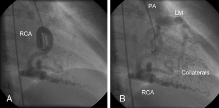
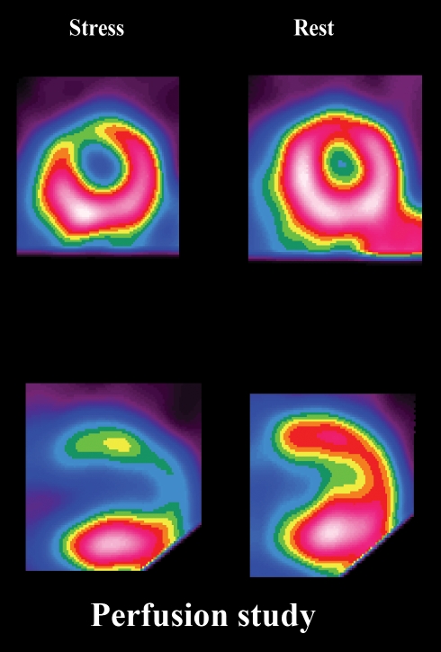
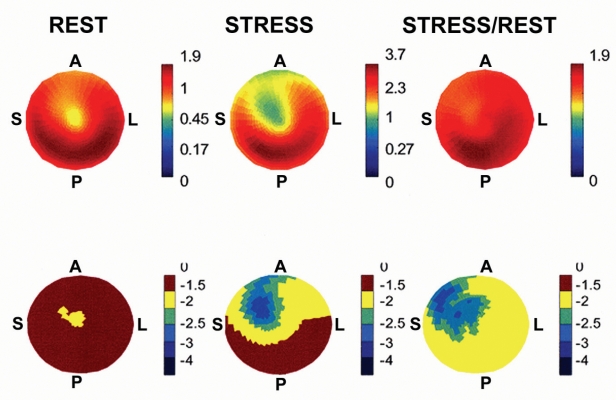
A 52-year-old woman presented with a two-year history of decreased exercise tolerance and palpitations. An echocardiogram demonstrated a markedly dilated and tortuous right coronary artery. The left main coronary artery could not be visualized. Coronary angiography showed an anomalous left coronary artery from the pulmonary artery (Figure 1). Blood flow to the left coronary system was entirely dependent on collaterals emanating from the right coronary artery. For a portion of the collateralized blood, there was a retrograde (‘upstream’) flow in the left epicardial system, with subsequent drainage into the pulmonary artery (‘myocardial-pulmonary steal’). This created a left-to-right shunt with a calculated pulmonary to systemic blood flow ratio of 1.5. On rubidium-82 positron emission tomography scanning, there was no perfusion abnormality at rest (Figure 2). This may have been possible due to chronic, compensatory arteriolar vasodilation in the distal bed of the left epicardial tree. This mechanism permitted the myocardium to ‘compete’ with the low pressure ‘sink’ of the pulmonary artery, thus allowing for a portion of the collateralized flow to pursue an antegrade (‘downstream’) course toward the left coronary microcirculation. On stress imaging with dipyridamole, there was a large defect in the left anterior descending artery area. Regional myocardial blood flow quantification, using rubidium-82 net retention, showed a marked reduction in coronary flow reserve in the left anterior descending artery area compared with that of the right coronary artery (Figure 3). This impaired flow reserve may be explained by limitations in further vasodilation at the site of the collateral network and/or the arteriolar circulation in the distal bed of the left anterior descending artery system. This setup creates the potential for intermittent ischemic episodes during stress and may explain the risk of sudden death reported in this patient population.
Figure 1.
Selective injection of the right coronary artery (RCA) in the 30° right anterior oblique projection. In the early phase of injection, a markedly dilated and tortuous RCA is noted (A). In the late phase of the injection, the left anterior descending artery tree is seen being filled with blood by an extensive collateral network (Collaterals) from the RCA (B). Contrast is seen as blood exits the left main (LM) coronary artery in a retrograde direction into the pulmonary artery (PA)
Figure 2.
Short and vertical long axis views of standard single photon emission computed tomography imaging with rubidium-82. Markedly diminished perfusion is seen in the left anterior descending artery area during dipyridamole infusion (stress), with normalization at rest
Figure 3.
Quantification method: Rest and stress rubidium-82 uptake plus stress/rest (flow reserve) polar maps. In the upper panel, calculated flows (in mL/g/min) are on the right of the colour bar. In the lower panel, each increment represents a degree of standard deviations below the mean of a normal population (less than 5% likelihood of coronary artery disease). The blue segments represent sectors greater than 2 SDs below the mean of a normal population. Note that the stress and stress/rest maps indicate impaired stress flow and flow reserve in the vascular bed of the left anterior descending artery. A Anterior; L Lateral; P Posterior; S Septal


