Abstract
Recent studies have revealed that eye gaze triggers reflexive shift of the observer's visuospatial attention to its direction even if it does not predict any events in the environment. To determine whether medial temporal structures are involved in this reflexive gaze processing, an experiment of the gaze-cuing paradigm was carried out in seven epileptic patients who had undergone unilateral temporal lobectomy and nine age- and IQ-matched epileptic controls who had not undergone any surgical treatments. Gaze cues were presented for 200 ms to the unilateral visual field, after which subjects were required to localize targets as quickly as possible. They were also instructed that gaze directions were not predictive of the location of the targets. When the gaze cues stimulated the intact hemisphere in lobectomized patients or either hemisphere in controls, reaction times for correct responses were significantly shorter when gaze directions were toward the targets than away from the targets. This cuing effect was not manifested following stimulation of the lesioned hemisphere in lobectomized patients. These findings suggest that the medial temporal structures, including the amygdala, play a crucial role in the reflexive shift of attention triggered by another person's gaze direction in humans.
Keywords: medial temporal structures, gaze direction, reflexive attentional shift, cuing paradigm, anterior temporal lobectomy, unilateral visual field presentation
The glance of another's gaze can induce an observer's gaze to follow the direction in which the observed person is looking. Through evolution, this gaze-following ability likely conferred an adaptive advantage for primates, allowing immediate detection of biologically significant stimuli in the environment (e.g. predatory animals) and appropriate collective responses to such stimuli (Emery, 2000). This ability also precedes the development of theory of mind in humans, the lack of which could severely impair social abilities (e.g. autism) (Baron-Cohen, 1995)
Psychological experiments using the cuing paradigm (Posner, 1980) have revealed that the shift of attention to the perceived gaze direction occurs rapidly (Friesen and Kingstone, 1998; Driver et al., 1999; Langton and Bruce, 1999). The studies have reported consistent findings using various types of gaze stimuli, such as photos of full faces with directional eye gaze (e.g. Driver et al., 1999) and the schematic drawing of isolated eye regions (e.g. Friesen and Kingstone, 1998). Because these attentional shifts by gaze can occur even when the gaze cues are unpredictive of the target location (e.g. Friesen and Kingstone, 1998), or counter-predictive of the target location (Driver et al., 1999; Friesen et al., 2004), it has been proposed that the gaze-triggered attentional shift is automatic (e.g. Langton and Bruce, 1999; however, see Ristic and Kingstone, 2005).
Some previous studies have explored the neural substrate for the gaze processing; they have been inconclusive. For example, a single-cell recording study (Perrett et al., 1985) and lesion studies (Campbell et al., 1990; Heywood and Cowey, 1992) in macaque monkeys suggested that specific cells in the anterior part of the superior temporal sulcus (STS) were responsive to specific gaze directions and devoted to their discrimination. In humans, studies using positron emission tomography (PET) (Wicker et al., 1998) and functional magnetic resonance imaging (fMRI) (Puce et al., 1998; Hoffman and Haxby, 2000; Kingstone et al., 2004) have examined the activity evoked by viewing gaze directions. These findings have consistently reported that the posterior part of the STS was involved in gaze perception in humans (Allison et al., 2000).
Other studies in humans have reported that other neural areas are also involved in gaze processing. For example, an fMRI study reported that not only the STS but also the fusiform gyrus was active while viewing gaze shifts (Pelphrey et al., 2003). Lesion studies reported that damage to the frontal lobe impaired the gaze-triggered attentional shift (Vecera and Rizzo, 2004, 2006).
In addition to these cortical areas, Kling and Brothers (1992) suggested the involvement of the amygdala in gaze perception in non-human primates. Defects in the discrimination of gaze direction were also reported in a patient with bilateral amygdala damage (Young et al., 1995). Using PET, Kawashima et al. (1999) demonstrated the activation of the amygdala in monitoring the direction of another person's gaze. Some fMRI studies have also shown activation of the amygdala in observing changes in gaze direction (Adams et al., 2003; Sato et al., 2004). Because previous neuroimaging and lesion studies have revealed that the amygdala is involved in automatic and rapid processing of social and/or emotional information (Whalen et al., 1998; Kubota et al., 2000; Anderson and Phelps, 2001), it is possible that gaze direction is also processed automatically and rapidly in the amygdala.
In the present study, we examined the involvement of the medial temporal structures, including the amygdala, in reflexive gaze processing. An experiment with a gaze-cuing paradigm was conducted in patients who had undergone unilateral temporal lobectomies, in which gaze cues were presented for short periods to the unilateral visual field. This experimental design allowed for comparison of performance following intact hemisphere stimulation with that after lesioned hemisphere stimulation as a within-subject factor. As eye gaze with and without facial context have been shown to have the capacity to trigger an attentional shift (e.g. Friesen and Kingstone, 1998), we tested both of these conditions.
MATERIALS AND METHODS
Subjects
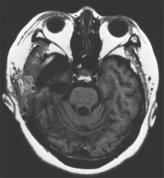
Seven subjects (three males and four females, aged 22–44 years) who had undergone unilateral temporal lobectomies for pharmacologically intractable seizures were investigated (Table 1). No patient had a visual field defect (see Procedure) or past history of ophthalmologic disease. Seizures were well controlled in all subjects, and all were mentally stable during the experiments. The patients had undergone standard anterior temporal lobectomies, left temporal lobectomies in five patients and right temporal lobectomies in two. The extent of temporal resection was 4–5 cm posterior to the temporal pole; two patients underwent resection below the superior temporal gyrus and five underwent resection below the middle temporal gyrus. Post-surgical MRIs were obtained for all patients to re-examine the extent of the resection (Figure 1). The procedures included removal of the anterior temporal neocortex, substantial portions of the amygdala, entorhinal cortex, perirhinal cortex and the anterior part of the hippocampal formation. The posterior STS was preserved in all patients. All seven patients were being maintained on one or more antiepileptic agents. The WAIS-R (Wechsler Adult Intelligence Scale-Revised) scores improved significantly after surgery (Table 1; dependent t-test, t = 3.53, P < 0.05).
Table 1.
Characteristics of patients who had undergone unilateral temporal lobectomies
| Patient | Age (years) | Gender | Handedness | WAIS-R (before surgery) | WAIS-R (after surgery) | Time from onset (years) | Time from surgery (years) | Resected side | Levels of resection |
|---|---|---|---|---|---|---|---|---|---|
| 1 | 44.0 | Female | Right | 73 | 77 | 43 | 2.8 | left | STG |
| 2 | 43.9 | Male | Right | 89 | 88 | 24 | 2.4 | right | MTG |
| 3 | 41.1 | Female | Right | 63 | 71 | 35 | 2.1 | right | MTG |
| 4 | 22.8 | Female | Right | 63 | 72 | 15 | 1.7 | left | MTG |
| 5 | 26.0 | Male | Right | 66 | 69 | 25 | 5.1 | left | MTG |
| 6 | 32.7 | Male | Right | 82 | 96 | 30 | 10.5 | left | MYG |
| 7 | 28.8 | Female | Right | 61 | 70 | 27 | 6.6 | left | STG |
WAIS-R = Wechsler adult intelligence scale-revised, STG = superior temporal gyrus, MTG = middle temporal gyrus.
Fig. 1.
Representative anatomical magnetic resonance imaging (MRI) image of a temporal-lobectomized patient who participated in the study.
As a control group, nine age- and IQ-matched epileptic patients who had undergone no surgical treatments (five males and four females; aged 20–43 years, independent t-test, t = 0.63, n.s.; IQ range 55–105, independent t-test, t = 0.06, n.s.) also participated in the experiment (Table 2). All control subjects were being maintained on one or more antiepileptic agents.
Table 2.
Characteristics of control subjects who had not undergone any surgical treatments
| Patient | Age (years) | Gender | Handedness | WAIS-R | Time from onset (years) | Types of epilepsy |
|---|---|---|---|---|---|---|
| 1 | 43.0 | Male | Right | 91 | 33 | Partial |
| 2 | 20.8 | Female | Right | 66 | 11 | Partial |
| 3 | 30.3 | Male | Right | 78 | 28 | Partial |
| 4 | 31.3 | Male | Right | 88 | 10 | Partial |
| 5 | 33.8 | Male | Right | 55 | 2 | Generalized |
| 6 | 34.0 | Male | Right | 66 | 3 | Generalized |
| 7 | 20.3 | Female | Right | 105 | 1 | Partial |
| 8 | 28.4 | Female | Right | 61 | 20 | Partial |
| 9 | 41.9 | Female | Right | 92 | 30 | Partial |
WAIS-R = Wechsler adult intelligence scale-revised.
All subjects in both groups were right-handed, as assessed using the Edinburgh Handedness Inventory (Oldfield, 1971). They had normal or corrected-to-normal visual acuity. All subjects gave written informed consent after the procedure was fully explained. Subjects were unaware of the goal of the experiments and of the nature of the experimental conditions. The testing time was approximately 45 min.
Apparatus
Stimuli were presented on a CRT monitor. The presentation of stimuli was controlled using SuperLab Pro software (ver.2.0, Cedrus, San Pedro, CA, USA). This software allowed for stimulus presentation within one screen refresh cycle (i.e. 10 ms) by setting up a new graphic page in the background of the screen. Reaction times (RTs) and accuracy measures were based on responses through a Cedrus RB-400 Response Box. Subjects were seated 57.3 cm from the monitor with a head-rest to keep their heads fixed, and the experimenter ensured that subjects were centered with respect to the monitor and keys of the switch box.
Stimulus materials
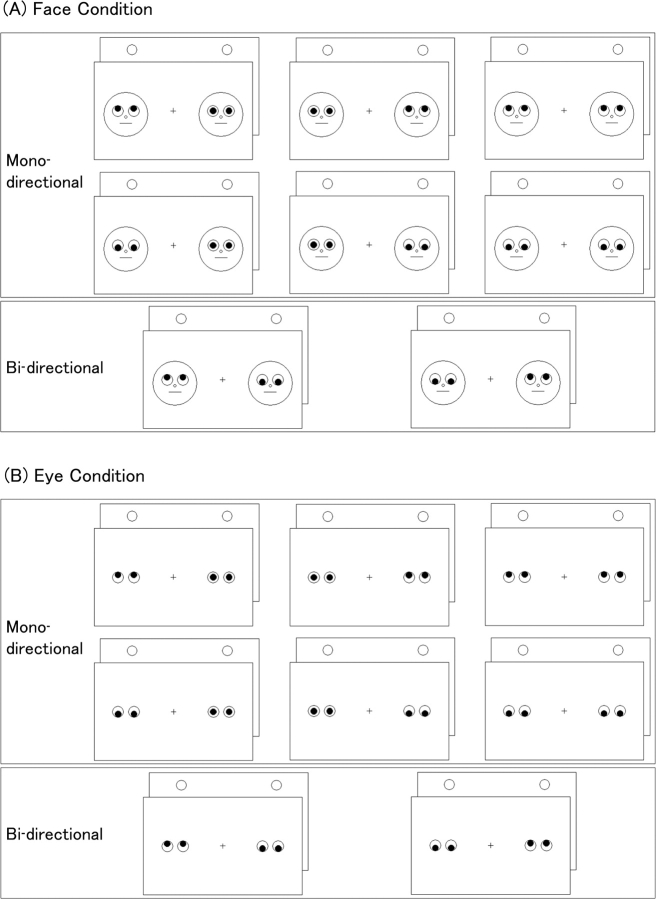
In the experiments, gazes were presented to each visual field as one of two types of stimuli: Face or Eye (Figure 2). Schematic stimuli of the face and eye were adopted to minimize extraneous complexities associated with real faces (e.g. face asymmetry, hair, gender) as in previous studies (Friesen and Kingstone, 1998; Kingstone et al., 2000).
Fig. 2.
Illustrated conditions of gaze directions and target locations: (A) Face display; and (B) Eye display. In each condition, presentation of cues and targets consisted of mono-directional and bi-directional cuing conditions. This figure shows the condition in which target circles were presented above the cues.
The Face display consisted of a white background with a black line drawing of two round faces subtending 3.6°, which were located 3.9° away from vertical axis of the screen (Figure 2). The Eye display consisted of a white background with two pairs of eyes. The eyes, pupils, fixation cross and targets were located in the same position as in the Face display.
Procedure
The experiments were performed individually. The subjects were seated in an armchair in a dark, quiet room at normal ambient temperature and instructed to look at the monitor in front of them.
Visual field examination
In the lobectomized-patient group, an assessment of possible visual field defects due to the temporal lobectomy was conducted using the same monitor. Subjects were instructed to look at a fixation cross in the center of the monitor. A target stimulus (black filled-in circle subtending 1.0°) was presented for 200 ms outside the area where the stimuli and targets were presented in test trials. The subjects were asked to point to the place where the target appeared.
Trial session
The start of a trial was signaled by a warning alarm and two faces on the CRT monitor. After 675 ms, the pupils of each face were randomly presented looking up or down. After 200 ms, the two faces were replaced by two target circles presented above or below the faces until a response was made. A stimulus onset asynchrony (SOA) of 200 ms was used for maximum stability in performance as predicted from the results of previous studies using different SOAs (Friesen and Kingstone, 1998; Driver et al., 1999; Langton and Bruce, 1999), and because this SOA was appropriate for the unilateral visual field presentation paradigm. The inter-trial interval was 675 ms. The procedure using the Eye display was identical to that using the Face display, except for the substitution of the Eye stimulus in place of the Face.
The subjects were instructed to indicate whether the targets appeared above or below the faces by pressing the upper or lower key on the switch box with either the left or right index finger. The positions of hands (i.e. fingers) for responses were switched between trial blocks, and the order of positioning was further counterbalanced among subjects. Reaction time was timed from the onset of target presentation, and measured in milliseconds.
The presentation of cues and targets was roughly divided into two conditions: mono-directional and bi-directional cuing conditions (Figure 2). The mono-directional cuing condition was characterized by a valid (i.e. gaze direction towards targets) or invalid (i.e. gaze direction away from targets) cue presented in the unilateral or bilateral visual fields. Presentation of a cue in a unilateral visual field had the effect of dominantly stimulating the opposing hemisphere. When the same directional gaze cue was presented in both visual fields, both hemispheres were equally stimulated. For the patient group, the mono-directional cuing condition was subdivided into three conditions according to the hemispheres stimulated: intact-, lesioned- and bilateral- hemispheric-stimulation. With the control group, the mono-directional cuing condition was subdivided into three conditions according to the sides of the hemispheres stimulated; left-, right- and bilateral-hemispheric-stimulation. The bi-directional cuing condition was defined as the presentation of a valid cue in one visual field and an invalid cue in the other visual field. For the lobectomized-patient group, the bi-directional cuing condition was subdivided into two conditions; ‘intact (valid)–lesioned (invalid)’, in which the intact hemisphere was stimulated by the valid cue and the lesioned hemisphere was stimulated by the invalid cue, and ‘intact (invalid)–lesioned (valid)’, in which the intact hemisphere was stimulated by the invalid cue and the lesioned hemisphere was stimulated by the valid cue. For the control group, the bi-directional cuing position was subdivided into two conditions; ‘left (valid)–right (invalid)’, in which the left hemisphere was stimulated by the valid cue and the right hemisphere was stimulated by the invalid cue, and ‘left (invalid)–right (valid)’, in which the left hemisphere was stimulated by the invalid cue and the right hemisphere was stimulated by the valid cue.
At the beginning of the experiments, the subjects were given 32 practice trials. After the practice trials, the first three blocks of 32 test trials were conducted. The subjects were then requested to exchange right and left hands for responding, and the second three blocks of 32 test trials were conducted (total of 192 test trials for each stimulus condition). The order of the test trials was randomized within each block. Short rests of about 15 s were interposed between blocks of test trials and long rests of several minutes were interposed after completing three blocks of test trials. The experiments were conducted in the following order: practice trials using the Face display, the first three blocks of test trials (session 1) using the Face display, the second three blocks of test trials (session 2) using the Face display, practice trials using the Eye display, the first three blocks of test trials (session 1) using the Eye display, and the second three blocks of test trials (session 2) using the Eye display.
Before beginning the test, the subjects were informed that it was important to fixate their eyes on the central fixation cross while it was presented, and that the gaze direction was not predictive of the location of the targets. The fixation of viewers’ eyes was verified by the experimenter during the task. They were also instructed to respond as quickly and as accurately as possible. Subjects were given an opportunity to ask questions regarding procedure before starting.
Data analysis
All data were analyzed using SPSS software (ver. 11.0J, SPSS Japan Inc., Tokyo, Japan). Incorrect responses were excluded from the analysis of RT. The median RT was calculated for each subject under each condition. For each group, the median RT was analyzed under mono-directional conditions using a 2 × 3 × 2 × 2 repeated-measures ANOVA performed with stimulus type (Face/Eye), stimulated hemisphere (Intact/Lesioned/Bilateral in the lobectomized-patient group and Left/Right/Bilateral in the control group), cue validity (Valid/Invalid) and session (first/second) as within-subject factors. Median RT findings under the bi-directional condition were analyzed using a 2 × 2 × 2 repeated-measures ANOVA with stimulus type (Face/Eye), cue validity [‘intact (valid)–lesioned (invalid)’/‘intact (invalid)–lesioned (valid)’ in the lobectomized-patient group; ‘left (valid)–right (invalid)’/‘left (invalid)–right (valid)’ in the control group] and session (first/second) as within-subject factors. Post hoc analyses were conducted using Ryan's method. Values were deemed statistically significant at P < 0.05. In cases where the assumption of sphericity was not met (P < 0.1, Mauchley's sphericity test), the Greenhouse–Geisser adjusted degrees of freedom was used (Greenhouse and Geisser, 1959).
Error analyses were conducted to confirm that the speed-accuracy trade-off phenomenon had no effect on RT. Wilcoxon's rank tests were conducted on the mean number of errors for the stimulated hemisphere and cue validity. Values were deemed significant at P < 0.05.
RESULTS
RT analysis
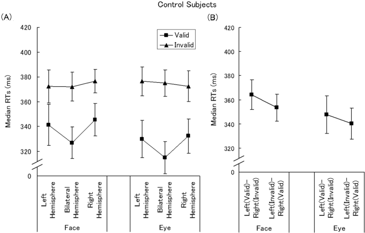
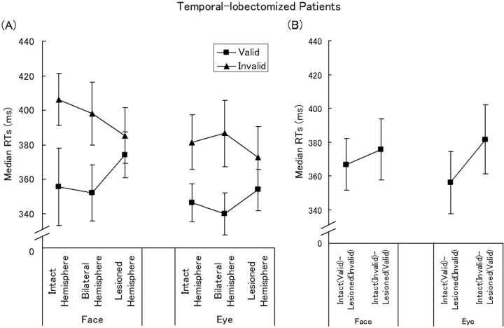
Median RTs under mono-directional conditions are shown in Figures 3A and 4A. The lobectomized-patients’ median RT data are also shown individually in Table 3. In the figures and tables, session factor was collapsed because analysis by session revealed no significant effect in the patient or control groups.
Fig. 3.
The means (±SEM) of median reaction times (RTs) in age- and IQ- matched control subjects: (A) mono-directional cuing condition; and (B) bi-directional cuing condition. Bars indicate standard errors.
Fig. 4.
The means (±SEM) of median reaction times (RTs) in temporal-lobectomized patients: (A) mono-directional cuing conditions; and (B) bi-directional cuing conditions. Bars indicate standard errors.
Table 3.
Median reaction times in temporal-lobectomized patients (ms)
| Patient | Face |
Eye |
||||||||||
|---|---|---|---|---|---|---|---|---|---|---|---|---|
| Intact |
Lesioned |
Bilateral |
Intact |
Lesioned |
Bilateral |
|||||||
| Valid | Invalid | Valid | Invalid | Valid | Invalid | Valid | Invalid | Valid | Invalid | Valid | Invalid | |
| 1 | 392 | 393 | 402 | 423 | 411 | 419 | 377 | 412 | 398 | 384 | 403 | 418 |
| 2 | 321 | 385 | 329 | 370 | 311 | 403 | 339 | 398 | 309 | 354 | 307 | 360 |
| 3 | 307 | 344 | 323 | 334 | 290 | 337 | 288 | 331 | 309 | 333 | 271 | 348 |
| 4 | 359 | 509 | 413 | 425 | 379 | 456 | 356 | 410 | 349 | 403 | 324 | 399 |
| 5 | 298 | 300 | 297 | 311 | 273 | 307 | 262 | 315 | 273 | 318 | 253 | 329 |
| 6 | 354 | 409 | 392 | 388 | 361 | 395 | 364 | 391 | 367 | 378 | 367 | 389 |
| 7 | 458 | 505 | 464 | 447 | 440 | 469 | 439 | 413 | 470 | 440 | 455 | 463 |
In the controls, there was a significant main effect of cue validity [F (1, 8) = 11.54, P < 0.01], indicating that RTs were significantly faster in the valid than invalid condition. Otherwise, there were no significant main effects or interactions.
In the lobectomized-patient group, there was a significant main effect of stimulus type [F (1, 6) = 6.27, P < 0.05], indicating that RTs were faster in the Eye than Face display condition. There was a significant main effect of cue validity [F (1, 12) = 13.98, P < 0.01] and significant interaction between the stimulated hemisphere and cue validity [F (2, 6) = 11.70, P < 0.005]. Follow-up analysis of this interaction revealed that the simple main effect of cue validity was significant only following stimulation of the intact hemisphere or bilateral hemispheres. That is, RTs were significantly faster under the valid than invalid condition, when the intact hemisphere was stimulated [F (1, 18) = 17.84, P < 0.001] or when bilateral hemispheres were stimulated [F (1, 18) = 20.80, P < 0.001], but not following stimulation of the lesioned hemisphere [F (1, 18) = 2.18, n.s.]. A significant simple main effect of the stimulated hemisphere was found under both the valid [F (2, 24) = 5.96, P < 0.01] and invalid conditions [F (2, 24) = 4.61, P < 0.05]. Post hoc multiple comparisons revealed that under the valid condition, RTs were significantly faster when the intact hemisphere was stimulated [t(24) = 3.34, P < 0.01] or when bilateral hemispheres were stimulated [t(24) = 2.42, P < 0.05], than when the lesioned hemisphere was stimulated. Under the invalid condition, RTs were significantly faster when the lesioned hemisphere was stimulated than when the intact hemisphere was stimulated [t(24) = 2.76, P < 0.05], or when bilateral hemispheres were stimulated [t(24) = 2.48, P < 0.05]. Under valid or invalid conditions, no significant difference was found between intact hemisphere stimulation and bilateral stimulation.
Median RTs under bi-directional cuing conditions are shown in Figures 3B and 4B. No significant main effects and interactions were found in the control group. In the lobectomized-patient group, a significant main effect with session [F (1, 6) = 7.33, P < 0.05] and a trend toward significance was found regarding the stimulated hemisphere [F (1, 6) = 4.49, P < 0.1].
Error analysis
In all groups, error rates were <2% under all conditions (0–1.94% in the control group; 0–0.95% in the lobectomized-patient group). In the control group, significantly more errors were noted following the presentation of invalid bilateral stimulation (Z = −2.3, P < 0.05) and right hemisphere stimulation (Z = −2.51, P < 0.05) in the Eye display conditions. In the patient group, significantly more errors were made following stimulation of the lesioned hemisphere by invalid cues using the Eye display (Z = 2.07, P < 0.05) and when bilateral hemispheres were stimulated (Z = 2.53, P < 0.05). These results suggest that the speed-accuracy trade-off phenomenon does not explain the RT findings.
DISCUSSION
The present study examined the role of the medial temporal structures in the reflexive shift of attention to another person's gaze direction.
In the controls, valid cues shortened the RT compared to invalid cues, regardless of the hemisphere stimulated. This suggests that the task-irrelevant gaze cues shifted the subjects’ attention reflexively to their directions. The findings on the unilateral visual field presentation are consistent with those of previous studies using the central visual field presentation paradigm (Friesen and Kingstone, 1998; Driver et al., 1999; Langton and Bruce, 1999).
Among temporal-lobectomized patients, valid cues shortened the RT compared to invalid cues when the gaze cues were presented to the intact hemisphere or bilateral hemispheres. This effect vanished, however, when the gaze cues stimulated the lesioned hemisphere. In the valid condition, RTs were significantly shorter following stimulation of the intact hemisphere or bilateral hemispheres compared with stimulation of the lesioned hemisphere. In the invalid condition, RTs were significantly longer following stimulation of the intact hemisphere or bilateral hemispheres than the lesioned hemisphere alone. That is, RTs were neither shortened following the presentation of valid cues nor prolonged after invalid cues when the lesioned hemisphere was stimulated. Additionally, under the bi-directional condition, RTs were faster when the intact hemisphere was stimulated by the valid cue (i.e. lesioned hemisphere stimulation by invalid cue) than when the lesioned hemisphere was stimulated by a valid cue (i.e. intact hemisphere stimulation by an invalid cue). Taken together, the effects of both valid and invalid cues appear to be weakened on presentation to the lesioned hemisphere, suggesting that reflexive shifts of attention to gaze directions are impaired by lesions in the medial temporal brain regions.
Some previous neuroimaging studies have reported the involvement of the posterior part of the STS in gaze perception in humans (Allison et al., 2000). In all subjects that participated in our study, the posterior regions of the STS were preserved, but the following areas had been removed: anterior temporal neocortex, substantial portions of the amygdala, entorhinal cortex, perirhinal cortex and the anterior part of the hippocampal formation. The human amygdala is a subcortical brain region involved in rapid processing of visual emotional non-social/social stimuli, including emotional facial expressions (Whalen et al. 1998; Kubota et al., 2000; Anderson and Phelps, 2001). It is likely that unilateral damage to the amygdala was a possible contributor in producing the lateralized attenuation of the cuing effect in our paradigm. Although previous lesion (Kling and Brothers, 1992; Young et al., 1995) and neuroimaging (Kawashima et al., 1999; Adams et al., 2003; Sato et al., 2004) studies have examined the role of the amygdala in gaze processing, no previous study has reported on behavioral consequences of amygdala damage in the reflexive shift of attention to another person's gaze direction. To our knowledge, this is the first study to suggest the involvement of medial temporal structures, including the amygdala, in the gaze-triggered reflexive shift of attention.
Previous studies have reported the involvement of the STS in gaze processing (Perrett et al., 1985; Puce et al., 1998; Wicker et al., 1998; Hoffman and Haxby, 2000). The STS and amygdala are connected reciprocally (Young et al., 1995). Thus, neural circuits consisting of the STS and amygdala may implement the processing of another individual's gaze direction. Moreover, a neuroimaging study (Hoffman and Haxby, 2000) has shown that the perception of another individual's averted gaze activates the intraparietal sulcus, which is directly connected to the STS and is involved in attentional orientation (Nobre et al., 1997). Based on these findings, it has been suggested that the amygdala may be involved in the reflexive shift of attention to perceived gaze directions by cooperating with other regions of the brain.
Previous studies have reported that the human amygdala is involved not only in gaze processing, but also in the processing of various types of communicative messages. For example, lesion and neuroimaging studies in humans have provided evidence that the amygdala is a key component in the recognition of emotions in facial expressions (Adolphs, 1999). Moreover, the STS is also involved in processing of gaze, facial expressions and biological motion (Allison et al., 2000). These findings suggest that the neuro-cognitive system constructed by the bi-directional connectivity between the STS and amygdala may process various types of social information for communication.
It should be noted, however, that resection was not restricted to the amygdala in the temporal-lobectomized patients. The resected brain regions also included the anterior temporal neocortex and the hippocampal formation. Although these brain regions have not been described as being important in gaze processing, it is possible that these regions may still be involved in gaze-triggered shifts of attention. To show that the amygdala is specifically involved in reflexive shifts of social attention, further studies are needed to examine whether amygdala-damaged patients, but not hippocampal-damaged patients, demonstrate a defect in social attention by gaze cues. Alternatively, functional neuroimaging studies of high spatial resolution may be able to address this.
The Eye and Face displays were used to clarify whether gaze processing in the amygdala depends on the processing of other parts of the face. It has previously been demonstrated that gaze directions without other face components triggers reflexive shifts of attention, and that this phenomenon is inhibited by the inversion of faces (Kingstone et al., 2000). That is, although the reflexive shift of attention to gaze directions can be affected by face processing, it is not dependent upon face processing (Kingstone et al., 2000). Although the RTs were significantly faster in the Eye than in the Face display condition, the cue effects were relatively similar between the two types of stimulus conditions. Although gaze processing may be affected by the surrounding facial features, the reflexive attentional shift to the gaze direction was absent following stimulation of the lesioned hemisphere under both the Face and Eye display conditions.
The use of gaze direction in social interactions is evident even in rats (Chance, 1962), suggesting that this communication mode may be commonly hard-wired in most mammals. In monkeys, it has been known that amygdala damage lead to changes in social responses (e.g. social isolation and taming) (Aggleton and Passingham, 1981). A more recent series of studies by Bachevalier (for review, see Bachevalier, 1994) reported that monkeys with early bilateral amygdala damage showed very few eye contact as well as reduced social communication, which persisted into adulthood. She also reported such findings were not observed in monkeys with lesions of inferior temporal cortex. These studies suggest a pivotal role of the amygdala in the regulation of eye contact and social behaviors, as well as a close relationship between them. It has been proposed that some amygdala functions related to biologically significant behaviors could be ubiquitous amongst mammals (LeDoux, 1996). The previous findings suggested that social cognition and gaze responses may be closely linked and that social communication via gaze direction in mammals may be universally implemented by the amygdala activity.
The gaze directions are more socially important for humans than for other mammals. It is found that the human eye morphology is uniquely characterized by the most widely exposed, white sclera, and the most horizontally elongated eye-outline among primates (Kobayashi and Kohshima, 2001). These morphological features are advantageous for sharing of attention with others. Baron-Cohen (1995) speculated that the development of sharing attention precedes that of the theory of mind, the ability to represent others’ thoughts and ideas.
Recently, Tomasello et al. (2005) proposed that shared attention may play a crucial role in human cultural activity. They proposed that in addition to the ability to read the intentions of others, the motivations to share psychological states with others, including both intentions and emotions, could lead to the collaborative activities that are specific to humans. Related to this hypothesis, a recent neuroimaging study indicated that the amygdala was involved in emotional elicitation while viewing others’ emotional facial expressions (Sato et al., 2004). Together with the present result indicating the involvement of the medial temporal structure in gaze-triggered attentional shift, the amygdala may play a special role in sharing intentions and emotions with other individuals, which may subsequently allow unique human collaborative interactions.
In summary, visuospatial attentional shifts were not observed when the lesioned hemispheres were stimulated by gaze cues in temporal-lobectomized patients. This suggests that the medial temporal structures, including the amygdala, play important roles in the reflexive shifts of attention triggered by the direction of another person's gaze.
Footnotes
Conflict of Interest
None declared.
REFERENCES
- Adams RB, Jr, Gordon HL, Baird AA, Ambady N, Kleck RE. Effects of gaze on amygdala sensitivity to anger and fear faces. Science. 2003;300:1536. doi: 10.1126/science.1082244. [DOI] [PubMed] [Google Scholar]
- Adolphs R. Social cognition and the human brain. Trends in Cognitive Sciences. 1999;3:469–79. doi: 10.1016/s1364-6613(99)01399-6. [DOI] [PubMed] [Google Scholar]
- Aggleton JP, Passingham RE. Syndrome produced by lesions of the amygdalain monkeys (Macaca mulatta) Journal of Comparative and Physiological Psychology. 1981;95:961–77. doi: 10.1037/h0077848. [DOI] [PubMed] [Google Scholar]
- Allison T, Puce A, McCarthy G. Social perception from visual cues: role of the STS region. Trends in Cognitive Sciences. 2000;4:267–78. doi: 10.1016/s1364-6613(00)01501-1. [DOI] [PubMed] [Google Scholar]
- Anderson AK, Phelps EA. Lesions of the human amygdala impair enhanced perception of emotionally salient events. Nature. 2001;411:305–9. doi: 10.1038/35077083. [DOI] [PubMed] [Google Scholar]
- Bachevalier J. Medial temporal lobe structures and autism: a review of clinical and experimental findings. Neuropsychologia. 1994;32:627–48. doi: 10.1016/0028-3932(94)90025-6. [DOI] [PubMed] [Google Scholar]
- Baron-Cohen S. Mindblindness: An Essay on Autism and Theory of Mind. London: MIT Press; 1995. [Google Scholar]
- Campbell R, Heywood CA, Cowey A, Regard M, Landis T. Sensitivity to eye gaze in prosopagnosic patients and monkeys with superior temporal sulcus ablation. Neuropsychologia. 1990;28:1123–42. doi: 10.1016/0028-3932(90)90050-x. [DOI] [PubMed] [Google Scholar]
- Chance M. Symposia of the Zoological Society of London. Vol. 8. London: Academic Press; 1962. An interpretation of some agonistic postures: the role of “cut-off” acts and postures; pp. 71–89. Evolutionary Aspects of Animal Communication. [Google Scholar]
- Driver J, Davis G, Ricciardelli P, Kidd P, Maxwell E, Baron-Cohen S. Gaze perception triggers reflexive visuospatial orienting. Visual Cognition. 1999;6:509–40. [Google Scholar]
- Emery NJ. The eyes have it: the neuroethology, function and evolution of social gaze. Neuroscience and Biobehavioral Reviews. 2000;24:581–604. doi: 10.1016/s0149-7634(00)00025-7. [DOI] [PubMed] [Google Scholar]
- Friesen CK, Kingstone A. The eyes have it! Reflexive orienting is triggered by nonpredictive gaze. Psychonomic Bulletin & Review. 1998;5:490–5. [Google Scholar]
- Friesen CK, Ristic J, Kingstone A. Attentional effects of counterpredictive gaze and arrow cues. Journal of Experimental Psychology. Human Perception and Performance. 2004;30:319–29. doi: 10.1037/0096-1523.30.2.319. [DOI] [PubMed] [Google Scholar]
- Greenhouse SW, Geisser S. On methods in the analysis of profile data. Psychometrika. 1959;24:95–112. [Google Scholar]
- Heywood CA, Cowey A. The role of the “face-cell” area in the discrimination and recognition of faces by monkeys. Philosophical Transactions of the Royal Society of London. Series B, Biological Sciences. 1992;335:31–8. doi: 10.1098/rstb.1992.0004. [DOI] [PubMed] [Google Scholar]
- Hoffman EA, Haxby JV. Distinct representations of eye gaze and identity in the distributed human neural system for face perception. Nature Neuroscience. 2000;3:80–4. doi: 10.1038/71152. [DOI] [PubMed] [Google Scholar]
- Kawashima R, Sugiura M, Kato T, et al. The human amygdala plays an important role in gaze monitoring. Brain: A Journal of Neurology. 1999;122:779–83. doi: 10.1093/brain/122.4.779. [DOI] [PubMed] [Google Scholar]
- Kingstone A, Friesen CK, Gazzaniga MS. Reflexive joint attention depends on lateralized cortical connections. Psychological Science: A Journal of the American Psychological Society. 2000;1:159–66. doi: 10.1111/1467-9280.00232. [DOI] [PubMed] [Google Scholar]
- Kingstone A, Tipper C, Ristic J, Ngan E. The eyes have it!: an fMRI investigation. Brain & Cognition. 2004;55:269–271. doi: 10.1016/j.bandc.2004.02.037. [DOI] [PubMed] [Google Scholar]
- Kling AS, Brothers LA. The amygdala and social behavior. In: Aggleton JP, editor. The Amygdala: Neurobiological Aspects of Emotion, Memory, and Mental Dysfunction. New York: Wiley-Liss; 1992. pp. 353–77. [Google Scholar]
- Kobayashi H, Kohshima S. Unique morphology of the human eye and its adaptive meaning: comparative studies on external morphology of the primate eye. Journal of Human Evolution. 2001;40:419–35. doi: 10.1006/jhev.2001.0468. [DOI] [PubMed] [Google Scholar]
- Kubota Y, Sato W, Murai T, Toichi M, Ikeda A, Sengoku A. Emotional cognition without awareness after unilateral temporal lobectomy in human. The Journal of Neuroscience: The Official Journal of the Society for Neuroscience. 2000;20:RC97(1–5). doi: 10.1523/JNEUROSCI.20-19-j0002.2000. [DOI] [PMC free article] [PubMed] [Google Scholar]
- Langton SRH, Bruce V. Reflexive visual orienting in response to the social attention to others. Visual Cognition. 1999;6:541–67. [Google Scholar]
- LeDoux JE. The Emotional Brain: The Mysterious Underpinnings of Emotional Life. New York: Simon and Schuster; 1996. [Google Scholar]
- Nobre AC, Sebestyen GN, Gitelman DR, Mesulam MM, Frackowiak RS, Frith CD. Functional localization of the system for visuospatial attention using positron emission tomography. Brain: A Journal of Neurology. 1997;120:515–33. doi: 10.1093/brain/120.3.515. [DOI] [PubMed] [Google Scholar]
- Oldfield RC. The assessment and analysis of handedness: the Edinburgh inventory. Neuropsychologia. 1971;9:97–113. doi: 10.1016/0028-3932(71)90067-4. [DOI] [PubMed] [Google Scholar]
- Pelphrey KA, Singerman JD, Allison T, McCarthy G. Brain activation evoked by perception of gaze shifts: the influence of context. Neuropsychologia. 2003;41:156–70. doi: 10.1016/s0028-3932(02)00146-x. [DOI] [PubMed] [Google Scholar]
- Perrett DI, Smith PA, Potter DD, et al. Visual cells in the temporal cortex sensitive to face view and gaze direction. 1985. Proceedings of the Royal Society of London. Series B, Containing Papers of a Biological Character. Royal Society (Great Britain), 223, 293–317. [DOI] [PubMed]
- Posner MI. Orienting of attention. The Quarterly Journal of Experimental Psychology. 1980;32:3–25. doi: 10.1080/00335558008248231. [DOI] [PubMed] [Google Scholar]
- Puce A, Allison T, Bentin S, Gore JC, McCarthy G. Temporal cortex activation in humans viewing eye and mouth movements. The Journal of Neuroscience: The Official Journal of the Society for Neuroscience. 1998;18:2188–99. doi: 10.1523/JNEUROSCI.18-06-02188.1998. [DOI] [PMC free article] [PubMed] [Google Scholar]
- Ristic J, Kingstone A. Taking control of reflexive attention. Cognition. 2005;94:B55–65. doi: 10.1016/j.cognition.2004.04.005. [DOI] [PubMed] [Google Scholar]
- Sato W, Yoshikawa S, Kochiyama T, Matsumura M. The amygdala processes the emotional significance of social expressions: an fMRI investigation using the interaction between expression and face direction. Neuroimage. 2004;22:1006–13. doi: 10.1016/j.neuroimage.2004.02.030. [DOI] [PubMed] [Google Scholar]
- Tomasello M, Carpenter M, Call J, Behne T, Moll H. Understanding and sharing intentions: the origins of cultural cognition. Behavioral and Brain Sciences. 2005;28:675–91. doi: 10.1017/S0140525X05000129. [DOI] [PubMed] [Google Scholar]
- Vecera SP, Rizzo M. What are you looking at?: impaired social attention’ following frontal-lobe damage. Neurospychologia. 2004;42:1657–65. doi: 10.1016/j.neuropsychologia.2004.04.009. [DOI] [PubMed] [Google Scholar]
- Vecera SP, Rizzo M. Eye gaze does not produce reflexive shifts of attention: evidence from frontal-lobe damage. Neuropsychologia. 2006;44:150–9. doi: 10.1016/j.neuropsychologia.2005.04.010. [DOI] [PubMed] [Google Scholar]
- Whalen PJ, Rauch SL, Etcoff NL, McInerney SC, Lee MB, Jenike MA. Masked presentations of emotional facial expressions modulate amygdala activity without explicit knowledge. The Journal of Neuroscience: The Official Journal of the Society for Neuroscience. 1998;18:411–18. doi: 10.1523/JNEUROSCI.18-01-00411.1998. [DOI] [PMC free article] [PubMed] [Google Scholar]
- Wicker B, Michel F, Henaff MA, Decety J. Brain regions involved in the perception of gaze: a PET study. NeuroImage. 1998;8:221–7. doi: 10.1006/nimg.1998.0357. [DOI] [PubMed] [Google Scholar]
- Young AW, Aggleton JP, Hellawell DJ, Johnson M, Broks P, Hanley JR. Face processing impairments after amygdalotomy. Brain: A Journal of Neurology. 1995;118:15–24. doi: 10.1093/brain/118.1.15. [DOI] [PubMed] [Google Scholar]


