Abstract
Urinary incontinence in young girls who have been toilet trained may be due to an ectopic ureter inserting below the urinary sphincter. This diagnosis is frequently delayed, is psychologically distressing, and may be missed at physical examination. Findings at ultrasound evaluation may be subtle and imaging with computed tomography or intravenous urography exposes young patients to ionizing radiation. We report two cases of girls with urinary incontinence where magnetic resonance (MR) urography revealed subtle renal duplication which implied the presence of an ectopic duplicated ureter with infrasphincteric insertion. These cases stress the importance of examining the kidneys, rather than the perineum, at MR, ultrasound and intravenous urogram evaluation, and show the value of MR urography as a safe alternative to computed tomography and intravenous urography for making this diagnosis.
Renal duplication with associated complete ureteral duplication can cause continuous incontinence in girls if the ureter draining the upper pole moiety inserts ectopically below the external urethral sphincter [1,2]. The classic presentation is that of lifelong dribbling of urine or wetness despite successful toilet training [2]. Ideally, diagnosis of this condition is made early without exposing the patient to ionizing radiation; however, when the upper moiety is small and dysplastic, diagnosis can be subtle and the condition can go unrecognized [2–4]. We present two cases where MR urography was helpful in diagnosing a small dysplastic upper pole moiety as the cause of continuous incontinence in young girls.
Case reports
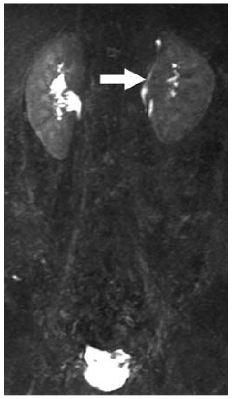
A 3-year-old girl presented with continuous urinary leakage despite successful toilet training. No ectopic ureteral opening was seen on physical examination. Renal sonography was reported as normal. MR urography demonstrated left renal duplication with a small dysplastic upper pole moiety, although the associated ureter was not seen (Fig. 1). Surgical exploration confirmed complete duplication of the left collecting system with the ureter of the small upper pole moiety draining ectopically just posterior to the external urethral meatus.
Fig. 1.
Three-year-old girl with perineal wetting. MR urogram shows a duplicated left kidney upper pole ureter (arrow) arising from a poorly enhancing upper pole moiety. The prior renal ultrasound (not shown) revealed no abnormality
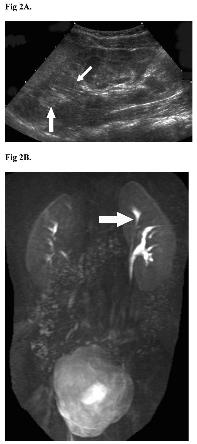
A 5-year-old girl presented with the same symptoms. No ectopic ureteral opening was seen on physical examination. Renal sonography was reported as showing only mild renal asymmetry (Fig. 2A), with a left renal length of 11 cm and a right renal length of 8 cm. MR urography demonstrated a duplicated left kidney with a small dysplastic upper pole moiety and pelvis (Fig. 2B).
Fig. 2.
Five-year-old girl with perineal wetting. (A) Ultrasound shows an enlarged left kidney. Subtle duplication of the renal collecting system with intervening renal parenchyma (small arrow) dividing the renal sinus fat (large arrow) into two compartments was not initially diagnosed. (B) MR urogram shows a duplicated upper pole ureter (arrow).
Surgical exploration confirmed complete ureteral duplication to at least the level of the bladder neck; the inferior extent was not dissected and no external opening was detected. Both patients underwent ureteroureterostomy with full symptomatic resolution.
Discussion
We report two cases where MR urography, a relatively novel imaging technique, was critical to diagnose duplicated ureters in girls with perineal wetting. In cases of duplicated collecting systems, MR urography has been shown to more clearly demonstrate the anatomy of the renal parenchyma, the renal collecting system, and the ureter and ureteral orifice, when compared to visualization on IVU and pelvic ultrasound [5]. This non-invasive technique may be useful when the findings at sonography or physical exam are subtle or overlooked, as may occur even in experienced centers. These examples also highlight the need for careful examination of the kidneys since the ureters may not be visualized due to limited excretion from dysplasia of the upper pole moiety in patients with suspected ectopic ureter [2]. An alternative imaging technique is the Tc-99m DMSA scan. Our second case indicates that the ectopic ureteral insertion may be occult even at surgery, but surgical repair should be performed if the appropriate constellation of clinical history and imaging findings is present. Correct recognition of the condition allows for straightforward surgical repair, with resolution of the distressing symptoms.
We conclude that MR urography may facilitate earlier diagnosis of subtle renal duplication with an infrasphinteric ectopic ureteral insertion in girls with persistent perineal wetting. Surgery may allow for rapid cure of the distressing and unpleasant symptoms.
Footnotes
Publisher's Disclaimer: This is a PDF file of an unedited manuscript that has been accepted for publication. As a service to our customers we are providing this early version of the manuscript. The manuscript will undergo copyediting, typesetting, and review of the resulting proof before it is published in its final citable form. Please note that during the production process errors may be discovered which could affect the content, and all legal disclaimers that apply to the journal pertain.
References
- 1.Berrocal T, Lopez-Pereira P, Arjonilla A, Gutierrez J. Anomalies of the distal ureter, bladder, and urethra in children: embryologic, radiologic, and pathologic features. Radiographics. 2002;22:1139–1164. doi: 10.1148/radiographics.22.5.g02se101139. [DOI] [PubMed] [Google Scholar]
- 2.Carrico C, Lebowitz RL. Incontinence due to an infrasphincteric ectopic ureter: why the delay in diagnosis and what the radiologist can do about it. Pediatr Radiol. 1998;28:942–949. doi: 10.1007/s002470050506. [DOI] [PubMed] [Google Scholar]
- 3.Braverman RM, Lebowitz RL. Occult ectopic ureter in girls with urinary incontinence: diagnosis by using CT. AJR Am J Roentgenol. 1991;156:365–366. doi: 10.2214/ajr.156.2.1898815. [DOI] [PubMed] [Google Scholar]
- 4.Gharagozloo AM, Lebowitz RL. Detection of a poorly functioning malpositioned kidney with single ecotopic ureter in girls with urinary dribbling: imaging evaluation in five patients. AJR Am J Roentgenol. 1995;164:957–961. doi: 10.2214/ajr.164.4.7726056. [DOI] [PubMed] [Google Scholar]
- 5.Riccabona M, Simbrunner J, Ring E, Ruppert-Kohlmayr A, Ebner F, Fotter R. Feasibility of MR urography in neonates and infants with anomalies of the upper urinary tract. Eur Radiol. 2002;12:1442–1450. doi: 10.1007/s00330-001-1180-6. [DOI] [PubMed] [Google Scholar]


