The dermatologist’s technique for sampling skin has remained relatively unchanged over the last 10 years. Exophytic lesions are typically shaven, while deeper processes are sampled with a punch biopsy. However, we have found that these two techniques are often impractical and cumbersome in their application to some cutaneous morphologies. We introduce a technique which employs a punch biopsy-pen to perform a shave or “scoop” excision. This method is ideally designed for those lesions in which a deep shave is required, but a straight blade is awkward to apply flush with the skin surface.
The potential disadvantage of shaving a flat lesion or plaque is the inability to achieve a sufficiently deep or representative sample. Oftentimes, placing a blade flush against the skin’s surface and cutting parallel makes it impossible to gain an adequate sample size. Alternatively, if one angles the blade at a 45-degree angle, a jagged or “ripped” sample may result in suboptimal histopathology and healing. The scoop ensures that adequate tissue sampling is achieved, thus making a histopathologic diagnosis readily available. The scoop also results in a smooth biopsy edge which results in less trauma and more rapid healing without scar. The scoop has the additional benefit of providing enough depth so as to make prognostication more accurate in cases of suspected malignancy.1
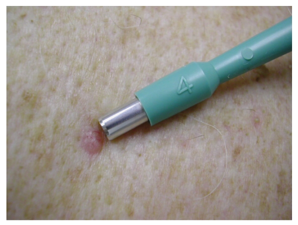
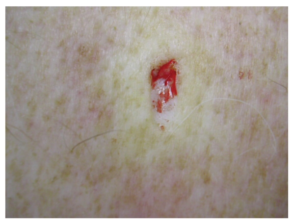
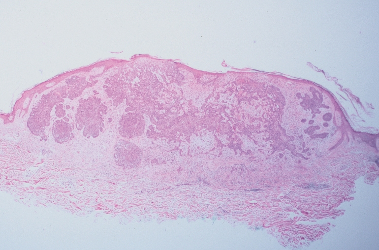
The site for sampling is chosen to ensure the highest yield. The scoop biopsy may be performed with a punch biopsy-pen of any width. The tools necessary for the scoop biopsy are similar to those required for a shave. Observing standard surgical techniques, the lesion is cleansed and locally anesthetized. Countertraction is applied with the nondominant hand, and the biopsy-pen is inserted into the skin in a pendulous manner. The punch tool scoops the skin like a pendulum (Figure 1 ▶). Once the tissue is removed, the subcutaneous tissue is visualized and a procoagulant, such as Monsel’s solution or Drysol, may be applied for hemostasis (Figure 2 ▶). Suturing of the resultant trough-shaped defect is not required. Healing, usually without scarring, can be expected in 1 to 2 weeks as with standard shave biopsy. A hematoxylin and eosin stain of the resultant scoop is shown in Figure 3 ▶. Note the ample sample size and smooth edges.
Figure 1.
Standard 4 mm punch biopsy-pen prior to scoop biopsy of a suspected basal cell carcinoma on a patient’s left chest.
Figure 2.
The resultant defect immediately following scoop biopsy. Monsel’s solution was applied for rapid hemostasis.
Figure 3.
Hematoxylin and eosin stain of the resultant scoop biopsy.
References
- 1.Ng PC, Barzilai DA, Ismail SA, Averitte RL Jr, Gilliam AC. Evaluating invasive cutaneous melanoma: is the initial biopsy representative of the final depth? J Am Acad Dermatol 2003;48:420–424. [DOI] [PubMed] [Google Scholar]


