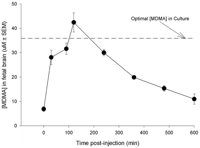
Figure 3.
The concentration of MDMA as a function of time in fetal brain after a 15 mg/kg s.c. injection of MDMA on E18 to the dam. The dam had been injected previously from E14 to E17 with 15 mg/kg MDMA (b.i.d). The dashed line represents the concentration of MDMA used in culture studies. Each time point represents three fetuses from a separate dam. These data demonstrate that the concentration of MDMA used for in vitro culture studies are within the physiologic range of a fetus exposed to a 15 mg/kg injection of MDMA to a drug-experienced dam.

