
Figure 2.
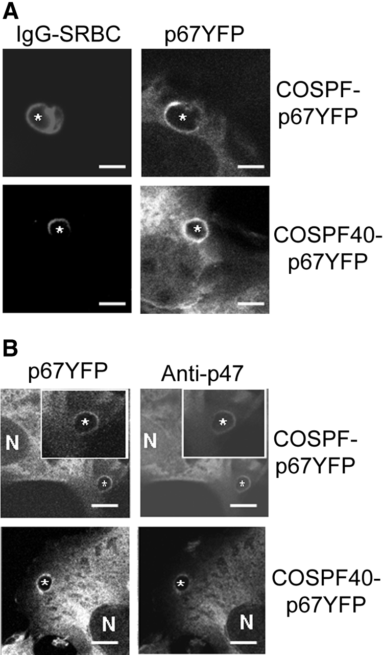
Translocation of p67phox-YFP and p47phox during phagocytosis of IgG-SRBCs by COS7 transgenic cells. IgG-SRBCs were fed to the indicated COSphox cells growing on coverslip-bottomed dishes. N shows the location of nucleus and asterisks indicate the IgG-SRBC phagosomes. Bar represents 5 μm. (A) Images of Alexa-633–labeled IgG-SRBCs and p67phox-YFP after fixation and analysis by confocal microscopy as described in “Immunofluorescence microscopy.” (B) Simultaneous imaging of p67phox-YFP and p47phox after immunofluorescent staining with anti-p47phox mAb and Alexa-555 goat anti–mouse IgG.