Abstract
H5N1 influenza A viruses are exacting a growing human toll, with more than 240 fatal cases to date. In the event of an influenza pandemic caused by these viruses, embryonated chicken eggs, which are the approved substrate for human inactivated-vaccine production, will likely be in short supply because chickens will be killed by these viruses or culled to limit the worldwide spread of the infection. The Madin-Darby canine kidney (MDCK) cell line is a promising alternative candidate substrate because it supports efficient growth of influenza viruses compared to other cell lines. Here, we addressed the molecular determinants for growth of an H5N1 vaccine seed virus in MDCK cells, revealing the critical responsibility of the Tyr residue at position 360 of PB2, the considerable requirement for functional balance between hemagglutinin (HA) and neuraminidase (NA), and the partial responsibility of the Glu residue at position 55 of NS1. Based on these findings, we produced a PR8/H5N1 reassortant, optimized for this cell line, that derives all of its genes for its internal proteins from the PR8(UW) strain except for the NS gene, which derives from the PR8(Cambridge) strain; its N1 NA gene, which has a long stalk and derives from an early H5N1 strain; and its HA gene, which has an avirulent-type cleavage site sequence and is derived from a circulating H5N1 virus. Our findings demonstrate the importance and feasibility of a cell culture-based approach to producing seed viruses for inactivated H5N1 vaccines that grow robustly and in a timely, cost-efficient manner as an alternative to egg-based vaccine production.
H5N1 influenza A viruses are exacting a growing human toll, with more than 240 fatal cases to date (http://www.who.int/csr/disease/avian_influenza/en/). The epidemic regions have expanded from Asia to Europe and Africa, raising concerns over a possible influenza pandemic (9). Currently, H5N1 prepandemic human vaccines are being stockpiled in many countries. These inactivated H5N1 vaccines are produced from viruses propagated in embryonated chicken eggs. In the event of a pandemic due to H5N1 viruses, however, it is highly likely that embryonated chicken eggs will be in short supply as H5N1 vaccine production will escalate but at the same time chickens will be killed by the viruses or culled in an effort to limit the worldwide spread of the virus infection. Therefore, an egg-free system should be considered as an alternative means of H5N1 vaccine production. Cell culture-based H5N1 vaccine production is a promising and safe approach that may meet this need (32).
Production of cell culture-based inactivate vaccines is in development in many countries, with some vaccines at the stage of clinical trials (8). This approach has considerable advantages over egg-based production: (i) it may lead to more-rapid and larger-scale vaccine production (6); (ii) it may avoid the potential for selecting variants adapted for embryonated chicken eggs (16), whose antigenicity would not longer match that of the circulating viruses; (iii) it may avoid contamination problems that have occurred with egg-based production; and (iv) it dispenses with the incorporation of the allergic component of eggs into vaccines (14). However, a limited number of cell lines are approved for cell culture-based vaccine production. One of them, the Madin-Darby canine kidney (MDCK) cell line, is a candidate for this purpose (8) because it efficiently supports the growth of influenza viruses compared to other cell lines (7).
A major concern of prepandemic H5N1 human vaccines without adjuvants is their limited immunogenicity, which requires higher concentrations of hemagglutinin (HA) antigens to provide protective immunity than are used for seasonal influenza vaccines (31). Therefore, to secure a large number of doses of effective H5N1 vaccine, the seed viruses are required to have high-level growth properties. Currently, prepandemic H5N1 vaccines, including the NIBRG-14-based vaccine recommended by the World Health Organization (WHO), are being stockpiled (2). These viruses comprise an HA gene whose cleavage site has been modified to an avirulent type (the mHA gene) and a neuraminidase (NA) gene, both derived from an H5N1 strain, and six remaining genes, derived from a high-growing donor virus (creating a so-called 6:2 reassortant). The WHO recommends A/Puerto Rico/8/34 (H1N1; PR8) as a donor strain for vaccine seed viruses because of its safety in humans and its high-level growth property in eggs (34). Previously, we found that the growth property of vaccine seed viruses in eggs depends on the genes encoding the internal proteins of the donor virus (10). Therefore, with respect to H5N1 vaccine production in MDCK cells, it is necessary to select a high-growth donor strain, whose internal proteins function efficiently in this cell line.
In this study, we found that the H5N1 6:2 reassortant viruses with our laboratory strain of PR8 [PR8(UW)] (10) as the background grew significantly better than NIBRG-14 with the PR8(Cambridge) background in MDCK cells. We therefore sought to determine the molecular basis of this difference in growth, which would contribute to the robustness of MDCK cell-based H5N1 vaccines, and tested whether the HA/NA functional balance regulates the growth of the vaccine seed viruses in MDCK cells, as it does in eggs (10). Our results should help to establish MDCK cell-based H5N1 vaccine production as a viable option for large-scale vaccine production in the event of a pandemic.
MATERIALS AND METHODS
Cells.
MDCK cells, maintained in our laboratory, were grown in minimal essential medium (MEM) with 5% newborn calf serum. 293T human embryonic kidney cells were maintained in Dulbecco's modified Eagle's MEM with 10% fetal calf serum. A72 canine tumor fibroblasts (13) were maintained in L15 medium (GIBCO-BRL) with 20% fetal calf serum. Cells were maintained at 37°C in 5% CO2.
Viruses.
The H5N1 A/Vietnam/UT30259/04 (VN30259), A/Vietnam/UT3030/04 (VN3030), A/Indonesia/UT3006/05 (Indo3006), and A/Anhui/2/05 (AH2) strains were propagated in 10-day-old embryonated chicken eggs at 37°C for 48 h, after which time the allantoic fluids containing viruses were harvested. All experiments with these infectious viruses were carried out in a biosafety level 3 containment laboratory. The WHO-recommended vaccine seed virus, NIBRG-14 (PR8/VN1194 6:2 reassortant), was a kind gift from J. Wood and J. Robertson at the National Institute for Biological Standards and Control, United Kingdom. Recombinant vesicular stomatitis virus (VSV) carrying the green fluorescent protein (GFP) gene instead of the G protein gene (VSV-ΔG*-GFP) (30) was kindly provided by M. A. Whitt at the University of Tennessee, Memphis, TN.
Construction of plasmids and reverse genetics.
To generate reassortants of influenza A viruses, we used plasmid-based reverse genetics (23). Viral RNA was extracted from the allantoic fluids by using a commercial kit (ISOGEN LS; Nippon Gene) and was converted to cDNA by using reverse transcriptase (SuperScript III; GIBCO-BRL) and primers containing the consensus sequences of the 3-prime ends of the RNA segments for the H5 viruses. The full-length cDNAs were then PCR amplified with ProofStart polymerase (Qiagen) and with segment-specific primer pairs and cloned into a plasmid under the control of the human polymerase I (PolI) promoter and the mouse RNA PolI terminator (PolI plasmids). By inverse PCR using back-to-back primer pairs followed by ligation, we altered the HA gene sequence that encodes the cleavage site of the wild-type VN30259, VN3030, Indo3006 (RERRRKKR), and AH2 (RERRRKR) viruses to create the avirulent-type sequence (RETR), as described elsewhere (11). We also constructed PolI-VN30259NA, PolI-VN3030NA, PolI-Indo3006NA, and pPolI-AH2NA plasmids containing the NA genes by using reverse transcription-PCR with N1-specific primers. We additionally constructed pPolI-VN1203FillNA with the insertion of a 20-amino-acid-encoding sequence derived from the A/goose/Guandong/1/96 NA stalk region into VN1203NA (VN1203FillNA). All of these constructs were sequenced to verify the absence of unwanted mutations. Primer sequences will be provided upon request.
We used our previously produced series of PolI constructs, derived from the PR8(UW) and PR8(Cambridge) strains, for reverse genetics (10, 11) and also used PolI plasmids containing mHA genes derived from VN1194 and VN1203 and NA genes derived from A/WSN/33 (H1N1; WSN), A/Hong Kong/213/03 (H5N1; HK213), and A/Kanagawa/173/2001 (H1N1; Kanagawa) (11, 17, 23).
Chimeric PB2 genes were constructed by switching the fragments at two BsmBI sites between pPolI-PR8(UW)PB2 and pPolI-PR8(Cambridge)PB2. To generate PB2 and NS mutants, PolI plasmids expressing the PB2 and NS genes of PR8 were used as templates for site-directed mutagenesis by the inverse PCR method. Plasmids expressing PR8 NP, PA, PB1, or PB2 under the control of the chicken β-actin promoter were used for all reverse genetics experiments. Briefly, PolI plasmids and protein expression plasmids were mixed with a transfection reagent, Trans-IT 293T (Panvera); incubated at room temperature for 15 min; and then added to 293T cells. Transfected cells were incubated in Opti-MEM I (GIBCO-BRL) for 48 h. Supernatants containing infectious viruses were harvested and propagated in 10-day-old embryonated chicken eggs at 37°C for 48 h, after which time the allantoic fluids containing virus were harvested and stored at −80°C until use.
Viral replication in MDCK cells.
Virus was inoculated into MDCK cell monolayers at a multiplicity of infection (MOI) of 0.01 PFU with MEM containing 0.3% bovine serum albumin and 0.8 μg/ml TPCK-trypsin and incubated at 33°C. Viruses in the culture supernatants were collected at a given number of hours postinfection (hpi) and then titrated by use of an MDCK plaque assay to determine the virus titers. The plaque sizes (at least 15 plaques for each virus) were measured by micrometer calipers after staining with crystal violet.
Luciferase assay-based assessment of viral polymerase activity.
The viral polymerase activity was measured as described previously (24). All experiments were independently performed in triplicate. Briefly, pPolIC250-NP(0)Fluc(0), a luciferase reporter plasmid under the control of the canine PolI promoter (22), was cotransfected into MDCK cells by using plasmids expressing PR8(UW) PB1, PA, and NP and either PB2 derived from PR8(UW) or PR8(Cambridge) or mutant PB2 under the control of the chicken β-actin promoter. The cells were subjected to a dual-luciferase reporter assay system (Promega, Madison, WI) on a microplate luminometer (Veritas; Turner Biosystems, Sunnyvale, CA) according to the manufacturer's instructions. pGL4.74[hRluc/TK] (Promega) was used as an internal control.
IFN bioassay.
Levels of interferon (IFN) secreted by virus-infected canine cells were determined as previously described (13, 25), with some modifications. Briefly, MDCK cells were infected with wild-type or NS1 mutant viruses at an MOI of 2. Supernatants from infected cells were harvested 24 hpi and then treated with UV light for 20 min to inactivate the infectivity of the virus. UV-treated supernatants were then added to A72 cells and incubated for 20 h before being infected with VSV-ΔG*-GFP at an MOI of 1. Adherent cells were detached by vigorous pipetting in phosphate-buffered saline at 12 hpi and then washed once with cold phosphate-buffered saline supplemented with 2% fetal calf serum and 0.1% sodium azide (wash buffer). Cells were washed again before the number of cells expressing GFP was counted by FACSCalibur with Cell Quest software (Becton Dickinson).
Statistical analysis.
All comparisons of the infectivity titers, relative luciferase activities, numbers of GFP-positive cells, and plaque diameters of each virus relied on Student's t test with two-tailed analysis to determine significant differences.
RESULTS
Growth of PR8/VN1194 6:2 reassortant viruses in MDCK cells.
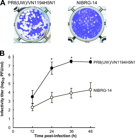
To assess the replication abilities of two PR8/VN1194 6:2 reassortants, PR8(UW)/VN1194H5N1 [PR8(UW) strain background] and NIBRG-14 [PR8(Cambridge) strain background], in MDCK cells, we examined the plaque-forming characteristics of these viruses and their growth kinetics. PR8(UW)/VN1194H5N1 formed larger plaques (Fig. 1A) and grew significantly better than NIBRG-14 (Fig. 1B). The peak viral titer reached 3.2 × 10⁷ ± 1.2 × 10⁷ PFU/ml at 36 hpi for PR8(UW)/VN1194H5N1, compared to 6.8 × 10⁴ ± 1.5 × 10⁴ PFU/ml at 48 hpi for NIBRG-14. These two viruses have identical HAs and NAs, indicating that our PR8(UW) strain is responsible for the superior growth kinetics relative to those for the PR8(Cambridge) strain background of H5N1 vaccine seed virus in MDCK cells.
FIG. 1.
Growth of PR8/H5N1 6:2 reassortant viruses in MDCK cells. (A) Plaque morphology of PR8(UW)/VN1194H5N1 and WHO-recommended NIBRG-14 on MDCK cells. (B) Viral titers of PR8(UW)/VN1194H5N1 and NIBRG-14 were determined at 12, 24, 36, and 48 hpi at an MOI of 0.01. The data are reported as mean titers with standard deviations for three independent experiments. Virus titers with significant growth enhancement relative to those of NIBRG-14 (P < 0.05; Student t test with two-tail analysis) are shown (*).
Growth of reassortants between the PR8(UW) and PR8(Cambridge) strains.
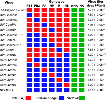
To identify the genes responsible for the superior growth of our 6:2 reassortant, we made a series of gene reassortants between the PR8(UW) and PR8(Cambridge) strains with VN1194 mHA and NA and measured their viral titers in MDCK cells (Fig. 2). Every reassortant possessing PR8(Cambridge) PB2 exhibited significantly poorer growth (more than 3 logs lower) than PR8(UW)/VN1194H5N1 (P < 0.01; Student's t test). Conversely, the reassortant Cam-UWPB2 virus that possesses PR8(UW) PB2 with PR8(Cambridge) as the background grew significantly better than NIBRG-14 (P < 0.01). These results indicated that PR8(UW) PB2 is responsible for the high growth of PR8(UW)/VN1194H5N1 in MDCK cells.
FIG. 2.
Growth of PR8/H5N1 reassortant viruses in MDCK cells. All viruses possess mHA and NA, both derived from VN1194 (green), and their remaining genes are from either PR8(UW) (red) or PR8(Cambridge) (blue). Cells were infected with each virus at an MOI of 0.01. Virus titers were determined in a plaque assay with MDCK cells at 36 hpi. The data are shown as mean titers with standard deviations (n ≥ 3). Titers significantly (P < 0.05) decreased (*) or increased (***) compared with that of PR8(UW)/VN1194H5N1 are shown. Titers significantly increased (**) compared with that of NIBRG-14 are also shown.
Interestingly, the reassortant UW-CamNS virus that possesses the PR8(Cambridge) NS gene with the PR8(UW) background grew significantly better than PR8(UW)/VN1194H5N1 (P < 0.01). This result indicates that the PR8(Cambridge) NS gene product enhanced viral growth in MDCK cells, although a negative effect was not observed when the NS gene of NIBRG-14 was replaced with that of the PR8(UW) (i.e., Cam-UWNS) strain.
These observations demonstrate that it is PB2 that primarily is responsible for the difference in virus growth in MDCK cells between PR8(UW)/VN1194H5N1 and NIBRG-14 but that NS also partially contributes to this difference.
PB2 is responsible for viral growth in MDCK cells.
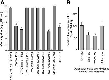
To determine the amino acid residues that determine virus growth in MDCK cells, we compared the PB2 amino acid sequence of PR8(UW) with that of PR8(Cambridge) and found six amino acid differences between the two strains (Table 1). To identify which residue(s) is responsible for virus growth, we generated two PB2 chimeric viruses whose other internal genes came from PR8(UW); chimera 1 possesses the amino-terminal portion (residues 1 to 387) of PR8(Cambridge) PB2 and the carboxyl-terminal portion (residues 388 to 759) of PR8(UW) PB2, and chimera 2 possesses the opposite configuration. When we tested the viral titers of chimeras 1 and 2 in MDCK cells, the titers were found to be 5.0 × 10³ PFU/ml and 3.9 × 10⁷ PFU/ml, respectively (Fig. 3A), revealing that the amino acid difference(s) in the amino-terminal portion is responsible for virus growth in MDCK cells. Therefore, we made an additional four PB2 mutant viruses with single-amino-acid substitutions at positions 105, 251, 299, and 360 of PR8(UW) PB2 and assessed the titer of each virus (Fig. 3A). Among these viruses, UW-Y360SUWPB2, which contains a Tyr-to-Ser substitution at position 360 of PB2, grew poorly, with a titer of 1.5 × 10³ PFU/ml, which was approximately 1,000 times lower than that of PR8(UW)/VN1194H5N1. Notably, this titer was significantly lower than that of NIBRG-14 (P < 0.05). Conversely, an NIBRG-14 mutant with a single-amino-acid substitution of Ser to Tyr at position 360 of Cambridge PB2 (NIB-S360YCamPB2) showed a viral titer comparable to that of PR8(UW)/VN1194H5N1. These results demonstrated that Tyr at position 360 of PR8(UW) PB2 gives rise to the high efficiency of viral growth in MDCK cells.
TABLE 1.
Amino acid differences between the PR8 strains for proteins other than HA and NA
| Protein | Position | Amino acid for indicated strain
|
|
|---|---|---|---|
| PR8(Cambridge) | PR8(UW) | ||
| PA | 158 | R | K |
| 550 | L | I | |
| PB1 | 175 | K | N |
| 205 | I | M | |
| 208 | R | K | |
| 216 | G | S | |
| 563 | R | I | |
| PB1-F2 | 59 | K | R |
| 60 | Q | R | |
| PB2 | 105 | M | I |
| 251 | K | R | |
| 299 | K | R | |
| 360 | S | Y | |
| 504 | V | I | |
| 702 | R | K | |
| NP | 353 | V | L |
| 425 | V | I | |
| 430 | T | N | |
| M2 | 27 | A | T |
| 39 | I | T | |
| NS1 | 55 | E | K |
| 101 | E | D | |
| NS2 | 89 | V | I |
FIG. 3.
Growth of PB2 mutant viruses and their polymerase activities. (A) MDCK cells were infected with PB2 mutant virus at an MOI of 0.01. UW- indicates a PR8(UW)/VN1194H5N1 background PB2 mutant, and NIB- indicates an NIBRG-14 background PB2 mutant [e.g., UW-I105MUWPB2 contains the I105M mutant of PR8(UW) PB2 in the PR8(UW)/VN1194H5N1 background]. Chimera 1 possesses the N-terminal portion of PR8(Cambridge)PB2 and the C-terminal portion of PR8(UW) PB2; chimera 2 possesses the opposite configuration. Virus titers were determined in a plaque assay with MDCK cells at 36 hpi. Titers significantly (P < 0.05) decreased (*) compared with that of the PR8(UW)/VN1194H5N1 virus are shown. (B) Polymerase activities with wild-type PR8(UW) PB2 and mutant PB2s were analyzed by use of a luciferase reporter assay. Wild-type or mutant PB2 was cotransfected into MDCK cells by using plasmids expressing PB1, PA, and NP of PR8(UW) plus a reporter plasmid expressing the firefly luciferase gene in the virus RNA under the control of the canine PolI promoter. At 24 h posttransfection, cells were subjected to the dual-luciferase assay. Polymerase activities, represented as ratios of firefly to Renilla luciferase (as an internal control) and normalized to levels for PR8(UW) PB2 transfected samples, are shown. Polymerase activities significantly (P < 0.05) decreased (*) compared with that of PR8(UW) PB2 are shown. The data are presented as mean values with standard deviations for three independent experiments.
To address the molecular basis for the importance of the amino acid at position 360 of PB2, we assessed the polymerase activity of the viral polymerase complex containing each PB2 in MDCK cells by using a polymerase activity assay with luciferase as a reporter (24). For this assay, plasmids expressing either PB2 of PR8(UW), PR8(Cambridge), or their mutants were cotransfected into MDCK cells by using four plasmids expressing PR8(UW) PB1, PA, and NP and a reporter plasmid expressing luciferase under the control of the canine PolI promoter (22). At 24 h posttransfection, the cells were lysed and the luciferase activity of the lysates was measured as an indicator of viral polymerase activity (Fig. 3B). The cell lysates transfected with the plasmid expressing PR8(UW) PB2 or PR8(Cambridge) S360Y mutant PB2 exhibited significantly higher luciferase activities than those of PR8(Cambridge) PB2 and PR8(UW) Y360S mutant PB2. These data indicate that the amino acid at position 360 of PB2 is responsible for viral polymerase activity in MDCK cells and is likely the impetus for the different growth rates among viruses.
The role of NS for viral growth in MDCK cells.
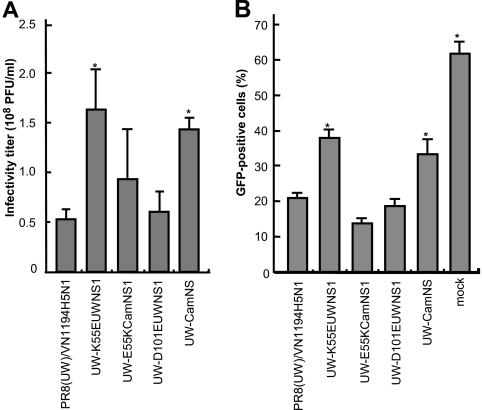
To address the role of NS for virus growth in MDCK cells, we compared the NS amino acid sequences of PR8(UW) and PR8(Cambridge) and found two amino acid differences in NS1 and one difference in NS2 between the two strains (Table 1). To determine which residue(s) is involved in viral growth kinetics, we generated NS1 mutant viruses with a single-amino-acid substitution either at position 55 or at position 101 of NS1 and assessed each viral titer (Fig. 4A). When a Lys-to-Glu substitution at position 55 was introduced into NS1 of PR8(UW)/VN1194H5N1 (UW-K55EUWNS1), the virus grew significantly better than its parent, PR8(UW)/VN1194H5N1 (P < 0.05). However, when a Glu-to-Lys substitution at the same position was introduced into PR8(Cambridge) NS1 of a PR8(UW)/VN1194H5N1-based mutant, UW-CamNS (see Fig. 2 for gene constellation) (UW-E55KCamNS1), the virus grew less well than its parent, UW-CamNS, and showed no significant difference relative to the growth of PR8(UW)/VN1194H5N1.
FIG. 4.
Growth of NS1 mutant viruses and their IFN antagonism. (A) MDCK cells were infected with NS1 mutant viruses at an MOI of 0.01. UW- indicates a PR8(UW)/VN1194H5N1 background NS mutant [e.g., UW-E55KcamNS1 contains the E55K mutant of PR8(Cambridge)NS1 in the PR8(UW)/VN1194H5N1 background]. Virus titers were determined in a plaque assay with MDCK cells (PFU/ml) at 36 hpi. The data are shown as mean titers with standard deviations (n ≥ 3). Titers significantly (P < 0.05) increased (*) compared with that of the PR8(UW)/VN1194H5N1 virus are shown. (B) A72 cells (13) were pretreated for 24 h with UV-inactivated supernatants from MDCK cells infected with the NS1 wild-type and mutant viruses. The pretreated A72 cells were then infected with VSV-ΔG*-GFP, and at 12 hpi, the number of GFP-positive cells was counted by fluorescence-activated cell sorting analysis. The data are shown as mean values with standard deviations (n = 3). Numbers of GFP-positive cells significantly (P < 0.05) increased (*) compared with that observed following treatment of the PR8(UW)/VN1194H5N1-infected cell supernatant are shown.
In addition, an Asn-to-Glu substitution at position 101 (UW-D101EUWNS1) did not yield a viral titer significantly different from that of PR8(UW)/VN1194H5N1. These results indicate that the amino acid at position 55 of NS1 affects virus growth in MDCK cells.
NS1 is known to mediate type I IFN antagonism to affect viral growth in host cells (5). We therefore analyzed the IFN antagonistic properties of each NS1 by use of an IFN bioassay to understand the molecular basis of the contribution of NS1 to the growth properties of virus in MDCK cells. In our IFN bioassay, the supernatant from MDCK cells infected with either virus possessing Glu at position 55 of NS1 (UW-K55EUWNS1 or UW-CamNS) was less able to inhibit VSV-ΔG*-GFP replication than was supernatant from cells infected with PR8(UW)/VN1194H5N1 (P < 0.05) (Fig. 4B). In contrast, inhibition levels were similar among viruses possessing Lys at position 55 of NS1 [PR8(UW)/VN1194H5N1, UW-E55KCamNS1, and UW-D101EUWNS1]. These results indicate that Glu at position 55 of NS1 is responsible for the enhanced type I IFN antagonistic property of PR8(Cambridge) NS1, leading to high growth in MDCK cells of viruses possessing this protein.
Alteration of the HA/NA functional balance enhances viral growth in MDCK cells.
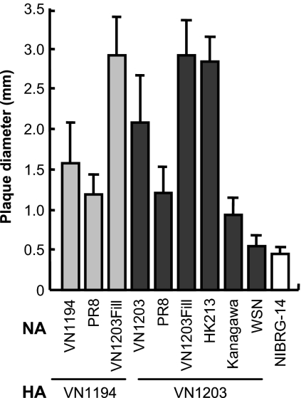
To see if we could further enhance viral growth in MDCK cells, we next determined the optimal functional balance between H5 NA and N1 NA. To this end, we produced a series of 6:2 (or 7:1) reassortants with the PR8(UW) background possessing H5 mHA derived from VN1194 or VN1203 and NAs derived from several N1 strains and compared the plaque sizes of each virus as an indicator of growth in MDCK cells (Fig. 5). The plaque size of PR8(UW)/VN1203H5N1 was 2.07 ± 0.59 mm (in diameter), which was significantly larger than that of PR8(UW)/VN1194H5N1 (1.57 ± 0.51 mm) (P < 0.01). We therefore used VN1203 mHA-bearing viruses for further assessments. Reassortant viruses bearing NAs from VN1203Fill or HK213 (containing a 20-amino-acid insertion in its stalk) formed significantly larger plaques (2.91 ± 0.42 or 2.83 ± 0.29 mm, respectively) than PR8(UW)/VN1203H5N1. In contrast, reassortants with NAs from the Kanagawa or PR8 strains formed smaller plaques (1.21 ± 0.31 or 0.93 ± 0.21 mm, respectively) than PR8(UW)/VN1203H5N1. Moreover, reassortants with NAs from WSN [PR8(UW)-VN1203H5/WSNN1] or NIBRG-14 formed even smaller plaques (0.53 ± 0.12 or 0.44 ± 0.09 mm, respectively). These results suggest that the HA/NA functional balance optimal for virus growth in MDCK cells is achieved by the combination of VN1203 mHA and NA with a longer stalk, such as HK213 NA or VN1203Fill NA.
FIG. 5.
Plaque sizes of PR8/H5N1 6:2 reassortant viruses. Plaque sizes of viruses containing mHA from either VN1194 or VN1203, NA from either H5N1 viruses (VN1194, VN1203, HK213, or VN1203Fill) or H1N1 viruses (PR8, Kanagawa, or WSN), and the remaining genes, which were from PR8(UW), were determined. The plaque size of NIBRG-14 is also shown. The data are reported as mean plaque diameters with standard deviations for >15 plaques with each virus.
Enhanced growth of the PR8/H5N1 6:2 reassortant in MDCK cells.
To test if the growth-enhancing activity of NS1 and HA/NA functional balance are additive, we generated 6:2 reassortant viruses possessing heterologous NS or NA with VN1203 mHA and assessed their growth in MDCK cells (Fig. 6A). Viruses bearing the long-stalk NA, PR8(UW)/VN1203H5/VN1203FillN1 and PR8(UW)/VN1203H5/HK213N1, grew to 1.3 × 10⁸ ± 0.5 × 10⁸ and 1.1 × 10⁸ ± 0.4 × 10⁸ PFU/ml, respectively, both of which titers were significantly higher than those of viruses possessing the short-stalk NA [PR8(UW) NA or VN1203 NA]. Replacement of the NS gene of these viruses with that from PR8(Cambridge) [PR8(UW)/VN1203H5/VN1203FillN1-CamNS or PR8(UW)/VN1203H5/HK213N1-CamNS, respectively] further enhanced viral growth significantly, compared with the levels for the parental viruses (2.33 × 10⁸ ± 0.27 × 10⁸ and 2.35 × 10⁸ ± 0.21 × 10⁸ PFU/ml, respectively).
FIG. 6.
Growth enhancement of a PR8/H5N1 reassortant mediated by NA and NS genes. (A) MDCK cells were infected with reassortant viruses containing mHA from either VN1194 or VN1203 and NA from a different N1 virus or NS from PR8(Cambridge) at an MOI of 0.01. Virus titers were determined in a plaque assay with MDCK cells at 36 hpi. The data are reported as mean titers with standard deviations for three independent experiments. Titers significantly (P < 0.05) increased (*) compared with that of the PR8(UW)/VN1194H5N1 virus are shown and compared with those of PR8(UW)/VN1203H5/VN1203FillN1 and PR8(UW)/VN1203H5/HK213N1 (**). (B) MDCK cells were infected with reassortant viruses containing H5 mHA from clade 1 and clade 2 strains with homologous NA or HK213 NA or PR8(Cambridge) NS instead of PR8(UW) NS at an MOI of 0.01. Virus titers were determined in a plaque assay with MDCK cells at 36 hpi. The data are reported as mean titers with standard deviations for three independent experiments. Titers significantly (P < 0.05) increased compared with those of the viruses possessing homologous NA (*) or compared with those of viruses possessing HK213 NA (**) are shown.
To authenticate the enhanced viral growth in MDCK cells mediated by NS and HA/NA functional balance, we produced reassortants with other H5 mHAs derived from clade 1 (VN30259 and VN3030), clade 2.1.3 (Indo3006), and clade 2.3.4 (AH2) H5N1 viruses with similar gene constellations (Fig. 6B). Each virus showed significantly enhanced growth with the introduction of the HK213 NA gene and even greater enhancement with the introduction of the PR8(Cambridge) NS gene. These results demonstrate the universal contribution of NS1 and HA/NA functional balance to the enhanced growth of H5N1 vaccine seed viruses in MDCK cells.
DISCUSSION
In this study, we addressed the molecular determinants for growth of H5N1 vaccine seed virus in MDCK cells, which are approved for human vaccine production. Seed viruses that provide robust growth in cell culture are needed to ensure an adequate supply of H5N1 vaccine either as a supplemental to or as an alternative method for egg-based vaccine production. The H5N1 influenza vaccine seed virus that we produced and optimized for MDCK cells was a PR8/H5N1 6:2 reassortant, the internal genes of which, with the exception of the NS gene, were derived from the PR8(UW) strain. Its NS gene was derived from the PR8(Cambridge) strain and its NA gene from the HK213 strain, and its mHA gene came from a circulating H5N1 virus.
We previously reported that seed viruses with our laboratory-maintained PR8(UW) strain as the background exhibit superior growth compared to the WHO-recommended NIBRG-14 seed virus in embryonated chicken eggs (four- to sevenfold enhancement) (10). In eggs, each polymerase and NP gene of the PR8(UW) strain enhanced virus growth slightly, but when combined, all four genes contributed to the superior growth of viruses possessing the PR8(UW) genes compared to those possessing the PR8(Cambridge) genes. In MDCK cells, however, the superior viral growth (more than 1,000-fold enhancement) was essentially determined by a single-amino-acid difference at position 360 of PB2, with an additional minor contribution from NS1 (Fig. 2 and 3). Therefore, the amino acid at position 360 of PB2 is a determinant for virus growth in MDCK cells but not in eggs. It may be that this amino acid is involved in host range determinants of avian and mammalian species, which are mediated by the host factors of each animal, as previously reported for the amino acids at positions 627 and 701 of PB2 (18, 29). Theoretically, an approximately 2-fold enhancement in polymerase activity caused by the amino acid difference at position 360 of PB2 could result in a >1,000-fold increase in overall viral growth with multiple replication cycles in an exponential manner; however, additional, unknown factors may also be involved in this growth enhancement.
Here, we showed that Glu at position 55 of NS1 mediates growth enhancement of vaccine seed viruses in MDCK cells via its IFN antagonistic action (Fig. 4). In addition, we found that the growth enhancement mediated by the PR8(Cambridge) NS gene was not observed in Vero cells, which lack the type I IFN genes (3) (data not shown). There are two possible explanations for this observation; one is that the K55E substitution of NS1 may enhance the productivity of this protein in this cell line, via its increased interaction with host-cell molecules, such as chaperons, which can precisely hold and rapidly transport NS1. The other is that this substitution may increase the intrinsic IFN antagonism of NS1. The region of NS1 from position 1 to 73 is believed to be an RNA-binding site (26) that exhibits IFN antagonism by binding host mRNA and reducing the signaling for IFN gene activation at a posttranscriptional level (20, 33). Given that position 55 of NS1 lies in this RNA-binding region, NS1 that possesses E55 may have a higher affinity for host mRNAs than NS1 with K55, resulting in the enhanced inhibition of IFN gene expression that we observed in MDCK cells.
There are several PR8 strains maintained in different laboratories. The present study, together with a database search, indicated that only the PR8(Cambridge) strain possesses S360 in PB2, whereas all the others possess Y360, as does the PR8(UW) strain. On the other hand, E55 in NS1 is not limited to the PR8(Cambridge) strain. For example, the PR8(Mt. Sinai) strain possesses E55 in NS1 as well as Y360 in PB2. Thus, comparative growth evaluations and selection of a vaccine donor virus among the PR8 strains would be valuable in the future for pandemic preparedness to increase vaccine productivity.
HA/NA functional balance is another important determinant for virus growth (19, 21). The stalk length of NA correlates with its biological activity (1, 4). The NAs of HK213 and VN1203Fill bear long stalks (44 amino acids in length), which probably enhance the functionality of NA compared to NAs bearing stalk lengths of 24 amino acids, which are observed in most H5N1 viruses. Our H5N1 seed viruses with long-stalk NAs grew better than those bearing homologous N1 NAs in MDCK cells, suggesting that the HA/NA functional balance of viruses with long-stalk NAs is superior to that of those viruses with short-stalk NAs for growth in this cell line. Recently, we reported that disrupting HA/NA functional balance by replacing the NA gene with PR8 NA enhances the growth of H5N1 vaccine seed viruses in eggs (10). In this study, optimized HA/NA functional balance in MDCK cells differed from that in eggs, possibly due to a difference in cell receptors between these two substrates.
In our previous report, we discussed, albeit to a limited extent, the possibility of a reduction in the protective immunity of the vaccine seeds due to the inclusion of heterologous N1 NAs (10). In this context, the seed virus with artificial VN1203Fill NA would fare better than that with HK213 NA. However, a recent report showed considerable cross-protective immunogenicity between N1s from human H1N1 and H5N1 viruses in a mouse model (28). Therefore, we believe that inclusion of heterologous N1 NAs in the seed viruses would provide an advantage for vaccine supply, which would likely offset the limited antigenic mismatch in this minor antigen.
It is well known that the HA antigenicities of human H3N2 and H1N1 influenza viruses readily change following propagation in eggs (15, 27). This occurs because of differences in the cell receptors expressed in eggs (12). However, MDCK cells contain both avian- and human-type receptors (12), which circumvents any HA antigenic changes in the H5N1 vaccine seed viruses.
In conclusion, we propose a seed virus for MDCK cell-based H5N1 vaccine production in the form of a 6:2 reassortant that possesses mHA from a circulating H5N1 virus, NA from HK213, and its remaining genes from the PR8(UW) strain, except for the NS gene, which comes from the PR8(Cambridge) strain. A cell culture-based strategy with this seed virus would allow increased production of prepandemic or pandemic inactivated H5N1 vaccines in a timely, cost-efficient manner as a supplement or an alternative to egg-based vaccine production.
Acknowledgments
We thank J. M. Wood and J. S. Robertson (National Institute for Biological Standards and Control, United Kingdom) for providing NIBRG-14 virus, Michael Whitt for providing VSV-ΔG*-GFP virus, and S. Watson for editing the manuscript.
This work was supported in part by grants-in-aid for Specially Promoted Research and for Scientific Research (B) from the Ministry of Education, Culture, Sports, Science, and Technology, Japan; by a grant for Scientific Research from the Ministry of Health, Labor, and Welfare, Japan; by CREST (Core Research for Evolutional Science and Technology), Japan; by a contract research fund for the Program of Founding Research Centers for Emerging and Reemerging Infectious Diseases from the Ministry of Education, Culture, Sports, Science and Technology, Japan; and by research grants from the U.S. Public Health Service, National Institute of Allergy and Infectious Diseases.
Footnotes
Published ahead of print on 3 September 2008.
REFERENCES
- 1.Castrucci, M., and Y. Kawaoka. 1993. Biologic importance of neuraminidase stalk length in influenza A virus. J. Virol. 67759-764. [DOI] [PMC free article] [PubMed] [Google Scholar]
- 2.Dennis, C. 2006. Flu-vaccine makers toil to boost supply. Nature 4401099. [DOI] [PubMed] [Google Scholar]
- 3.Diaz, M., S. Ziemin, M. Le Beau, P. Pitha, S. Smith, R. Chilcote, and J. Rowley. 1988. Homozygous deletion of the alpha- and beta 1-interferon genes in human leukemia and derived cell lines. Proc. Natl. Acad. Sci. USA 855259-5263. [DOI] [PMC free article] [PubMed] [Google Scholar]
- 4.Els, M. C., G. M. Air, K. G. Murti, R. G. Webster, and W. G. Laver. 1985. An 18-amino acid deletion in an influenza neuraminidase. Virology 142241-247. [DOI] [PubMed] [Google Scholar]
- 5.García-Sastre, A., and C. Biron. 2006. Type 1 interferons and the virus-host relationship: a lesson in détente. Science 312879-882. [DOI] [PubMed] [Google Scholar]
- 6.Genzel, Y., M. Fischer, and U. Reichl. 2006. Serum-free influenza virus production avoiding washing steps and medium exchange in large-scale microcarrier culture. Vaccine 243261-3272. [DOI] [PubMed] [Google Scholar]
- 7.Govorkova, E., S. Kodihalli, I. Alymova, B. Fanget, and R. Webster. 1999. Growth and immunogenicity of influenza viruses cultivated in Vero or MDCK cells and in embryonated chicken eggs. Dev. Biol. Stand. 9839-51, 73-74. [PubMed] [Google Scholar]
- 8.Halperin, S., B. Smith, T. Mabrouk, M. Germain, P. Trépanier, T. Hassell, J. Treanor, R. Gauthier, and E. Mills. 2002. Safety and immunogenicity of a trivalent, inactivated, mammalian cell culture-derived influenza vaccine in healthy adults, seniors, and children. Vaccine 201240-1247. [DOI] [PubMed] [Google Scholar]
- 9.Horimoto, T., and Y. Kawaoka. 2005. Influenza: lessons from past pandemics, warnings from current incidents. Nat. Rev. Microbiol. 3591-600. [DOI] [PubMed] [Google Scholar]
- 10.Horimoto, T., S. Murakami, Y. Muramoto, S. Yamada, K. Fujii, M. Kiso, K. Iwatsuki-Horimoto, Y. Kino, and Y. Kawaoka. 2007. Enhanced growth of seed viruses for H5N1 influenza vaccines. Virology 36623-27. [DOI] [PMC free article] [PubMed] [Google Scholar]
- 11.Horimoto, T., A. Takada, K. Fujii, H. Goto, M. Hatta, S. Watanabe, K. Iwatsuki-Horimoto, M. Ito, Y. Tagawa-Sakai, S. Yamada, H. Ito, T. Ito, M. Imai, S. Itamura, T. Odagiri, M. Tashiro, W. Lim, Y. Guan, M. Peiris, and Y. Kawaoka. 2006. The development and characterization of H5 influenza virus vaccines derived from a 2003 human isolate. Vaccine 243669-3676. [DOI] [PubMed] [Google Scholar]
- 12.Ito, T., Y. Suzuki, A. Takada, A. Kawamoto, K. Otsuki, H. Masuda, M. Yamada, T. Suzuki, H. Kida, and Y. Kawaoka. 1997. Differences in sialic acid-galactose linkages in the chicken egg amnion and allantois influence human influenza virus receptor specificity and variant selection. J. Virol. 713357-3362. [DOI] [PMC free article] [PubMed] [Google Scholar]
- 13.Iwata, A., N. M. Iwata, T. Saito, K. Hamada, Y. Sokawa, and S. Ueda. 1996. Cytopathic effect inhibition assay for canine interferon activity. J. Vet. Med. Sci. 5823-27. [DOI] [PubMed] [Google Scholar]
- 14.James, J., R. Zeiger, M. Lester, M. Fasano, J. Gern, L. Mansfield, H. Schwartz, H. Sampson, H. Windom, S. Machtinger, and S. Lensing. 1998. Safe administration of influenza vaccine to patients with egg allergy. J. Pediatr. 133624-628. [DOI] [PubMed] [Google Scholar]
- 15.Katz, J., C. Naeve, and R. Webster. 1987. Host cell-mediated variation in H3N2 influenza viruses. Virology 156386-395. [DOI] [PubMed] [Google Scholar]
- 16.Katz, J., and R. Webster. 1992. Amino acid sequence identity between the HA1 of influenza A (H3N2) viruses grown in mammalian and primary chick kidney cells. J. Gen. Virol. 731159-1165. [DOI] [PubMed] [Google Scholar]
- 17.Kobasa, D., A. Takada, K. Shinya, M. Hatta, P. Halfmann, S. Theriault, H. Suzuki, H. Nishimura, K. Mitamura, N. Sugaya, T. Usui, T. Murata, Y. Maeda, S. Watanabe, M. Suresh, T. Suzuki, Y. Suzuki, H. Feldmann, and Y. Kawaoka. 2004. Enhanced virulence of influenza A viruses with the haemagglutinin of the 1918 pandemic virus. Nature 431703-707. [DOI] [PubMed] [Google Scholar]
- 18.Li, Z., H. Chen, P. Jiao, G. Deng, G. Tian, Y. Li, E. Hoffmann, R. Webster, Y. Matsuoka, and K. Yu. 2005. Molecular basis of replication of duck H5N1 influenza viruses in a mammalian mouse model. J. Virol. 7912058-12064. [DOI] [PMC free article] [PubMed] [Google Scholar]
- 19.Lu, B., H. Zhou, D. Ye, G. Kemble, and H. Jin. 2005. Improvement of influenza A/Fujian/411/02 (H3N2) virus growth in embryonated chicken eggs by balancing the hemagglutinin and neuraminidase activities, using reverse genetics. J. Virol. 796763-6771. [DOI] [PMC free article] [PubMed] [Google Scholar]
- 20.Ludwig, S., X. Wang, C. Ehrhardt, H. Zheng, N. Donelan, O. Planz, S. Pleschka, A. García-Sastre, G. Heins, and T. Wolff. 2002. The influenza A virus NS1 protein inhibits activation of Jun N-terminal kinase and AP-1 transcription factors. J. Virol. 7611166-11171. [DOI] [PMC free article] [PubMed] [Google Scholar]
- 21.Mitnaul, L., M. Matrosovich, M. Castrucci, A. Tuzikov, N. Bovin, D. Kobasa, and Y. Kawaoka. 2000. Balanced hemagglutinin and neuraminidase activities are critical for efficient replication of influenza A virus. J. Virol. 746015-6020. [DOI] [PMC free article] [PubMed] [Google Scholar]
- 22.Murakami, S., T. Horimoto, S. Yamada, S. Kakugawa, H. Goto, and Y. Kawaoka. 2008. Establishment of canine RNA polymerase I-driven reverse genetics for influenza A virus: its application for H5N1 vaccine production. J. Virol. 821605-1609. [DOI] [PMC free article] [PubMed] [Google Scholar]
- 23.Neumann, G., T. Watanabe, H. Ito, S. Watanabe, H. Goto, P. Gao, M. Hughes, D. Perez, R. Donis, E. Hoffmann, G. Hobom, and Y. Kawaoka. 1999. Generation of influenza A viruses entirely from cloned cDNAs. Proc. Natl. Acad. Sci. USA 969345-9350. [DOI] [PMC free article] [PubMed] [Google Scholar]
- 24.Ozawa, M., K. Fujii, Y. Muramoto, S. Yamada, S. Yamayoshi, A. Takada, H. Goto, T. Horimoto, and Y. Kawaoka. 2007. Contributions of two nuclear localization signals of influenza A virus nucleoprotein to viral replication. J. Virol. 8130-41. [DOI] [PMC free article] [PubMed] [Google Scholar]
- 25.Park, M. S., M. L. Shaw, J. Muñoz-Jordan, J. F. Cros, T. Nakaya, N. Bouvier, P. Palese, A. García-Sastre, and C. F. Basler. 2003. Newcastle disease virus (NDV)-based assay demonstrates interferon-antagonist activity for the NDV V protein and the Nipah virus V, W, and C proteins. J. Virol. 771501-1511. [DOI] [PMC free article] [PubMed] [Google Scholar]
- 26.Qian, X., C. Chien, Y. Lu, G. Montelione, and R. Krug. 1995. An amino-terminal polypeptide fragment of the influenza virus NS1 protein possesses specific RNA-binding activity and largely helical backbone structure. RNA 1948-956. [PMC free article] [PubMed] [Google Scholar]
- 27.Robertson, J., J. Bootman, R. Newman, J. Oxford, R. Daniels, R. Webster, and G. Schild. 1987. Structural changes in the haemagglutinin which accompany egg adaptation of an influenza A(H1N1) virus. Virology 16031-37. [DOI] [PubMed] [Google Scholar]
- 28.Sandbulte, M., G. Jimenez, A. Boon, L. Smith, J. Treanor, and R. Webby. 2007. Cross-reactive neuraminidase antibodies afford partial protection against H5N1 in mice and are present in unexposed humans. PLoS Med. 4e59. [DOI] [PMC free article] [PubMed] [Google Scholar]
- 29.Subbarao, E., W. London, and B. Murphy. 1993. A single amino acid in the PB2 gene of influenza A virus is a determinant of host range. J. Virol. 671761-1764. [DOI] [PMC free article] [PubMed] [Google Scholar]
- 30.Takada, A., C. Robison, H. Goto, A. Sanchez, K. Murti, M. Whitt, and Y. Kawaoka. 1997. A system for functional analysis of Ebola virus glycoprotein. Proc. Natl. Acad. Sci. USA 9414764-14769. [DOI] [PMC free article] [PubMed] [Google Scholar]
- 31.Treanor, J., J. Campbell, K. Zangwill, T. Rowe, and M. Wolff. 2006. Safety and immunogenicity of an inactivated subvirion influenza A (H5N1) vaccine. N. Engl. J. Med. 3541343-1351. [DOI] [PubMed] [Google Scholar]
- 32.Ulmer, J., U. Valley, and R. Rappuoli. 2006. Vaccine manufacturing: challenges and solutions. Nat. Biotechnol. 241377-1383. [DOI] [PubMed] [Google Scholar]
- 33.Wang, X., M. Li, H. Zheng, T. Muster, P. Palese, A. Beg, and A. García-Sastre. 2000. Influenza A virus NS1 protein prevents activation of NF-κB and induction of alpha/beta interferon. J. Virol. 7411566-11573. [DOI] [PMC free article] [PubMed] [Google Scholar]
- 34.Wood, J., and J. Robertson. 2004. From lethal virus to life-saving vaccine: developing inactivated vaccines for pandemic influenza. Nat. Rev. Microbiol. 2842-847. [DOI] [PubMed] [Google Scholar]


