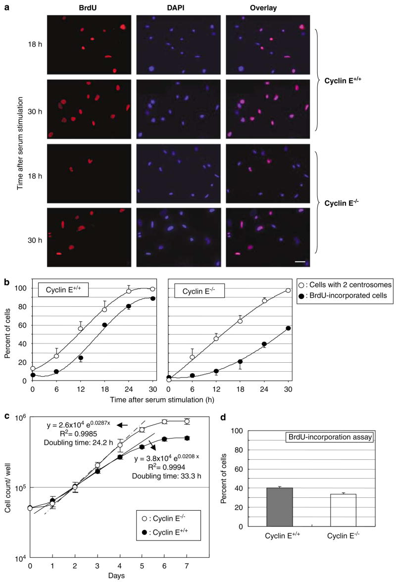
Figure 1.
Analysis of the DNA and centrosome duplication kinetics in cyclins E+/+ and E−/− mouse embryonic fibroblasts (MEFs) expressing DNp53. Cyclins E+/+ and E−/− MEFs expressing DNp53 were serum-starved for 48 h, and then serum-stimulated with the medium containing 20% fetal bovine serum (FBS) and bromodeoxyuridine (BrdU). At indicated time points, cells were fixed and immunostained for incorporated BrdU and γ-tubulin. Representative images of immunofluorescence assay for BrdU incorporation are shown in (a). Scale bar: 40 μm. The rates of BrdU incorporation and centrosome duplication in cyclins E+/+ and E−/− cells were plotted in the graph as average±standard error from three independent experiments (b). For the analysis of centrosomes, >200 γ-tubulin immunostained cells were examined for each time point. In this analysis, cells with amplified (⩾3) centrosomes were excluded from the analysis; it is technically difficult to differentiate unduplicated or duplicated centrosomes in the cells with amplified centrosomes. Determining the rate of centrosome duplication by examining only the cells with one or two centrosomes in the synchronized cells is possible, because the newly duplicated centrosomes do not reduplicate until acquiring the duplication competency. The time course period after serum stimulation in this experiment, there is not sufficient time for the duplicated centrosomes to reestablish the duplication competency. The percentages of the number of cells with two centrosomes among the total number of cells with either one or two centrosomes are plotted in the graph (b) as average±standard error from three independent experiments. Similar results were obtained with the procedure to immunostain centrioles (see Materials and methods section) (data not shown). (c) Determination of doubling times of cyclins E+/+ and E−/− MEFs. Cyclins E+/+ and E−/− cells were plated in the cell culture wells, and the number of cells in the well was counted at every 24 h for 7 days, and plotted in the graph as average±standard error from three independent experiments. (d) Cyclins E+/+ and E−/− MEFs under an optimal growth condition were incubated with BrdU for 1 h, and the percent of cells that had incorporated BrdU was determined, and shown in the graph as average±standard error from three experiments.

