Abstract
The genome of the gram-positive bacterium Enterococcus faecalis contains the genes that encode the citrate lyase complex. This complex splits citrate into oxaloacetate and acetate and is involved in all the known anaerobic bacterial citrate fermentation pathways. Although citrate fermentation in E. faecalis has been investigated before, the regulation and transcriptional pattern of the cit locus has still not been fully explored. To fill this gap, in this paper we demonstrate that the GntR transcriptional regulator CitO is a novel positive regulator involved in the expression of the cit operons. The transcriptional analysis of the cit clusters revealed two divergent operons: citHO, which codes for the transporter (citH) and the regulatory protein (citO), and upstream from it and in the opposite direction the oadHDB-citCDEFX-oadA-citMG operon, which includes the citrate lyase subunits (citD, citE, and citF), the soluble oxaloacetate decarboxylase (citM), and also the genes encoding a putative oxaloacetate decarboxylase complex (oadB, oadA, oadD and oadH). This analysis also showed that both operons are specifically activated by the addition of citrate to the medium. In order to study the functional role of CitO, a mutant strain with an interrupted citO gene was constructed, causing a total loss of the ability to degrade citrate. Reintroduction of a functional copy of citO to the citO-deficient strain restored the response to citrate and the Cit+ phenotype. Furthermore, we present evidence that CitO binds to the cis-acting sequences O1 and O2, located in the cit intergenic region, increasing its affinity for these binding sites when citrate is present and allowing the induction of both cit promoters.
Enterococcus faecalis, a gram-positive catalase-negative coccus, is a natural member of the human and animal microflora. This ubiquitous microorganism has numerous fields of interest due to its importance as a cause of nosocomial infections and to its utilization in the food industry. With respect to the latter, the bacteria are involved in the ripening process and in aroma development of diverse cheeses. These positive effects have been attributed to specific biochemical traits such as lipolytic activity and citrate utilization. Citrate metabolism has been extensively studied in bacteria, both from a basic and applied point of view, since citrate fermentation plays an important role during the production of diverse kinds of drinks and foods (1-3, 7, 8, 12, 15, 16, 21, 28, 29, 31-33, 48-51).
Citrate fermentation was recently investigated in E. faecalis, and the contribution to aroma development was characterized (18, 46, 47). In this microorganism, the first steps of the citrate degradative pathway are shared with other citrate-fermenting bacteria. Initially, citrate is converted to oxaloacetate and acetate by the enzyme citrate lyase. Then, oxaloacetate is decarboxylated to pyruvate. This compound is subsequently degraded to acetate, CO2, and formate with the generation of ATP by the acetate kinase enzyme. In addition, small quantities of lactate, acetoin, and ethanol are produced, suggesting a low activity of lactate dehydrogenase, acetolactate synthase, and alcohol dehydrogenase enzymes (46, 47). Although the genes involved in the citrate fermentation pathway have been identified, the regulation of citrate metabolism remains unclear in this microorganism.
To date, two different mechanisms for sensing citrate and controlling its utilization have been described in bacteria. These mechanisms ensure that selected genes are expressed under the appropriate conditions and at the right time. The expression of the specific transporters and the citrate lyase complex, required for citrate fermentation, are regulated through the control of transcription initiation by the two-component sensor-regulator system CitA/B (6, 37) and by the transcriptional activator protein CitI (34).
The CitA/CitB two-component system was initially described in enterobacteria (6-8, 37). CitA functions as a sensor of extracellular citrate, whereas CitB acts as a transcriptional activator of the citrate-specific fermentation genes (cit operons). In Klebsiella pneumoniae, the citS-oadGAB-citAB operon encodes the citrate carrier CitS, the three subunits of the oxaloacetate decarboxylase (OadA, OadB, and OadG), and the CitA/B two-component system (6, 7, 37, 41). The divergent operon citCDEFG encodes the citrate lyase complex (CitD, CitE, and CitF) and the auxiliary proteins CitC and CitG (7, 8, 48, 49). The expression of the citrate fermentation genes requires not only citrate but also anoxic conditions and Na+ ions (8, 36).
Recently, the transcriptional factor CitI from Weissella paramesenteroides was reported to function as a switch of the cit genes, which are directly activated by citrate (34). CitI, a member of DeoR family, is highly conserved in different lactic acid bacteria such as W. paramesenteroides, Leuconosctoc mesenteroides, Lactococcus lactis, and Lactobacillus plantarum (31-34).
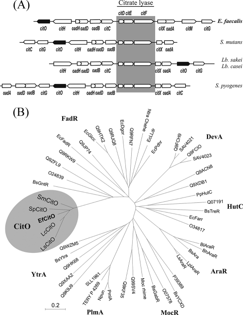
Interestingly, the presence of an alternative transcriptional factor associated with citrate breakdown emerged from gene context analysis of citrate fermentation gene clusters. In fact, we found that the genetic organization of cit operons was different in E. faecalis, Streptococcus mutants, Lactobacillus casei, Lactobacillus sakei, and Streptococcus pyogenes (Fig. 1A). In this group of microorganisms, citO is a common element. It is predicted to encode a GntR family transcriptional regulator. The members of this family consist of a conserved N-terminal helix-turn-helix DNA binding domain, and at the C terminus, there is usually a variable effector-binding/oligomerization domain (19, 43).
FIG. 1.
(A) Genetic organization of cit genes involved in citrate utilization in E. faecalis, S. mutans, L. sakei, L. casei, and S. pyogenes. The shaded box indicates the organization of the highly conserved citDEF genes in the different cit clusters. (B) Phylogenetic tree corresponding to proteins of the GntR family. A multiple sequence alignment was computed using MEGA3. GntR-like regulators were classified in seven subfamilies according to the clusters of branches that emerged from the constructed tree. The shaded ellipse shows the new subfamily constituted by citO homologs.
Another important feature of the cit operons from this group is the presence of the alternative citrate transporter named CitH (Fig. 1A). In a previous work, we reported the functional characterization of the citrate transporter, CitH, from E. faecalis. This protein was expressed in Escherichia coli and functionally characterized in membrane vesicles prepared from these cells. Citrate uptake was dependent on the presence of divalent metal ions (Ca2+, Sr2+, Mn2+, Cd2+, and Pb2+) (5).
In this report, we demonstrate that the citO gene encodes a novel transcriptional factor necessary for the activation of the citrate fermentation pathway in E. faecalis. When citrate is present, CitO induces the transcription of the citHO and oadHDB-citCDEFX-oadA-citMG operons by binding to the intergenic region (IR) between them. In this way, CitO allows E. faecalis cells to coordinate the expression of the genes involved in synthesizing the enzymes necessary for citrate transport and degradation.
MATERIALS AND METHODS
Bacterial strains and growth conditions.
The E. faecalis ATCC 29212 strain was selected based on its ability to metabolize citrate detected by using Kempler and McKay medium (25). E. faecalis JH2-2 was utilized for genetic manipulation since the E. faecalis ATCC 29212 strain has been refractory to the usual techniques of transformation. E. faecalis ATCC 29212, JH2-2, and V583 conserve the cit locus, as estimated by PCR and sequence reactions. Cultures of E. faecalis were grown at 37°C without shaking in 100-ml sealed bottles containing 20 to 50 ml of Luria-Bertani medium (LB) (45) supplemented with 33 mM trisodium citrate (LBC medium); initial pH was 7.0.
E. coli strain DH5α was used as an intermediate host for cloning, E. coli BL21(DE3) was used for overproduction of CitO with a six-His tag (His6-CitO), and E. coli EC101 was used as host for pGh9 constructs. E. coli strains were routinely grown aerobically at 37°C in LB medium and transformed as previously described (45). Growth was monitored by measuring absorbance at 600 nm in a Beckman DU640 spectrophotometer. Aerobic growth was achieved by gyratory shaking at 250 rpm. Ampicillin (100 μg/ml), erythromycin (150 μg/ml), or kanamycin (50 μg/ml) was included in the medium to select cells harboring ampicillin-, erythromycin-, or kanamycin-resistant plasmids. 5-Bromo-4-chloro-3-indolyl-β-d-galactopyranoside (20 μg/ml) was used to identify recombinant plasmids with DNA insertions that impaired β-galactosidase activity in the DH5α strain induced with 0.5 mM isopropyl-β-d-thiogalactopyranoside (IPTG).
Construction of the E. faecalis JH2-2 CitO mutant strain.
The E. faecalis JH2-2 strain was constructed by interrupting the citO gene by a single recombination event using the thermosensitive vector pGh9 (30). An internal fragment of citO was amplified by PCR using chromosomal DNA of E. faecalis ATCC 29212 as a template. The forward primer fcitOU introduced an EcoRI site, and the reverse primer fcitOL introduced an HindIII site (see Table S1 in the supplemental material). The PCR product was digested with these two enzymes and ligated into the corresponding sites of the pGh9 vector. The resulting plasmid, named pmCitO (Table 1), was introduced into E. coli EC101, isolated, and then electroporated into the E. faecalis JH2-2 strain as described elsewhere (14). The transformant strain was grown overnight at a permissive temperature of 30°C in LB medium plus glucose with 5 μg/ml erythromycin for plasmid maintenance. The saturated culture was diluted 500-fold into fresh medium and incubated at the restrictive temperature of 37°C at which plasmid replication is disabled. When the culture reached an optical density at 600 nm (OD660) of 0.5, serial dilutions were plated on LB medium plus glucose and antibiotic. Next, the colonies were transferred to LB plates with or without 33 mM citrate. Colonies with no differential growth in these media were further analyzed for a citrate utilization phenotype. The resultant citO-deficient strain was named JHB1 (Table 2). The interruption of citO was confirmed by PCR and Southern blotting.
TABLE 1.
Plasmids used in this study
| Plasmid | Comments | Oligonucleotides | Reference or source |
|---|---|---|---|
| pGh9 | Thermosensitive plasmid carrying erythromycin resistance | 30 | |
| pGEMT-easy | Promega | ||
| pCR-Blunt II-TOPO | Invitrogen | ||
| pET28a | Novagen | ||
| pBM02 | pUC18 derivative carrying CRL264 replicon, Pcit (promoter) and chloramphenicol resistance | B. Marelli et al. | |
| pTCV-lac | Promoterless vector which allows lacZ fusion construction | 40 | |
| pmCitO | pGh9 derivative carrying a 500 bp citO internal fragment | fcitOU, fcitOL | This work |
| pCitO | pBM02 derivative for expressing CitO in E. faecalis | This work | |
| pET-CitO | pET28a derivative expressing His6-CitO | CitOU, CitOL | This work |
| pTCV-PcitHO | EfHpromU, EfDpromL | This work | |
| pTCV-PcitHO1 | Efint2U, EfDpromL | This work | |
| pTCV-PcitHO2 | EfHpromU, Efint3L | This work | |
| pTCV-PcitHO3 | EfHpromU, Efint4L | This work | |
| pTCV-PcitCL | EfHpromU, EfDpromL | This work | |
| pTCV-PcitCL1 | EfHpromU, EfDpromL | This work | |
| pTCV-PcitCL2 | EfbsintU, EfDpromL | This work | |
| pTCV-PcitCL3 | EfbsPoadHU, EfDpromL | This work | |
| pTO1O2 | pTOPO derivative carrying a DNA fragment including the O1 and O2 binding sites | Efint2U, EfbsoadHL | This work |
| pCO1O2 | pBM02 derivative carrying a DNA fragment including the O1 and O2 binding sites | This work |
TABLE 2.
E. faecalis strains used in this study
| Strain | Genotype or description | Source or reference |
|---|---|---|
| ATCC 29212 | cit+ | ATCC |
| JH2-2 | cit+ | 11, 24 |
| JHB1 | JH2-2 citO::pmCitO | This work |
| JHB2 | JH2-2 (pTCV-PcitHO) | This work |
| JHB3 | JH2-2 (pTCV-PcitHO1) | This work |
| JHB4 | JH2-2 (pTCV-PcitHO2) | This work |
| JHB5 | JH2-2 (pTCV-PcitHO3) | This work |
| JHB6 | JH2-2 (pTCV-PcitCL) | This work |
| JHB7 | JH2-2 (pTCV-PcitCL1) | This work |
| JHB8 | JH2-2 (pTCV-PcitCL2) | This work |
| JHB9 | JH2-2 (pTCV-PcitCL3) | This work |
| JHB10 | JH2-2 (pTCV-lac) | This work |
| JHB11 | JHB1 (pCitO) | This work |
| JHB12 | JHB1 (pBM02) | This work |
| JHB13 | JHB1 (pTCV-PcitHO) | This work |
| JHB14 | JHB1 (pTCV-PcitCL) | This work |
| JHB15 | JHB1 (pTCV-PcitHO) (pCitO) | This work |
| JHB16 | JHB1 (pTCV-PcitCL) (pCitO) | This work |
| JHB17 | JHB2 (pBM02) | This work |
| JHB18 | JHB2 (pCO1O2) | This work |
| JHB19 | JHB6 (pBM02) | This work |
| JHB20 | JHB6 (pCO1O2) | This work |
Construction of plasmids with Pcit-lacZ transcriptional fusions.
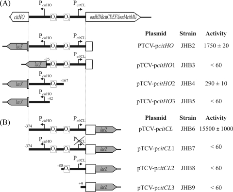
The plasmids bearing the promoter-lacZ transcriptional fusions, listed in Table 1 and Fig. 4, are all derivatives of the pTCV-lac vector (40), and the oligonucleotides used in their construction are given in Table S1 in the supplemental material. To construct the pTCV-lac-derivative plasmids, DNA fragments from the promoter regions of the citHO and oadHDB-citCDEFX-oadA-citMG operons were obtained by PCR using chromosomal DNA of E. faecalis ATCC 29212 as the template and cloned into the pCR-Blunt II-TOPO vector (Invitrogen). pTOPO-derivative plasmids were digested with EcoRI, and each released fragment was ligated into the corresponding site of pTCV-lac vector. The desired orientation of the fragments was determined by PCR.
FIG. 4.
Schematic representation of the pTCV-lac-derived plasmids. The promoter regions of the citHO (A) and oadHDB-citCDEFX-oadA-citMG (B) operons are depicted. The CitO binding sites identified in vitro (O1 and O2) are represented by boxes. The end of each DNA fragment is labeled relative to the transcriptional start site. The levels of accumulated β-galactosidase activity were measured in cell extracts from cultures grown in LBC medium for 8 h.
To mutate the −10 box of the PcitCL promoter, the oligonucleotides EfHpromU-EfPCLmutL and EfPCLmutU-EfDpromL (see Table S1 in the supplemental material) were used for the amplification of two overlap extension PCRs. A mixture of these PCR products was used as a DNA template for another PCR using the oligonucleotides EfHpromU and EfDpromL (see Table S1 in the supplemental material). The amplification product was cloned into pCR-Blunt II-TOPO vector and then subcloned into pTCV-lac vector as described above, yielding plasmid pTCV-PcitCL1 (Table 1).
Plasmid pCO1O2 used for the competence assay was obtained by subcloning from the pTO1O2 plasmid the DNA fragment (obtained with the primers Efint2U and EfbsoadHL) (see Table S1 in the supplemental material) which contains the binding sites (designated O1 and O2) of CitO. pTO1O2 was digested with EcoRI, and the released fragment was ligated into the same restriction site of pBM02 (B. Marelli et al., unpublished results) (Table 1).
The cloned fragments were checked by sequencing at the University of Maine DNA sequencing facility.
β-Galactosidase assays.
Cells were grown overnight in LB broth containing 0.5% glucose and kanamycin (1,000 μg/ml). Overnight cultures were diluted to an OD660 of 0.08 and grown in LB medium supplemented with kanamycin until the cells were harvested in early stationary phase. When indicated in the figure legends, different carbon sources were added to the growth medium at specified concentrations. β-Galactosidase was assayed as described by Israelsen et al. (23), except that the cells were permeabilized by treatment with 20 U/ml of mutanolysin (Sigma) for 10 min at 37°C.
Citrate lyase activity.
Citrate lyase activity was determined at 25°C in a coupled spectrophotometric assay with malate dehydrogenase as previously described (33). Briefly, the assay mixture contained, in a final volume of 1 ml, 100 mM potassium phosphate buffer (pH 7.2), 5 mM trisodium citrate, 3 mM MgCl2, 0.23 mM NADH, 25 U of malate dehydrogenase, and 20 μg of total protein from different cell extracts. The oxidation of NADH was measured in a spectrophotometer at 340 nm. One unit of enzyme activity is defined as 1 pmol of citrate converted to acetate and oxaloacetate per min under the conditions used. For chemical acetylation of citrate lyase, 20 μg of protein was incubated for 5 min at 25°C with 5 mM acetic anhydride and then used immediately for determination of citrate lyase activity.
RNA isolation and analysis.
For Northern blotting and primer extension analysis, total RNA was isolated from E. faecalis cells grown for 9 h in LB or LBC medium, as previously described (33). The RNAs were checked for their integrity and yield of the rRNA in all samples. The patterns of the rRNAs were similar in all preparations. Total RNA concentration was determined by UV spectrophotometry and by gel quantification with Gel Doc 1000 (Bio-Rad). Primer extension analysis was performed as previously described (33). The primers used for detection of the start sites of citH, oadH, citC, and citM were pecitH, pecitH2, peoadH, peoadH2, pecitC, and pecitM (see Table S1 in the supplemental material), respectively. One picomole of primer was annealed to 15 μg of RNA. Primer extension reactions were performed by incubation of the annealing mixture with 200 U of Moloney murine leukemia virus reverse transcriptase (Promega) at 42°C for 60 min. Determination of the size of the reaction products was carried out in 6% polyacrylamide gels containing 8 M urea. Extension products were detected in a GE Healthcare Life Sciences Phosphorimager.
For Northern blot analysis, samples containing 20 μg of total RNA were separated in a 1.2% agarose gel. Transfer of nucleic acids to nylon membranes and hybridization with radioactive probes were performed as previously described (33). The double-stranded probes hybridizing with citH, citEF, and citM were labeled by incorporation of [α-32P]dATP in the PCR amplification using oligonucleotides citHU-citHL, citEFU-citEFL, and citMU-citML, respectively (see Table S1 in the supplemental material). mRNA molecular sizes were estimated by using a 0.5- to 9-kb RNA ladder (NEB).
Cloning of citO.
The gene encoding the putative regulator CitO was amplified by PCR using genomic DNA of E. faecalis ATCC 29212 as the template, following a standard protocol. The forward primer citOU introduced an NdeI site around the initiation codon of the citO gene, and the reverse primer citOL introduced a BamHI site downstream of the stop codon (see Table S1 in the supplemental material). The PCR product was double digested and ligated into the corresponding restriction sites of vector pET-28a(+) (Novagen). The resulting plasmid, named pET-CitO, codes for His6-CitO at the N terminus (Table 1). The sequence of the insert was confirmed, and the plasmid was subsequently introduced into E. coli BL21(DE3) for citO overexpression.
pET-CitO was then digested with NdeI and EcoRI. The released citO fragment was ligated into the corresponding sites of the pBM02 vector (B. Marelli et al., unpublished). The resulting plasmid, named pCitO, has the citO gene downstream of a promoter that allows the expression of CitO in trans in E. faecalis (Table 1).
Purification of His6-CitO and gel mobility shift assays.
E. coli BL21(DE3) harboring pET-CitO was grown in 500 ml of LB medium at 37°C until an OD600 of 0.6. Then, CitO expression was subsequently induced by addition of 0.5 mM IPTG. After 5 h, the culture was harvested by centrifugation and resuspended in ice-cold Tris-HCl buffer (100 mM, pH 8.0), containing 1 mM phenylmethylsulfonyl fluoride, 1 mM EDTA, 300 mM NaCl, and 5% glycerol. The cells were disrupted by passing them three times through a French pressure cell at 10,000 lb/inch2. The suspension was centrifuged at 30,000 × g for 25 min to remove cell debris, and the supernatant was mixed with nickel-nitrilotriacetic acid agarose (Novagen) and incubated on ice for 1 h. The matrix was washed with buffer (100 mM Tris-HCl buffer, pH 8.0,1 mM phenylmethylsulfonyl fluoride, 1 mM EDTA, 300 mM NaCl, and 5% glycerol) containing 25 mM imidazole. The proteins were eluted with the same buffer, containing 100 mM imidazole. The purified His6-CitO was then dialyzed against binding buffer (25 mM Tris-HCl, pH 6.6) containing 150 mM NaCl and 10% glycerol and stored at −80°C for further studies. Protein purity was estimated at >90% as judged by sodium dodecyl sulfate-polyacrylamide gel electrophoresis, and its concentration was determined by the Bradford method (9).
Gel mobility shift assays were performed as follows. A DNA fragment including the citH and oadH promoter regions was radiolabeled by incorporation of [α-32P]dATP in a PCR mixture, for which the EfbscitHU and EfbsPoadH (see Table S1 in the supplemental material) primers were used. The amplicon was purified from a 5% polyacrylamide gel prior to its use for binding reactions, which were performed in a 10-μl reaction mixture containing 25 mM Tris-HCl, pH 6.6, 150 mM NaCl, 1 mM CaCl2, 0.05% Tween 20, and 10% glycerol. DNA fragments (0.5 nM) and proteins (75 to 800 nM) were incubated for 15 min at 30°C. The samples were applied to a 5% polyacrylamide gel, which had been prerun for 1 h at 4°C in 45 mM Tris-borate (pH 8.0)-1 mM EDTA. Gels were dried onto Whatman 3MM paper and exposed to a storage phosphor screen, and band patterns were detected in a GE Healthcare Life Sciences Phosphorimager.
DNase I footprinting.
For DNase I footprinting, labeled target DNA fragments (0.7 nM) were incubated in the presence of different amounts of CitO in 70 μl of binding buffer consisting of 25 mM Tris-HCl (pH 6.6), 150 mM NaCl, 1 mM CaCl2, 0.05% Tween 20, and 10% glycerol with a 100-fold excess of competitor DNA [poly(dI-dC)]. After 15 min of binding at 30°C, 9 μl of 27.5 mM CaCl2 was added to each tube. RQ1 RNase-free DNase (Promega) was added at a final concentration of 0.003 U ml−1. Digestion was allowed for 2 min at room temperature and quenched by the addition of 125 μl of reaction stop solution (200 mM NaCl, 20 mM EDTA, pH 8.0, 0.25 mg ml−1 tRNA). After phenol-chloroform extraction, DNA fragments were precipitated with ethanol and dissolved in 10 mM Tris-HCl (pH 8.0), 20 mM EDTA (pH 8.0), 0.05% (wt/vol) bromophenol blue, 0.05% (wt/vol) xylene cyanol, and 95% (vol/vol) formamide. Fragments were then denatured at 95°C for 2 min and fractionated in a polyacrylamide-6% urea (wt/vol) gel. Gels were dried onto Whatman 3MM paper and exposed to a storage phosphor screen, and band patterns were detected in a GE Healthcare Life Sciences Phosphorimager.
Cloning of Cit cluster genes.
In order to sequence the cit cluster from E. faecalis ATCC 29212, we used the annotated sequence of E. faecalis V583 (www.tigr.org) to design oligonucleotide primers. A series of pGEM-T easy (Promega)-derived plasmids (pBV01 to pBV07) was constructed by cloning the product of the PCR amplification using the oligonucleotide pairs Efs1 to Efs7 indicated in Table S1 in the supplemental material. The complete DNA sequence of the cit cluster was determined by sequencing these plasmids through the automated DNA sequencing instrumentation at the University of Maine DNA sequencing facility.
RESULTS
Transcription of the cit divergon from E. faecalis is induced by citrate.
The ability of different E. faecalis strains to metabolize citrate was evaluated using Kempler and McKay differential medium. E. faecalis ATCC 29212 strain was selected to begin with the genetic characterization due to a strong positive reaction observed in this medium. After that, we determined the complete nucleotide sequence of a 12.5-kb region comprising the cit locus present in E. faecalis ATCC 29212. A sequence analysis showed that this locus possesses 99.3% identity with the corresponding E. faecalis V583 genes (www.tigr.org) (see Materials and Methods). On the basis of their homology we identified 13 genes belonging to the cit locus (Fig. 1A). In a central position, we found the citD, citE, and citF genes, which encode the three citrate lyase subunits, γ (acyl carrier protein [ACP]), β (citryl-ACP oxaloacetate lyase), and α (acetyl-ACP:citrate ACP-transferase), respectively. In addition, in this region we identified the citC, citX, and citG accessories genes, which encode the acetate:SH-citrate lyase ligase, apo-citrate lyase phosphoribosyl-dephospho-coenzyme A transferase, and triphosphoribosyl-dephospho-coenzyme A synthase, respectively (7, 8, 48-50).
In this cluster, we also identified a putative β-subunit of a membrane oxaloacetate decarboxylase (oadB). Moreover, we found two related open reading frames (ORFs), OadA and OadD, with a high degree of similarity to the N-terminal domain (carboxyltransferase activity) and the C-terminal domain (biotin carrier protein) of the OAD α-subunit from K. pneumoniae, respectively (12, 51). Upstream from oadD, an ORF named oadH was found that encodes a protein that did not show homology with any protein of known function. However, it is conserved in the group of bacteria in which the cit cluster is present (Fig. 1A). Since the gene coding for a homolog of the K. pneumoniae OAD complex γ-subunit is missing in these cit clusters and since neither was found elsewhere on the genome of this group of lactic acid bacteria (Fig. 1A), we propose that the oadH gene encodes a protein with functional properties analogous to the product of the oadG gene from K. pneumoniae.
Moreover, an additional ORF is present in the cit cluster, CitM, which is homologous to the characterized soluble oxaloacetate decarboxylase from L. lactis (52). Upstream from oadH and on the complementary strand, we found two ORFs, CitH, encoding the citrate transporter (5), and CitO, which shows a high degree of similarity to transcriptional regulatory proteins (Fig. 1A).
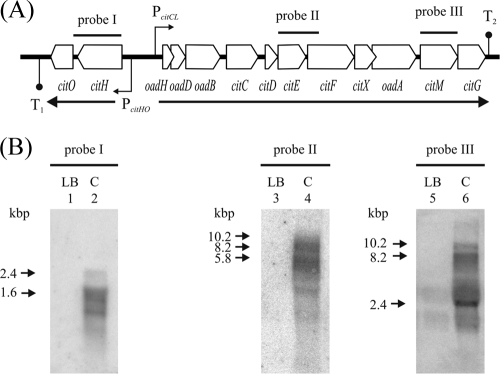
In order to determine the transcriptional pattern of the cit cluster in E. faecalis, Northern blot analysis was performed. Total RNA was isolated from cultures of the E. faecalis ATCC 29212 strain grown in LB or LBC medium. Total RNA was hybridized against α-32P-labeled DNA probes: probe I consists of a 1,400-bp fragment covering the citH gene, probe II covers the citEF region (1,200 bp), while probe III corresponds to citM gene (1,200 bp) (Fig. 2A). Northern blot analysis revealed that the citrate fermentation gene cluster in E. faecalis is transcribed in two different units (Fig. 2B). The citHO operon (2,400 nucleotides [nt]) and the oadHDB-citCDEFX-oadA-citMG operon (10,200 nt). Both operons were clearly induced when cells were grown in the presence of citrate (Fig. 2B, lanes 2, 4, and 6). In fact, there were no detectable transcripts of cit operons when cells were grown in LB medium (Fig. 2B, lanes 1, 3, and 5). In addition to the 2,400-nt citHO mRNA, smaller mRNA species were detected with probe I, which could be the result of the larger transcript processing. A putative secondary structure was found in the IR between both genes (cauuuuGUCUUuuCUugcGUUAUaUUUUcAUuuuUAAUAuuauauuUAUUAucuaUAAAAAUAACcuauAGGGAGGuGCcucaua, with a ΔG of −8.3 kcal/mol; here and below, uppercase letters correspond to nucleotides involved in the complementary interaction, and lowercase letters represent internal loops) using the Mfold program (35, 58). The transcriptional start site of the citHO operon was determined by primer extension experiments using two independent oligonucleotides (pecitH and pecitH2) (see Table S1 in the supplemental material). The transcript starts with a cytosine residue located 134 nt upstream from ATG of citH. The promoter is situated upstream of this sequence, with the −10 (TATATT) and −35 (TATTTA) putative boxes separated by 17 nt (Fig. 3A, lane 1, and C). A putative Rho-independent terminator sequence was identified in the citO 3′ region (gcAA AAAAGACCUGCAGUcGCucguGCaACUGCAGGUCUUU UUUcauuuuuuuu, with a ΔG of −27.1 kcal/mol) (35, 58) (Fig. 2A).
FIG. 2.
Transcriptional analysis of the cit operons in E. faecalis. (A) Schematic representation of the citHO and oadHDB-citCDEFX-oadA-citMG operons. PcitHO and PcitCL indicate the promoters of the cit operons. The secondary structures T1 and T2 represent putative Rho-independent transcriptional terminators. (B) Northern blot analysis of E. faecalis ATCC 29212 strain. Cells were cultivated in LB or LBC (C) medium. Total RNA was hybridized against specific probes: citH (probe I), citEF (probe II), and citM (probe III). The major RNA species observed in the Northern blot are indicated by arrows.
FIG. 3.
Primer extension analysis of the transcriptional start sites of citHO (A) and oadHDB-citCDEFX-oadA-citMG operons (B). The images show primer extension experiments performed with RNA extracted from strain ATCC 29212 grown in LBC medium. Lanes A, C, G, and T show unrelated sequencing ladders. The length of the fragments is indicated on the left side of the sequence ladder. (A) Lane 1 corresponds to detection of the 5′ end of citHO mRNA using the pecitH oligonucleotide. (B) Lane 1 corresponds to detection of the 5′ end of oadHDB-citCDEFX-oadA-citMG mRNA using the peoadH2 oligonucleotide. (C) Nucleotide sequence of the citH-oadH promoter regions. Transcriptional start sites are indicated (+1); −10 and −35 regions are shown by boxes. The directions of transcription and translation are indicated by horizontal arrows. The coding sequences for CitH and OadH are shown in bold. The sequences protected by CitO in DNase I footprinting experiments on the top and bottom DNA strands are underlined. The direct repeat sequence, which is possibly involved in CitO binding, is in boldface.
DNA probes II and III revealed the presence of a larger mRNA transcript that could correspond to a 10,200-nt mRNA starting upstream of oadH and ending downstream of citG. We identified a putative Rho-independent terminator sequence in the citG 3′ region (uAAAAGGCGGCAuuCGUUcaaaAACGgcUGUCGCCUUUUuuuaugaaauuuuuuu, with a ΔG of −19.8 kcal/mol) (35, 58) (Fig. 2A). The differential pattern observed with both probes suggested a specific processing of the larger transcript. In the blots hybridized with probes II and III, we identified smaller products that probably correspond to transcripts processed in three putative secondary structures. The first putative secondary structure was identified in the 3′ region of the oadH gene (GUUGCcauuauuGCAGC, with a ΔG of −4.5 kcal/mol), the second one was found in the oadB-citC IR (UGUUAGCUuuuUGGUUGGGGCGGCGuuuuCGUUGUUUUAuuuAUCAuuuAUC UAGC, with a ΔG of −15.6 kcal/mol), and the third was found between oadA and citM (GAAGCAAGGGUUUGAGCGACgauaacgGUUGUUCAAACCCUUGCUU, with a ΔG of −34.6 kcal/mol). Therefore, in addition to the citHO (2.4 kbp) and the oadHDB-citCDEFX-oadA-citMG (10.2 kbp) transcripts, smaller mRNA species were observed, suggesting posttranscriptional regulation, as previously reported for other microorganisms (3, 6, 32, 33). Thus, RNA processing could be used as a mechanism to ensure that the catabolic citrate lyase complex (citDEF) and the decarboxylases (oadHDB, oadA, and citM) are synthesized in a differential manner. Similarly, this mode of processing could be necessary to maintain the balance between the levels of expression of the citrate transporter (citH) and the regulatory protein (citO).
Next, primer extension experiments using two independent oligonucleotides (peoadH and peoadH2) (see Table S1 in the supplemental material), showed that the transcriptional start site of the oadHDB-citCDEFX-oadA-citMG operon corresponds to a guanosine residue located 32 nt upstream of the assumed oadH start codon. A putative promoter was detected with −10 (TTCTAT) and a −35 (TTCACT) hexamers (Fig. 3B and C). The location of the promoter was confirmed by reverse transcription-PCR (not shown) and by transcriptional fusion analysis (see below).
To validate the data observed by Northern blotting, the intergenic promoter region was studied by using the promoterless vector pTCV-lac (40), which allows lacZ fusion construction. Since attempts to transform E. faecalis ATCC 29212 failed, the laboratory strain JH2-2 was used for genetic manipulation. β-Galactosidase activity was determined in cell extracts of E. faecalis JH2-2 harboring plasmid pTCV-PcitHO or pTCV-PcitCL (Fig. 4) (strains JHB2 and JHB6, respectively) grown under the same conditions described for Northern blotting. The β-galactosidase activities measured in E. faecalis JHB2 and JHB6 grown in LBC were 1,750 ± 20 Miller units and 15,500 ± 1,000 Miller units, respectively. No significant β-galactosidase activity (less than 60 MU) was found in E. faecalis JHB10 (harboring pTCV-lac empty vector) cell extracts grown in LBC medium or in JHB2, JHB6, and JHB10 cell extracts grown in LB medium.
Deletion of PcitHO promoter (strain JHB3) (Fig. 4A) resulted in no significant β-galactosidase activity, implying that this is the only promoter that directs the transcription of citHO operon. In addition, the PcitCL promoter −10 box TTCTAT was mutated to TGGCGC (strain JHB7) (Fig. 4B), producing no significant β-galactosidase activity in E. faecalis JHB7 cell extracts. This indicates that PcitCL is the only promoter driving the transcription of the oadHDB-citCDEFX-oadA-citMG operon.
Hence, from these experiments we conclude that the expression of the cit divergon is controlled by PcitHO and PcitCL promoters and is induced at the transcriptional level by the presence of citrate.
A new cluster of GntR-like transcriptional regulators.
CitO belongs to the GntR-like transcriptional regulator family, which includes more than 5,000 proteins annotated in the Swiss-Prot database. Members of this family of regulators are distributed among very diverse bacterial groups and regulate a variety of biological processes. GntR-like proteins were classified into four major subfamilies (FadR, MocR, HutC, and YtrA) and into three minor subfamilies (DevA, PlmA, and AraR) (20, 27, 43) (Fig. 1B). A multiple alignment of 51 full-length GntR sequences (NCBI) was constructed using the native implementation of CLUSTAL W in the alignment explorer tool of MEGA3 (26). Phylogenetic trees were constructed with MEGA3 by using the neighbor-joining method (44). The resulting trees were visualized using the tree explorer tool of MEGA3. The phylogenetic tree of the family reveals that the five members associated with the citrate fermentation pathways of S. mutants, S. pyogenes, L. casei, L. sakei, and E. faecalis are on a separate branch of the FadR subfamily (Fig. 1B). We infer from this result that the CitO cluster arose from an ancestral sequence shared with the FadR subfamily that diverged through a replacement process in the effector binding domain. Even though several FadR subfamily members were characterized and reported to be involved in the regulation of aspartate (AnsR) (53), pyruvate (PdhR) (38), glycolate (GlcC) (39), lactate (LldR) (17), and gluconate (GntR) (42) metabolism, no evidence concerning to the role of the CitO transcriptional factor cluster was found.
CitO is the positive activator of the cit divergon.
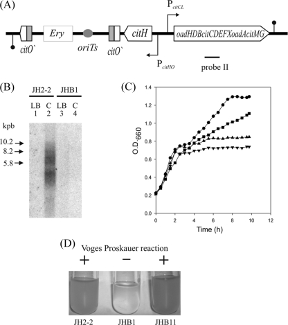
In order to investigate the role of CitO in the regulation of the cit operons, we constructed a citO mutant by interrupting this gene using single-crossover chromosomal integration of the pmCitO plasmid (Fig. 5A). The appropriate disruption of citO was confirmed by Southern blot hybridization (data not shown). The resulting strain, JHB1, was tested in its ability to use citrate. While the parental E. faecalis JH2-2 strain showed a citrate lyase activity of 1.5 ± 0.2 U μg−1 in a protein cell extract obtained from cells grown in LBC medium, the citO mutant strain JHB1 was unable to express this activity under the same conditions.
FIG. 5.
Construction and characterization of an E. faecalis citO-defective strain (JHB1). (A) Schematic representation of citO inactivation by insertion of the pmCitO plasmid. The fragments labeled citO′ are parts of the citO gene; ery and ori(Ts) are the erythromycin cassette and the thermosensitive replication origin of pGh9 plasmid, respectively. (B) Northern blot analysis in citO+ (JH2-2) and citO mutant (JHB1) strains. Cells were grown in LB (lanes 1 and 3) or LBC (lanes 2 and 4) medium. The assay was performed as described in the legend of Fig. 2 but using only probe II. Transcripts were detected only for strain JH2-2 grown in the presence of citrate (lane 2) and not in the mutant strain (lane 4) or in the absence of citrate (lanes 1 and 3). (C) Growth curves in LBC medium of E. faecalis JH2-2 and derivative strains, citO+ (JH2-2; •), citO mutant(JHB1; ▾), citO mutant harboring the plasmid pCitO (JHB11; ▪) and citO mutant harboring the plasmid pBM02 (JHB12; ▴). (D) Voges-Proskauer assays. Strains JH2-2, JHB1, and JHB11 were grown in LBC medium. A positive reaction was obtained in the wild type (JH2-2) and in the complemented strain (JHB11).
We subsequently performed a Northern blot assay using probe II (Fig. 5A) with total RNA obtained from cells grown as described above. As expected, the presence of transcript was detected in the lane corresponding to RNA from JH2-2 grown in LBC medium, and no signal was observed in the samples corresponding to JHB1 (Fig. 5B). In agreement with this transcriptional evidence, growth curves showed that citrate metabolism allowed the parental strain to reach a final OD660 of 1.3 in LBC medium while strain JHB1 reached an OD660 of only 0.75 (Fig. 5C).
Finally, the ability to degrade citrate was evaluated by the production of acetoin through the Voges-Proskauer test. Liquid cultures (LBC medium) of wild-type JH2-2 produced large quantities of acetoin, whereas the level of acetoin was undetectable in the supernatant of the strain JHB1 analyzed in the same conditions (Fig. 5D).
To confirm the role of CitO as the activator of the pathway, we proceeded to complement the deficient strain JHB1 by expressing CitO in trans using the pCitO plasmid (see Materials and Methods) (Table 1). JHB1 cells harboring pCitO (JHB11) recovered the ability to metabolize citrate, which was evidenced by the increase in the final OD660 for cells grown in LBC medium (Fig. 5C) (OD660 of 1.0), the detection of citrate lyase activity in extracts of cells grown in LBC medium (1.1 ± 0.1 U μg−1 of protein), and the production of C4 compounds measured by the Voges-Proskauer reaction (Fig. 5D).
Moreover, we analyzed the PcitHO and PcitCL lacZ fusions in the citO mutant strains (JHB13 and JHB14, respectively). The β-galactosidase levels in these strains were less than 60 MU, even when cells were grown in the presence of citrate, but normal activity was restored in the JHB15 and JHB16 complemented strains (harboring pTCV-PcitHO and pTCV-PcitCL, respectively) when cells were grown in LBC medium (see also Fig. 8B and C).
FIG. 8.
(A) Effect of diverse citrate analogs on DNase I protection of the cit promoters by CitO. The oadHDB-citCDEFX-oadA-citMG coding strand was employed as target DNA. The CitO protein was used at a concentration of 1.5 μM. The different citrate analogs were used at a final concentration of 1.1 mM each. The presence (+) or absence (−) of the compounds and of CitO is shown in the top panel. The boxes indicate the extent of the protected regions. Influence of diverse citrate analogs on the expression of PcitHO-lacZ (B) and PcitCL-lacZ (C) fusions. Strain JHB15 (JH2-2 citO mutant harboring plasmids pCitO/pTCV-PcitHO) and strain JHB16 (JH2-2 citO mutant harboring plasmids pCitO/pTCV-PcitCL) were grown in LB medium and LB medium supplemented with initial concentrations of 30 mM citrate, malate, succinate, α-ketoglutarate, fumarate, acetate, or pyruvate or 10 mM isocitrate. The levels of accumulated β-galactosidase activity were measured 8 h after inoculation. Error bars represents the standard deviation of triplicate measurements.
Binding of CitO to the promoter regions.
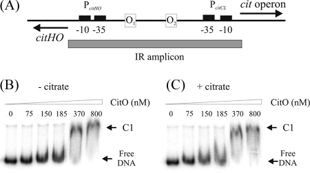
To address whether the regulatory effect of the citO gene product involved a direct interaction with the citH and oadH promoter regions, a His6-CitO fusion protein was constructed and expressed in E. coli. This fusion protein was subsequently used in gel mobility shift assays using a DNA fragment corresponding to the citH-oadH IR (Fig. 6A). The assay was performed by incubating the IR amplicon (427 bp) with increasing concentrations of purified CitO. As a result, a CitO-IR complex was identified (Fig. 6B, C1). Binding specificity was confirmed by determining that CitO did not bind to an unrelated 174-bp fragment belonging to the B. subtilis acp promoter (data not shown).
FIG. 6.
Binding of CitO to the citHO-oadHDB-citCDEFX-oadA-citMG IR. (A) Schematic representation of the cit operon IR. PcitHO and PcitCL promoters are indicated. The thick line on the top indicates the location of the 427-bp DNA probe (IR amplicon) used in the gel shift experiments. (B and C) Image of gel shift assay performed with 0.5 nM IR amplicon and increasing concentrations of CitO without (B) and with (C) 1.1 mM citrate. Arrows indicate the positions of the retarded complex C1 and free DNA.
The results presented to this point indicate that induction of cit genes occurs upon growth of E. faecalis in LB medium in the presence of citrate. This led us to think that citrate could interact with CitO, modifying its binding capability to the DNA. To confirm this hypothesis, we evaluated the effect of citrate in a gel mobility shift assay, using this compound at a final concentration of 1.1 mM and increasing concentrations of CitO. With this approach a subtle effect of citrate presence was detected. Figure 6C shows that with 185 nM CitO, a small amount of labeled probe is retarded, while in the absence of citrate all the DNA is in the free form (Fig. 6B). These experiments suggest that the presence of citrate might increase the affinity of the regulator for its binding sites. This assumption was confirmed by DNase I footprinting experiments (see below).
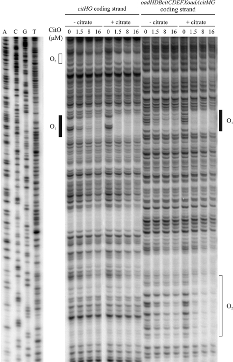
The localization of the CitO binding sites within the cit promoters was determined by DNase I footprinting on both strands of the DNA using a 100-fold excess of competitor DNA (Fig. 7). A 427-bp fragment containing the promoter regions of the citHO and oadHDB-citCDEFX-oadA-citMG operons was used as target DNA in the experiments. This fragment carries all the sequences necessary for CitO to exert its regulation on the cit divergon, as was demonstrated by transcriptional fusions (Fig. 4). Two binding sites were detected in the promoter regions of the cit operons when citrate was present. In the citHO operon coding strand (Fig. 7), CitO protects the region between positions −41 and −65 (binding site O1) while in the oadHDB-citCDEFX-oadA-citMG operon coding strand (Fig. 7), CitO protects the region between positions −51 and −74 (binding site O2). The same sequences are protected in the noncoding strand. The exact position of O2 was also determined using a different 247-bp fragment that allowed a better resolution of the protected nucleotides (not shown). The position of the binding sites relative to the cit operon promoters is depicted in Fig. 3C.
FIG. 7.
DNase I protection experiments of the operons promoters by CitO. A 427-bp DNA fragment including the citHO-oadHDB-citCDEFX-oadA-citMG operon promoter regions was end labeled with [γ-32P]ATP and used as target DNA in the assays. Each strand was labeled in separate experiments. Unrelated sequencing reactions were run at the side with the DNase I footprinting reactions. Protected regions (O1 and O2) are indicated at the sides of the gel lanes.
To further demonstrate that CitO specifically recognizes the IR in vivo, we cloned a 247-bp DNA fragment containing the O1 and O2 sites into plasmid pBM02 to generate the pCO1O2 plasmid (Table 1). Both pBM02 and pCO1O2 were used to transform the JHB2 and JHB6 strains (harboring pTCV-PcitHO and pTCV-PcitCL, respectively). Transformation with the empty vector resulted in normal transcriptional induction of both strains (JHB17 and JHB19), while transformation with plasmid pCO1O2 (strains JHB18 and JHB20) reduced the level of β-galactosidase activity 4.3- and 5-fold, respectively. We concluded that the presence in trans of the O1 and O2 regions titrates the intracellular CitO molecules, leading to a failure in the induction of the cit operons in the presence of equal amounts of inducer.
Citrate specifically induces binding of CitO to the O1 and O2 DNA sequences.
The results presented in Fig. 6 suggest that the presence of citrate could modify the interaction of CitO with its binding sites. To confirm this hypothesis, we tested the effect of citrate addition in a DNase I footprinting assay (using this compound at a final concentration of 1.1 mM) and increasing concentrations of CitO (Fig. 7). The experiment showed that at higher concentrations (8 to 16 μM), CitO is able to bind to DNA even in the absence of citrate (as observed in the electrophoretic mobility shift assay), while in the presence of 1.1 mM citrate the protection pattern of the binding sites (O1 and O2) is already observed with the lowest concentration of CitO (1.5 μM), indicating that citrate increases the affinity of CitO for its binding sequences. Further analysis of the protected regions showed that CitO has a higher affinity for the O1 site since full protection is observed with 1.5 μM, whereas for O2 only partial protection is observed when 16 μM CitO is employed.
Moreover, the effect of citrate analog compounds (malate, succinate, and isocitrate, among others) was tested (Fig. 8A). No effect on the affinity of CitO for its binding regions was observed with these compounds, indicating that citrate is the specific inducer that modulates CitO binding to DNA.
Furthermore, we determined the specific substrate/effector for CitO in vivo by using strains JHB15 and JHB16 (derivatives of the JH2-2 citO mutant strain and carrying the pCitO plasmid and pTCV-PcitHO or pTCV-PcitCL plasmid, respectively). Cell extracts from these two strains grown in LBC medium were able to express the lacZ gene present in the pTCV-lac-derived vector, while cells growing with citrate-analogous compounds, such as malate, succinate, α-ketoglutarate, fumarate, pyruvate, or isocitrate, were not (Fig. 8B and C).
It is worth noting that although CitO is expressed from the pCitO plasmid in strains JHB15 and JHB16, under these conditions induction of the PcitHO and PcitCL promoters is detected only in the presence of citrate. These results suggest that while CitO is able to bind to DNA in the absence of citrate (Fig. 6 and 7), it might be incapable of activating the transcription of the cit genes without interaction with the specific inducer.
Deletion analysis of the promoter region of the cit divergon.
To define the role of CitO binding regions in the mechanism of transcription regulation mediated by CitO, a set of DNA fragments covering different regions of the cit promoters were PCR amplified and fused to the promoterless lacZ reporter gene of pTCV-lac vector (Fig. 4). The plasmids harboring the cit-lacZ transcriptional fusions were electroporated into the E. faecalis JH2-2 strain. Then, the level of accumulated β-galactosidase activity of the resulting strains was subsequently examined in vivo in the presence of the inducer citrate, and the results are shown in Fig. 4.
Deletion of the CitO binding site O2 on the PcitHO promoter (strain JHB4) resulted in a sixfold decrease in lacZ activity relative to the full promoter fusion. When both sites (O1 and O2) were absent (strain JHB5), no significant activity was detected. On the other hand, deletion of O1 or both sites on the PcitCL promoter fusion (strains JHB8 or JHB9, respectively) results in no significant activity of this promoter.
In conclusion, the two CitO binding sites identified in vitro are very likely to be the fundamental cis-acting elements for the regulation of the citrate metabolic operon promoters by CitO in vivo. Furthermore, the lower level of induction or the total loss of activity observed in a promoter with only one of the binding sites present suggests a regulatory mechanism dependent on cooperative binding of the regulatory protein to its DNA targets.
DISCUSSION
Citrate metabolism is widespread among microorganisms, and it constitutes an alternative carbon and energy source in prokaryotes. In addition, citrate metabolism is an important industrial feature due to its contribution to the synthesis of aroma compounds such as acetoin, diacetyl, and butanediol (21). Moreover, citrate metabolism by Enterococcus spp. has recently received increasing attention (13, 47) since many Mediterranean cheeses naturally contain high numbers of enterococci, which are also being used as starter cultures (13). Although citrate metabolism has been extensively explored in enterobacteria and other microorganisms, this is the first time the transcriptional and regulatory mechanisms have been described in E. faecalis.
First, citrate fermentation genes were characterized in E. faecalis by means of Northern blot assays, which showed the presence of two polycistronic units constituted by the citHO and oadHDB-citCDEFX-oadA-citMG genes. Both mRNA transcripts were specifically processed in smaller species. This posttranscriptional regulation could allow cells to coordinate the level of different activities such as regulatory (CitO), transport (CitH), and degradative (citrate lyase complex or membrane or soluble oxaloacetate decarboxylase) activities. This kind of processing was also described in K. pneumoniae (36), L. mesenteroides (3), W. paramesenteroides (32), and L. lactis (33). We then identified two promoter regions which drive the expression of the cit operons, PcitHO and PcitCL.
Our work showed that expression of the citHO and oadHDB-citCDEFX-oadA-citMG operons in bacteria grown in the presence of citrate was mediated by the transcriptional activator CitO. A citO mutant strain was constructed that was unable to metabolize citrate. Furthermore, this mutant strain was also unable to produce C4 compounds, suggesting a relationship between the production of these compounds and citrate fermentation. The Cit+ phenotype was restored when citO was complemented in trans. Moreover, the expression of a wild-type copy of citO in this strain enabled the production of C4 compounds.
E. faecalis CitO belongs to the FadR subfamily of the GntR family of transcriptional regulators that includes activators, repressors, and molecules which both activate and repress a wide range of bacterial operons (43). Members of the GntR family, which was first described by Haydon and Guest (19), share similar N-terminal DNA-binding domains but show heterogeneous C-terminal effector-binding and oligomerization domains. FadR from E. coli is the only member of the FadR group whose crystal structure has been determined (22, 54, 55, 57). FadR plays a dual role in transcriptional regulation of the enzymes involved in fatty acid metabolism. Its function as a repressor or an activator is readily explained by the location of its binding sites within the promoter regions. When the binding site either overlaps the −10 or −35 regions or lies just downstream of the −10 sequence, FadR represses transcription. On the other hand, when the binding sites are located immediately upstream of the −35 region of the promoter, FadR binding activates transcription, presumably by promoting the binding or action of RNA polymerase. Consensus sequence for the cis-acting element recognized by the FadR family members was used as a tool to predict a putative operator sequence. No palindromic sequences related to the consensus sequence (NyGTNxACNy, where x 4p = 3 nucleotides and y = 5 nucleotides) were found in the citH-oadH intergenic region. Although most members of the GntR family recognize palindromic or pseudopalindromic sequences, transcriptional activators of the MocR subfamily of the GntR family, GabR (ATACCA) (4) and TauR (CTGGACYTAA) (Y is T or C) (56), are able to recognize direct repeat sequences in the promoter regions. Even though none of these GntR consensus sequences was found in the cit intergenic region, our genetic and biochemical data clearly indicated that CitO activates the gene expression by binding to DNA at the PcitHO and PcitCL promoter regions. In fact, DNase I protection studies determined two CitO binding sites which were positioned at −41 through −65 relative to the transcription start site of citHO ( site O1) and −51 through −74 relative to the transcription start site of citCL operon (site O2). The O1 sequence on the coding strand was 5′-TGT TTT TTT GAT GTT CTT TTT GTT T-′3, while the O2 protected sequence on the coding strand was 5′-ACA AAA TAA ACA ATA AAA GAA CAA TC-′3. The O1 binding site is located only 5 bp from the −35 box of the PcitHO, while the O2 site is at 14 bp from the −35 box of the PcitCL. Thus, CitO bound at these positions promotes the transcriptional activity of the bound RNA polymerase by protein-protein interactions. These interactions most commonly include contacts to the α-subunit C-terminal domain of RNA polymerase but can also involve contacts to the N-terminal domain of the α-, σ-, β-, or β′-subunit (10). Nucleotide sequence comparison between the O1 and O2 sites shows that these regions are direct repeat sequences, where it is possible to identify two similar short putative binding sites, AAAARAAC (R is A or G) in each protected region (Fig. 3). Short runs of repeated adenine or thymine residues, such as AAAA or TTTT, which are known to lead to intrinsic DNA bending are also present in the intergenic region. However, in the DNase I protection assays, we could not observe hypersensitive sites. Site-directed mutagenesis of the O1 and O2 sites will be necessary to determine the role of the relevant sequences and analyze if bending is involved in regulation.
The results presented in this work led us to hypothesize that CitO could be able to interact with intracellular citrate, increasing in this way CitO affinity for its binding sites; this fact is supported by electrophoretic mobility shift assay (Fig. 6) and DNase I protection assays (Fig. 7). Deletion experiments indicated that the O1 DNA region is required for the expression of PcitCL, suggesting cooperativity between the O1 and O2 sites. Moreover, the O1 binding site seems to be occupied at lower CitO concentrations than the O2 site (Fig. 7). This could imply that the citHO operon is transcriptionally activated at lower concentrations of CitO and citrate.
All together, these results suggest a model for the activation of the cit divergon in E. faecalis. When the enterococcal cells are grown in the absence of citrate, both cit operons are switched off, and there is no basal transcription of these genes. The presence of citrate in the medium induces the activation of the citHO operon. Then, it is possible that in the presence of citrate the intracellular concentrations of CitO (activator) and the inducer (citrate) increase, reaching the level at which CitO can bind to O2, which results in the full expression of the degradative pathway. Clearly, more experiments will be necessary to understand the fine-tuning of this activation mechanism.
In conclusion, this is the first time a transcriptional activator belonging to GntR family has been studied in E. faecalis, offering an attractive system to explore the molecular basis of the specific interactions of the CitO activator to DNA and providing different regulatory models for other industrial and pathogenic microorganisms.
Supplementary Material
Acknowledgments
We thank Lilian Guibert for improving the use of English in the manuscript and two anonymous reviewers for their comments on a previous version of the manuscript.
This work was supported by grants from the Agencia Nacional de Promoción Científica y Tecnológica (ANPCyT, contract 15-38025, Argentina) and a European Union grant (BIAMFood, contract KBBE-211441). V.B. and G.R are fellows of CONICET (Argentina), C.S. is a fellow of ANPCyT (Argentina), and C.M. is a Career Investigator from CONICET (Argentina).
Footnotes
Published ahead of print on 19 September 2008.
Supplemental material for this article may be found at http://jb.asm.org/.
REFERENCES
- 1.Bandell, M., M. E. Lhotte, C. Marty-Teysset, A. Veyrat, H. Prevost, V. Dartois, C. Divies, W. N. Konings, and J. S. Lolkema. 1998. Mechanism of the citrate transporters in carbohydrate and citrate cometabolism in Lactococcus and Leuconostoc species. Appl. Environ. Microbiol. 641594-1600. [DOI] [PMC free article] [PubMed] [Google Scholar]
- 2.Bekal, S., J. Van Beeumen, B. Samyn, D. Garmyn, S. Henini, C. Divies, and H. Prevost. 1998. Purification of Leuconostoc mesenteroides citrate lyase and cloning and characterization of the citCDEFG gene cluster. J. Bacteriol. 180647-654. [DOI] [PMC free article] [PubMed] [Google Scholar]
- 3.Bekal-Si Ali, S., C. Divies, and H. Prevost. 1999. Genetic organization of the citCDEF locus and identification of mae and clyR genes from Leuconostoc mesenteroides. J. Bacteriol. 1814411-4416. [DOI] [PMC free article] [PubMed] [Google Scholar]
- 4.Belitsky, B. R. 2004. Bacillus subtilis GabR, a protein with DNA-binding and aminotransferase domains, is a PLP-dependent transcriptional regulator. J. Mol. Biol. 340655-664. [DOI] [PubMed] [Google Scholar]
- 5.Blancato, V. S., C. Magni, and J. S. Lolkema. 2006. Functional characterization and Me2+ ion specificity of a Ca2+-citrate transporter from Enterococcus faecalis. FEBS J. 2735121-5130. [DOI] [PubMed] [Google Scholar]
- 6.Bott, M. 1997. Anaerobic citrate metabolism and its regulation in enterobacteria. Arch. Microbiol. 16778-88. [PubMed] [Google Scholar]
- 7.Bott, M., and P. Dimroth. 1994. Klebsiella pneumoniae genes for citrate lyase and citrate lyase ligase: localization, sequencing, and expression. Mol. Microbiol. 14347-356. [DOI] [PubMed] [Google Scholar]
- 8.Bott, M., M. Meyer, and P. Dimroth. 1995. Regulation of anaerobic citrate metabolism in Klebsiella pneumoniae. Mol. Microbiol. 18533-546. [DOI] [PubMed] [Google Scholar]
- 9.Bradford, M. M. 1976. A rapid and sensitive method for the quantitation of microgram quantities of protein utilizing the principle of protein-dye binding. Anal. Biochem. 72248-254. [DOI] [PubMed] [Google Scholar]
- 10.Browning, D. F., and S. J. Busby. 2004. The regulation of bacterial transcription initiation. Nat. Rev. Microbiol. 257-65. [DOI] [PubMed] [Google Scholar]
- 11.Clewell, D. B., P. K. Tomich, M. C. Gawron-Burke, A. E. Franke, Y. Yagi, and F. Y. An. 1982. Mapping of Streptococcus faecalis plasmids pAD1 and pAD2 and studies relating to transposition of Tn917. J. Bacteriol. 1521220-1230. [DOI] [PMC free article] [PubMed] [Google Scholar]
- 12.Dimroth, P., P. Jockel, and M. Schmid. 2001. Coupling mechanism of the oxaloacetate decarboxylase Na(+) pump. Biochim. Biophys. Acta 15051-14. [DOI] [PubMed] [Google Scholar]
- 13.Foulquie Moreno, M. R., P. Sarantinopoulos, E. Tsakalidou, and L. De Vuyst. 2006. The role and application of enterococci in food and health. Int. J. Food Microbiol. 1061-24. [DOI] [PubMed] [Google Scholar]
- 14.Friesenegger, A., S. Fiedler, L. A. Devriese, and R. Wirth. 1991. Genetic transformation of various species of Enterococcus by electroporation. FEMS Microbiol. Lett. 63323-327. [DOI] [PubMed] [Google Scholar]
- 15.Garcia-Quintans, N., C. Magni, D. de Mendoza, and P. Lopez. 1998. The citrate transport system of Lactococcus lactis subsp. lactis biovar diacetylactis is induced by acid stress. Appl. Environ. Microbiol. 64850-857. [DOI] [PMC free article] [PubMed] [Google Scholar]
- 16.Garcia-Quintans, N., G. Repizo, M. Martin, C. Magni, and P. Lopez. 2008. Activation of the diacetyl/acetoin pathway in Lactococcus lactis subsp. lactis bv. diacetylactis CRL264 by acidic growth. Appl. Environ. Microbiol. 741988-1996. [DOI] [PMC free article] [PubMed] [Google Scholar]
- 17.Georgi, T., V. Engels, and V. F. Wendisch. 2008. Regulation of l-lactate utilization by the FadR-type regulator LldR of Corynebacterium glutamicum. J. Bacteriol. 190963-971. [DOI] [PMC free article] [PubMed] [Google Scholar]
- 18.Giraffa, G. 2003. Functionality of enterococci in dairy products. Int. J. Food Microbiol. 88215-222. [DOI] [PubMed] [Google Scholar]
- 19.Haydon, D. J., and J. R. Guest. 1991. A new family of bacterial regulatory proteins. FEMS Microbiol. Lett. 63291-295. [DOI] [PubMed] [Google Scholar]
- 20.Hoskisson, P. A., S. Rigali, K. Fowler, K. C. Findlay, and M. J. Buttner. 2006. DevA, a GntR-like transcriptional regulator required for development in Streptomyces coelicolor. J. Bacteriol. 1885014-5023. [DOI] [PMC free article] [PubMed] [Google Scholar]
- 21.Hugenholtz, J. 1993. Citrate metabolism in lactic acid bacteria. FEMS Microbiol. Rev. 12165-178. [Google Scholar]
- 22.Iram, S. H., and J. E. Cronan. 2005. Unexpected functional diversity among FadR fatty acid transcriptional regulatory proteins. J. Biol. Chem. 28032148-32156. [DOI] [PubMed] [Google Scholar]
- 23.Israelsen, H., S. M. Madsen, A. Vrang, E. B. Hansen, and E. Johansen. 1995. Cloning and partial characterization of regulated promoters from Lactococcus lactis Tn917-lacZ integrants with the new promoter probe vector, pAK80. Appl. Environ. Microbiol. 612540-2547. [DOI] [PMC free article] [PubMed] [Google Scholar]
- 24.Jacob, A. E., and S. J. Hobbs. 1974. Conjugal transfer of plasmid-borne multiple antibiotic resistance in Streptococcus faecalis var. zymogenes. J. Bacteriol. 117360-372. [DOI] [PMC free article] [PubMed] [Google Scholar]
- 25.Kempler, G., and L. McKay. 1980. Improved medium for detection of citrate-fermenting Streptococcus lactis subsp. diacetylactis. Appl. Environ. Microbiol. 39926-927. [DOI] [PMC free article] [PubMed] [Google Scholar]
- 26.Kumar, S., K. Tamura, and M. Nei. 2004. MEGA3: integrated software for molecular evolutionary genetics analysis and sequence alignment. Brief. Bioinform. 5150-163. [DOI] [PubMed] [Google Scholar]
- 27.Lee, M. H., M. Scherer, S. Rigali, and J. W. Golden. 2003. PlmA, a new member of the GntR family, has plasmid maintenance functions in Anabaena sp. strain PCC 7120. J. Bacteriol. 1854315-4325. [DOI] [PMC free article] [PubMed] [Google Scholar]
- 28.Lopez de Felipe, F., C. Magni, D. de Mendoza, and P. Lopez. 1996. Transcriptional activation of the citrate permease P gene of Lactococcus lactis biovar diacetylactis by an insertion sequence-like element present in plasmid pCIT264. Mol. Gen. Genet. 250428-436. [DOI] [PubMed] [Google Scholar]
- 29.Magni, C., D. de Mendoza, W. N. Konings, and J. S. Lolkema. 1999. Mechanism of citrate metabolism in Lactococcus lactis: resistance against lactate toxicity at low pH. J. Bacteriol. 1811451-1457. [DOI] [PMC free article] [PubMed] [Google Scholar]
- 30.Maguin, E., H. Prevost, S. D. Ehrlich, and A. Gruss. 1996. Efficient insertional mutagenesis in lactococci and other gram-positive bacteria. J. Bacteriol. 178931-935. [DOI] [PMC free article] [PubMed] [Google Scholar]
- 31.Martin, M., M. A. Corrales, D. de Mendoza, P. Lopez, and C. Magni. 1999. Cloning and molecular characterization of the citrate utilization citMCDEFGRP cluster of Leuconostoc paramesenteroides. FEMS Microbiol. Lett. 174231-238. [DOI] [PubMed] [Google Scholar]
- 32.Martin, M., C. Magni, P. Lopez, and D. de Mendoza. 2000. Transcriptional control of the citrate-inducible citMCDEFGRP operon, encoding genes involved in citrate fermentation in Leuconostoc paramesenteroides. J. Bacteriol. 1823904-3912. [DOI] [PMC free article] [PubMed] [Google Scholar]
- 33.Martin, M. G., P. D. Sender, S. Peiru, D. de Mendoza, and C. Magni. 2004. Acid-inducible transcription of the operon encoding the citrate lyase complex of Lactococcus lactis biovar diacetylactis CRL264. J. Bacteriol. 1865649-5660. [DOI] [PMC free article] [PubMed] [Google Scholar]
- 34.Martin, M. G., C. Magni, D. de Mendoza, and P. Lopez. 2005. CitI, a transcription factor involved in regulation of citrate metabolism in lactic acid bacteria. J. Bacteriol. 1875146-5155. [DOI] [PMC free article] [PubMed] [Google Scholar]
- 35.Mathews, D. H., J. Sabina, M. Zuker, and D. H. Turner. 1999. Expanded sequence dependence of thermodynamic parameters improves prediction of RNA secondary structure. J. Mol. Biol. 288911-940. [DOI] [PubMed] [Google Scholar]
- 36.Meyer, M., P. Dimroth, and M. Bott. 1997. In vitro binding of the response regulator CitB and of its carboxy-terminal domain to A+T-rich DNA target sequences in the control region of the divergent citC and citS operons of Klebsiella pneumoniae. J. Mol. Biol. 269719-731. [DOI] [PubMed] [Google Scholar]
- 37.Meyer, M., P. Dimroth, and M. Bott. 2001. Catabolite repression of the citrate fermentation genes in Klebsiella pneumoniae: evidence for involvement of the cyclic AMP receptor protein. J. Bacteriol. 1835248-5256. [DOI] [PMC free article] [PubMed] [Google Scholar]
- 38.Ogasawara, H., Y. Ishida, K. Yamada, K. Yamamoto, and A. Ishihama. 2007. PdhR (pyruvate dehydrogenase complex regulator) controls the respiratory electron transport system in Escherichia coli. J. Bacteriol. 1895534-5541. [DOI] [PMC free article] [PubMed] [Google Scholar]
- 39.Pellicer, M. T., J. Badia, J. Aguilar, and L. Baldoma. 1996. glc locus of Escherichia coli: characterization of genes encoding the subunits of glycolate oxidase and the glc regulator protein. J. Bacteriol. 1782051-2059. [DOI] [PMC free article] [PubMed] [Google Scholar]
- 40.Poyart, C., and P. Trieu-Cuot. 1997. A broad-host-range mobilizable shuttle vector for the construction of transcriptional fusions to beta-galactosidase in gram-positive bacteria. FEMS Microbiol. Lett. 156193-198. [DOI] [PubMed] [Google Scholar]
- 41.Reinelt, S., E. Hofmann, T. Gerharz, M. Bott, and D. R. Madden. 2003. The structure of the periplasmic ligand-binding domain of the sensor kinase CitA reveals the first extracellular PAS domain. J. Biol. Chem. 27839189-39196. [DOI] [PubMed] [Google Scholar]
- 42.Reizer, A., J. Deutscher, M. H. Saier, and J. Reizer. 1991. Analysis of the gluconate (gnt) operon of Bacillus subtilis. Mol. Microbiol. 51081-1089. [DOI] [PubMed] [Google Scholar]
- 43.Rigali, S., A. Derouaux, F. Giannotta, and J. Dusart. 2002. Subdivision of the helix-turn-helix GntR family of bacterial regulators in the FadR, HutC, MocR, and YtrA subfamilies. J. Biol. Chem. 27712507-12515. [DOI] [PubMed] [Google Scholar]
- 44.Saitou, N., and M. Nei. 1987. The neighbor-joining method: a new method for reconstructing phylogenetic trees. Mol. Biol. Evol. 4406-425. [DOI] [PubMed] [Google Scholar]
- 45.Sambrook, J., E. F. Fritsch, and T. Maniatis. 1989. Molecular cloning: a laboratory manual, 2nd ed. Cold Spring Harbor Laboratory Press, Cold Spring Harbor, NY.
- 46.Sarantinopoulos, P., G. Kalantzopoulos, and E. Tsakalidou. 2001. Citrate metabolism by Enterococcus faecalis FAIR-E 229. Appl. Environ. Microbiol. 675482-5487. [DOI] [PMC free article] [PubMed] [Google Scholar]
- 47.Sarantinopoulos, P., L. Makras, F. Vaningelgem, G. Kalantzopoulos, L. De Vuyst, and E. Tsakalidou. 2003. Growth and energy generation by Enterococcus faecium FAIR-E 198 during citrate metabolism. Int. J. Food Microbiol. 84197-206. [DOI] [PubMed] [Google Scholar]
- 48.Schneider, K., P. Dimroth, and M. Bott. 2000. Identification of triphosphoribosyl-dephospho-CoA as precursor of the citrate lyase prosthetic group. FEBS Lett. 483165-168. [DOI] [PubMed] [Google Scholar]
- 49.Schneider, K., P. Dimroth, and M. Bott. 2000. Biosynthesis of the prosthetic group of citrate lyase. Biochemistry 399438-9450. [DOI] [PubMed] [Google Scholar]
- 50.Schneider, K., C. N. Kastner, M. Meyer, M. Wessel, P. Dimroth, and M. Bott. 2002. Identification of a gene cluster in Klebsiella pneumoniae which includes citX, a gene required for biosynthesis of the citrate lyase prosthetic group. J. Bacteriol. 1842439-2446. [DOI] [PMC free article] [PubMed] [Google Scholar]
- 51.Schwarz, E., D. Oesterhelt, H. Reinke, K. Beyreuther, and P. Dimroth. 1988. The sodium ion translocating oxalacetate decarboxylase of Klebsiella pneumoniae. Sequence of the biotin-containing alpha-subunit and relationship to other biotin-containing enzymes. J. Biol. Chem. 2639640-9645. [PubMed] [Google Scholar]
- 52.Sender, P. D., M. G. Martin, S. Peiru, and C. Magni. 2004. Characterization of an oxaloacetate decarboxylase that belongs to the malic enzyme family. FEBS Lett. 570217-222. [DOI] [PubMed] [Google Scholar]
- 53.Sun, D., and P. Setlow. 1993. Cloning and nucleotide sequence of the Bacillus subtilis ansR gene, which encodes a repressor of the ans operon coding for l-asparaginase and l-aspartase. J. Bacteriol. 1752501-2506. [DOI] [PMC free article] [PubMed] [Google Scholar]
- 54.van Aalten, D. M., C. C. DiRusso, and J. Knudsen. 2001. The structural basis of acyl coenzyme A-dependent regulation of the transcription factor FadR. EMBO J. 202041-2050. [DOI] [PMC free article] [PubMed] [Google Scholar]
- 55.van Aalten, D. M., C. C. DiRusso, J. Knudsen, and R. K. Wierenga. 2000. Crystal structure of FadR, a fatty acid-responsive transcription factor with a novel acyl coenzyme A-binding fold. EMBO J. 195167-5177. [DOI] [PMC free article] [PubMed] [Google Scholar]
- 56.Wiethaus, J., B. Schubert, Y. Pfander, F. Narberhaus, and B. Masepohl. 2008. The GntR-like regulator TauR activates expression of taurine utilization genes in Rhodobacter capsulatus. J. Bacteriol. 190487-493. [DOI] [PMC free article] [PubMed] [Google Scholar]
- 57.Xu, Y., R. J. Heath, Z. Li, C. O. Rock, and S. W. White. 2001. The FadR DNA complex: transcriptional control of fatty acid metabolism in Escherichia coli. J. Biol. Chem. 27617373-17379. [DOI] [PubMed] [Google Scholar]
- 58.Zuker, M. 2003. Mfold web server for nucleic acid folding and hybridization prediction. Nucleic Acids Res. 313406-3415. [DOI] [PMC free article] [PubMed] [Google Scholar]
Associated Data
This section collects any data citations, data availability statements, or supplementary materials included in this article.


