Abstract
Background
In tennis, injuries to the elbow and wrist occur secondary to the repetitive nature of play and are seen at increasingly young ages. Isokinetic testing can be used to determine muscular strength levels, but dominant/non‐dominant and agonist/antagonist relations are needed for meaningful interpretation of the results.
Objectives
To determine whether there are laterality differences in wrist extension/flexion (E/F) and forearm supination/pronation (S/P) strength in elite female tennis players.
Methods
32 elite female tennis players (age 12 to 16 years) with no history of upper extremity injury underwent bilateral isokinetic testing using a Cybex 6000 dynamometer. Peak torque and single repetition work values for wrist E/F and forearm S/P were measured at speeds of 90°/s and 210°/s, with random determination of the starting extremity. Repeated measures analysis of variance was used to determine differences between extremities for peak torque and single repetition work values.
Results
Significantly greater (p<0.01) dominant arm wrist E/F and forearm pronation strength was measured at both testing speeds. Significantly less (p<0.01) dominant side forearm supination strength was measured at both testing speeds.
Conclusions
Greater dominant arm wrist E/F and forearm pronation strength is common and normal in young elite level female tennis players. These strength relations indicate sport specific muscular adaptations in the dominant tennis playing extremity. The results of this study can guide clinicians who work with young athletes from this population. Restoring greater dominant side wrist and forearm strength is indicated after an injury to the dominant upper extremity in such players.
Keywords: strength testing, wrist, forearm, tennis
Injuries to the elbow and wrist in elite junior tennis players have been reported to occur at an incidence of around 19–25%.1 Various factors can contribute to the high injury rates in the dominant upper extremity. Changes in the modern game have resulted in the use of very powerful serves and ground strokes, with approximately 75% of all strokes comprised of forehands and serves. The use of the western and semiwestern grips allows the player to produce greater amounts of topspin and has altered the position of the wrist and forearm during the execution of the stroke.2 Additionally, the repetitive training required for skill acquisition and development can lead to overuse injury.
The upper extremity movement pattern required during the service motion also places increased valgus stress upon the medial aspect of the elbow.3 This valgus extension overload occurs following maximal glenohumeral joint external rotation. It necessitates dynamic stabilisation of the wrist and forearm musculature to prevent injury to the medial ulnar collateral ligament and bony structures, as in the case of overhead throwing motion.3,4
Rehabilitation and preventive conditioning of the distal upper extremity of the elite tennis player often involve an evaluation of wrist and forearm strength.5 Grip strength testing using hand grip dynamometry has identified significantly greater dominant arm strength (by 10–15%) in elite level tennis players.5,6,7 In highly skilled adult tennis players, significantly greater dominant arm wrist flexion and extension, as well as forearm pronation strength, was measured by Ellenbecker.8
Our aim in this study was to determine whether significantly greater dominant‐side wrist and forearm strength is present in elite female junior tennis players. These data are important to allow the correct interpretation of isokinetic test results in both healthy and injured tennis players for performance enhancement, injury prevention, and rehabilitation.
Methods
Before data collection all subjects and their guardians completed an informed consent form. Approval for this experiment was granted by the USTA Player Development Department. Subjects were 32 female elite junior tennis players, age 12 to 16 years (mean (SD) age, 13.7 (1.2)), who had either National or Sectional rankings in the United States Tennis Association (USTA). Subjects were free from any upper extremity injury at the time of testing as well as in the year before data collection. Additionally, subjects were excluded if there was any history of fracture or surgery to either upper extremity.
The testing procedure consisted of a five minute warm up on a Cybex Upper Body Ergometer (UBE) (Cybex Inc, Ronkonkoma, New York, USA) using the clockwise direction (relative to right side crank view) at 900 kpm. This was followed by two separate isokinetic tests carried out in a randomised order: wrist flexion/extension and forearm pronation/supination. Further randomisation was followed with respect to the starting extremity to minimise the effects of learning bias.9
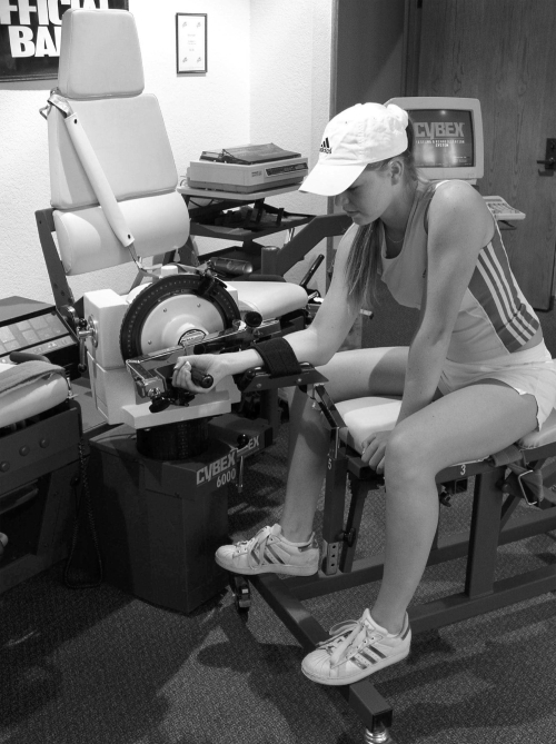
A calibrated Cybex 6000 isokinetic dynamometer (Cybex Inc) was used for all testing. For testing the movement pattern of wrist flexion and extension, the dynamometer input axis was aligned with the diagonal axis of the distal radius and ulna. Subjects were seated in a straddled fashion on the upper body testing table and placed their forearm in a supinated (palm up) position in the forearm “V” pad for stabilisation throughout testing (fig 1). A strap was used to stabilise the forearm, along with manual stabilisation from the examiner during testing to minimise the contribution of elbow flexion and extension during wrist flexion/extension testing.8,9 Range of motion stops were applied to ensure that all subjects used identical ranges of motion bilaterally for the development of the descriptive profile. The range of motion used in this testing protocol consisted of 0–55° of wrist flexion and 0–35° of wrist extension.8 Testing began in full wrist flexion. Four gradient submaximal repetitions of wrist extension and flexion were carried out before actual data collection. Five maximal repetitions of wrist extension/flexion were used for data generation and analysis. Each subject was tested at speeds of 90°/s and 210°/s. The test speed was not randomised to enhance the quality of the data acquisition.10 Subjects were tested at 90°/s first, followed by 210°/s. Thirty seconds' rest was given between test speeds with identical testing procedures undertaken bilaterally.
Figure 1 Isokinetic testing set‐up for wrist flexion/extension with the Cybex 6000 dynamometer. Informed consent was obtained for publication of this figure.
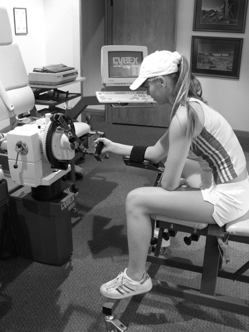
Testing for forearm pronation/supination again used the Cybex upper body testing table (fig 2). The subject was positioned in a straddled position with the forearm stabilised in the forearm “V” pad as recommended by the manufacturer. The dynamometer input axis was directly aligned between the third and fourth finger. A testing range of motion of 0–50° of pronation and 0–50° of supination was targeted using the range of motion stops on the dynamometer. Testing began in forearm supination with four gradient warm‐up repetitions followed by five maximal repetitions of forearm pronation/supination for data collection. Testing speeds of 90°/s and 210°/s were again used, with 30 seconds of rest allowed between testing speeds.
Figure 2 Isokinetic testing set‐up for forearm pronation/supination with the Cybex 6000 dynamometer. Informed consent was obtained for publication of this figure.
Peak torque and single repetition work values were recorded from the Cybex 6000. Print‐outs were generated and the values transferred to spreadsheets for data analysis. Data for descriptive profiling consisted of both peak torque and single repetition work to body weight ratios and were therefore expressed relative to body weight. Both peak torque and single repetition work values were subjected to statistical analysis. SPSS (SPSS Inc, Chicago, Illinois, USA) was used for data analysis. A repeated measures analysis of variance (ANOVA) was used to identify the main effect differences between the dominant and non‐dominant extremities in the four movement patterns tested. The α = 0.01 level of significance was used to determine whether significant differences existed between the dominant and non‐dominant arms.
Results
Tables 1 and 2 give descriptive data for wrist flexion/extension and forearm pronation/supination peak torque and single repetition work to body weight ratios for the 32 subjects. Significantly greater (p<0.01) wrist flexion and extension as well as forearm pronation peak torque and single repetition work strength values were measured on the dominant extremity compared with the contralateral non‐dominant extremity at both testing speeds in these players. Significantly less (p<0.01) forearm supination strength at both testing speeds was found on the dominant side compared with the non‐dominant side. Unilateral strength ratios for wrist extension/flexion and forearm supination/pronation are given in table 3 for descriptive purposes and were not subjected to statistical analysis.
Table 1 Peak torque/body weight ratios for wrist flexion/extension and forearm pronation/supination in elite female junior tennis players.
| Movement pattern/speed | Dominant | Non‐dominant |
|---|---|---|
| Wrist extension 90° | 3.81 (1.20)* | 3.34 (1.15) |
| Wrist extension 210° | 2.94 (1.13)* | 2.25 (1.01) |
| Wrist flexion 90° | 5.00 (1.74)* | 3.87 (1.26) |
| Wrist flexion 210° | 3.59 (1.29)* | 2.75 (1.07) |
| Forearm pronation 90° | 3.78 (1.05)* | 2.33 (1.03) |
| Forearm pronation 210° | 3.26 (0.90)* | 1.74 (0.99) |
| Forearm supination 90° | 2.15 (0.60)† | 2.22 (0.64) |
| Forearm supination 210° | 1.85 (0.66)† | 2.00 (0.55) |
Values are mean (SD).
All speeds are expressed in degrees/second. Torque is expressed in foot pounds relative to body weight in pounds.
*Significantly greater than the non‐dominant side, p<0.01.
†Significantly less than the non‐dominant side, p<0.01.
Table 2 Single repetition work/body weight ratios for wrist flexion/extension and forearm pronation/supination in elite female junior tennis players.
| Movement pattern/speed | Dominant | Non‐dominant |
|---|---|---|
| Wrist extension 90° | 3.78 (1.33)* | 3.28 (1.19) |
| Wrist extension 210° | 2.65 (1.23)* | 2.03 (0.96) |
| Wrist flexion 90° | 5.31 (1.99)* | 3.96 (1.57) |
| Wrist flexion 210° | 3.63 (1.49)* | 2.43 (1.31) |
| Forearm pronation 90° | 4.33 (1.35)* | 2.29 (1.20) |
| Forearm pronation 210° | 3.56 (1.08)* | 1.48 (0.97) |
| Forearm supination 90° | 2.14 (1.02)† | 2.55 (0.84) |
| Forearm supination 210° | 1.51 (0.75)† | 2.00 (0.73) |
Values are mean (SD).
All speeds expressed in degrees/second. Single repetition work is expressed in foot pounds relative to body weight in pounds.
*Significantly greater than the non‐dominant side, p<0.01.
†Significantly less than the non‐dominant side, p<0.01.
Table 3 Peak torque and single repetition work unilateral strength ratios in elite female junior tennis players.
| Movement ratio/speed | Dominant | Non‐dominant | |
|---|---|---|---|
| Peak torque | |||
| Wrist extension/flexion 90° | 80.96 (24.35) | 89.93 (27.15) | |
| Wrist extension/flexion 210° | 83.03 (21.84) | 87.56 (23.57) | |
| Forearm supination/pronation 90° | 65.25 (25.10) | 104.10 (32.56) | |
| Forearm supination/pronation 210° | 64.59 (24.11) | 125.62 (53.92) | |
| Single repetition work | |||
| Wrist extension/flexion 90° | 78.40 (37.43) | 87.56 (23.57) | |
| Wrist extension/flexion 210° | 77.93 (25.41) | 96.84 (44.78) | |
| Forearm supination/pronation 90° | 59.59 (33.70) | 127.59 (65.90) | |
| Forearm supination/pronation 210° | 53.88 (27.91) | 152.74 (86.24) |
Values are mean (SD).
All speeds expressed in degrees/second. Ratios are expressed as per cent.
Discussion
These data show selective sport‐specific muscular development in the distal upper extremity of elite female junior tennis players. The data show similar dominant arm increases in flexion/extension strength at the wrist and in forearm pronation to those found in highly skilled adult tennis players and in professional baseball pitchers.8,11
Morris et al12 has outlined the EMG activity of the wrist and forearm musculature during the tennis serve and ground strokes. They report high levels of muscular activity during the acceleration phase of the ground stroke as well as during late cocking and acceleration of the service motion in the wrist flexors and extensors and the forearm pronators. It is important to note that while research has demonstrated high levels of muscular activation in these muscles, they are not used as primary power sources.2,7,13 The use of these segments as part of the kinetic chain is acknowledged, however, and estimates in published reports show that as much as 54% of the power derived in the tennis service motion comes from the lower extremities and trunk.13,14
Repetitive loading of the wrist and forearm musculature can result in the development of humeral epicondylitis, and in the adolescent and prepubescent athlete, growth plate injury at the medial epicondyle.6,11 Preventative evaluation of the elbow, forearm, and wrist often consists of the measurement of joint range of motion and estimation of muscular strength through the use of a static hand grip measurement with a dynamometer.7,11 Isokinetic testing can be used to measure wrist and forearm strength dynamically and provide a more detailed estimate of muscular strength and agonist/antagonist muscle balance.9,15 The data presented in this study can serve to provide a descriptive profile of distal upper extremity strength in these elite tennis players, with the interpretation guiding both sports medicine and strength and conditioning professionals in the development of sport‐specific training programmes for performance enhancement.
Additionally, these data can be used during the rehabilitation of the injured tennis player. The data generated clearly indicate that greater dominant extremity wrist and forearm strength is present in the healthy player, and a failure to return increased dominant arm wrist flexion/extension and forearm pronation strength levels to an elite player following elbow or wrist injury would represent incomplete rehabilitation. Returning the elite player to a level of muscular strength actually exceeding the contralateral extremity in these sport‐specific patterns is indicated to ensure the full return of dynamic muscular stabilisation.
Previous research has identified a pattern of upper extremity dominance in the internal rotators,8,16,17,18 shoulder extensors,8 and elbow extensors19 with no difference between extremities in shoulder external rotation.8,16,17,18 In contrast, isokinetic descriptive studies for the lower extremity muscle groups have shown bilateral symmetry in elite players.20 This study parallels the findings reported by Ellenbecker8 in older, more mature players, which showed significantly greater wrist flexion/extension and forearm pronation strength on the player's dominant side. It is evident that these sport‐specific patterns of muscular development occur during a player's developing years, and that monitoring of these important strength levels is indicated to ensure proper development and injury prevention.
Conclusion
Isokinetic testing of wrist flexion/extension and forearm pronation/supination has identified significantly greater dominant arm wrist flexion and extension strength and forearm pronation strength in healthy elite female junior tennis players. The results of this study can be used to aid in the interpretation of isokinetic strength tests for both performance enhancement and injury prevention and rehabilitation of elite junior tennis players.
What is already known on this topic
Sport‐specific muscular adaptations of the upper extremity have been reported in elite junior tennis players
Shoulder internal rotation, shoulder extension, and elbow extension have all been shown to be significantly stronger in the dominant extremity in such players
Significantly greater wrist flexion/extension and forearm pronation strength has been measured in the dominant extremity of adult tennis players who are highly skilled
What this study adds
Sport‐specific adaptations in distal upper extremity strength occur in elite junior tennis players
These adaptations are important for evaluation for performance enhancement and injury prevention, as well as for rehabilitation of the injured tennis player
Footnotes
Informed consent was obtained for publication of figures 1 and 2.
Competing interests: none declared
References
- 1.Safran M R, Hutchinson M R, Moss R.et al A comparison of injuries in elite boys and girls tennis players. Transactions of the 9th Annual Meeting of the Society of Tennis Medicine and Science. California: Indian Wells, 1999
- 2.Roetert E P, Groppel J.World class tennis technique. Champaign IL: Human Kinetics Publishers, 2001
- 3.Marshall R N, Noffal G J, Legnani G.Simluation of the tennis serve. Factors affecting elbow torques related to medial epicondylitis. Paris: Biomechanics ISB, 1993
- 4.Dillman C J, Schultheis J M, Hintermeister R A.et al What do we know about body mechanics involved in tennis skills? In: Krahl H, Pieper H, Kibler WB, et al, editors. Tennis: sports medicine and science . Auglage: Society for Tennis Medicine and Science,6–11.
- 5.Ellenbecker T S. Rehabilitation of shoulder and elbow injuries in tennis players. Clin Sports Med 19951487–110. [PubMed] [Google Scholar]
- 6.Nirschl R P, Sobel J. Conservative treatment of tennis elbow. Phys Sportsmed 1981943–54. [DOI] [PubMed] [Google Scholar]
- 7.Roetert E P, Ellenbecker T S.Complete conditioning for tennis. Champaign IL: Human Kinetics Publishers, 1998
- 8.Ellenbecker T S. A total arm strength isokinetic profile of highly skilled tennis players. Isokinet Exerc Sci 199119–21. [Google Scholar]
- 9.Davies G J. A compendium of isokinetics in clinical usage. LaCrosse, WI: S & S Publishers, 1992
- 10.Wilhite M R, Cohen E R, Wilhite S C. Reliability of concentric and eccentric measurements of quadriceps performance using the Kin‐Com dynamometer. The effects of testing order for three different speeds. J Orthop Sports Phys Ther 199215175–182. [DOI] [PubMed] [Google Scholar]
- 11.Ellenbecker T S, Mattalino A J.The elbow in sport. Champaign IL: Human Kinetics Publishers, 1997
- 12.Morris M, Jobe F W, Perry J.et al Electromyographic analysis of elbow function in tennis players. Am J Sports Med 198917241–247. [DOI] [PubMed] [Google Scholar]
- 13.Kibler W B. Clinical biomechanics of the elbow in tennis: implications for evaluation and diagnosis. Med Sci Sports Exerc 1994261203–1206. [PubMed] [Google Scholar]
- 14.Ellenbecker T S, Roetert E P. Velocity of a tennis serve and measurement of isokinetic muscular performance. Brief review and comment. Percept Motor Skills 2004981368–1370. [DOI] [PubMed] [Google Scholar]
- 15.Brown L E.Isokinetics in human performance. Champaign, IL: Human Kinetics Publishers, 2000
- 16.Chandler T J, Kibler W B, Stracener E C.et al Shoulder strength, power, and endurance in college tennis players. Am J Sports Med 199220455–458. [DOI] [PubMed] [Google Scholar]
- 17.Ellenbecker T S. Shoulder internal and external rotation strength and range of motion in highly skilled tennis players. Isokinet Exerc Sci 199221–8. [Google Scholar]
- 18.Ellenbecker T S, Roetert E P. Age specific isokinetic glenohumeral internal and external rotation strength in elite junior tennis players. J Sci Med Sport 2003663–70. [DOI] [PubMed] [Google Scholar]
- 19.Ellenbecker T S, Roetert E P. Isokinetic profile of elbow flexion and extension strength in elite junior tennis players. J Orthop Sports Phys Ther 20033379–84. [DOI] [PubMed] [Google Scholar]
- 20.Ellenbecker T S, Roetert E P. Concentric isokinetic quadriceps and hamstring strength in elite junior tennis players. Isokinet Exerc Sci 199553–6. [Google Scholar]


