

Abstract
Starting with 5-iodo-2'-deoxyuridine, a series of 5-alkynyl-2'-deoxyuridines (with n-propyl, cyclopropyl, 1-hydroxycyclohexyl, p-tolyl, p-tert-butylphenyl, p-pentylphenyl, and trimethylsilyl alkyne substituents) have been synthesized via the palladium-catalyzed (Sonogashira) coupling reaction followed by a simplified isolation protocol (76–94% yield). The cytotoxic activity of modified nucleosides against MCF-7 and MDA-MB-231 human breast cancer cells has been determined in vitro. 5-Ethynyl-2'-deoxyuridine, the only nucleoside in the series containing a terminal acetylene, is the most potent inhibitor with IC50 (μM) 0.4 ± 0.3 for MCF-7 and 4.4 ± 0.4 for MDA-MB-231.
Keywords: Nucleosides, Alkynes, 5-Alkynyl-2'-deoxyuridines, Antitumor chemotherapy, Anticancer, Anti-proliferative agents, Human breast cancer cells, MCF-7, MDA-MB-231
1. Introduction
Modified nucleosides have acquired an important role as therapeutics.1–3 Cytotoxic nucleoside analogues were among the first chemotherapeutic agents to be introduced for the medical treatment of cancer.3 This family of compounds has grown to include a variety of purine and pyrimidine nucleoside derivatives with activity in both solid tumors and hematological malignancies. These agents behave as antimetabolites, compete with physiologic nucleosides, and consequently, interact with a large number of intracellular targets to induce cytotoxicity.4
Alkynyl modifications of uridines have been explored for diverse biochemical studies. A common approach is based upon the attachment of the active units via the ethynyl arm at position C-5 of the uracil base. Altered in such a way uridine nucleosides facilitate blue-to-red energy transfer (absorption-fluorescence emission), as well as when incorporated into oligonucleotides.5 1-Ethynylpyrene derivatives help to organize the helical-stacked arrangements along the major groove of duplex DNA.6 Alkynyl-modified dU:dA base pairs may stabilize duplexes and reinforce helices,7 or serve as tools for charge transfer excited-state dynamics DNA duplex studies,8 to name a few.
Medicinal-related activity of various 5-alkynyl pyrimidine analogues has been investigated for some time, 9,10 including their oligonucleotides.11 Recently potent inhibitory properties of mycobacteria were reported.12a Although broad antiviral studies have been conducted, anticancer investigations of 5-alkynyl uridine analogues have been limited.1,12b
Various synthetic routes to 5-alkynyl uridines have been reported,13,14 including a reaction of 5-trifluoromethanesulfonyloxy pyrimidine nucleosides with alkynes.15 The classical, effective, and most frequently used synthesis of 5-alkynyl uridines involves palladium-catalyzed (Sonogashira)16 coupling of unprotected 5-iodo-2'-deoxyuridine (I).14,17–19 This reaction is carried out under precisely controlled conditions, since an elevated temperature and the presence of metal/amine lead to a cyclization reaction yielding 2,3-dihydrofuro-[2,3-d]pyrimidin-2-one nucleoside (furopyrimidine).13c,20 Following the reported effect of additives,21 we elaborated a room temperature coupling procedure that includes triphenylphosphine and tetrabutylammonium iodide.22,23 Currently, for a somewhat larger scale, due to material economy and purification effectiveness a procedure with no additives was applied.
2. Results and discussion
2.1. Synthesis and characterization
The series of known and new 5-alkynyl-2'-deoxyuridines (2) was synthesized via Sonogashira coupling, as visualized in Scheme 1 (4.0 equiv of acetylene, 0.1 equiv of Pd(PPh3)4 and Cul, 2.0 equiv of Et3N, DMF, 55 °C, 17 h). To provide a starting position for more detailed structure-activity relationship studies, diverse alkyne substituents R were selected, which include a linear alkyl chain (2a, R = n-propyl),13c cycloalkyl (2b, R = cyclopropyl),23 tertiary cycloalkanol (2c, R = 1-hydroxy-cyclohexyl), aromatic ring substituted with linear (2d,e R = p-tolyl,22 p-pentylphenyl) and branched alkyl groups (2f, R = p-tert-pentylphenyl),23 and silicon (2g, R = trimethylsilyl).15 The nucleoside containing a terminal alkyne (2h, R = H)24 was prepared by desilylation of 2g with K2CO3/MeOH13c,25 or TBAF/THF.15
Scheme 1.
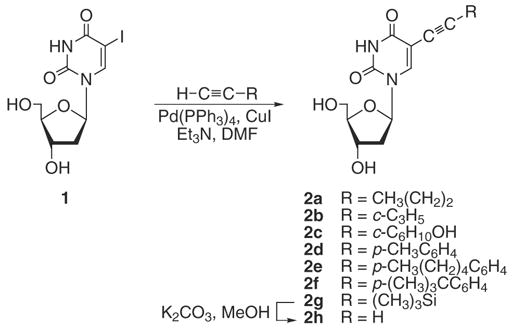
Synthesis of 5-alkynyl-2'-deoxyuridines 2a–h.
Practical preparative comments are shared below. TLC observation of the reaction under a UV 254 nm lamp (to determine completion) can be misleading due to the low fluorescence of the 5-iodo-2'-deoxyuridine 1. Careful monitoring by 'H NMR (H-6 signal) helped to establish reaction progress and revealed that conversion of 1 to its alkynyl derivatives 2 proceeded essentially in a quantitative manner. 5-Alkynyl uridines with unprotected hydroxyl groups are reportedly difficult to purify,26 thus the yields are moderate.27 Separation from ammonium salt is usually accomplished by protection/chromatography/deprotection,10a,26 quenching with ion-exchange resin,27,28 or with careful column chromatography. Thus, a convenient isolation protocol has significant practical value.29 The alkynyl nucleosides 2a–g were separated with good analytical purity from spent and unused reagents through a workup procedure that includes two subsequent crystallizations. After thorough removal of DMF, a post-reaction mixture was treated with methanol, to precipitate phosphine/palladium spent reagents.30 Subsequently, after drying under vacuum, addition of chloroform precipitated the nucleoside whereas the ammonium salt (Et3NHI) remained in solution.31 This protocol provided material without any detectable triethylammonium- or triphenylphosphine-derived impurities when analyzed by 1H NMR. This workup procedure allows for the synthesis to be easily carried out on a somewhat larger scale (0.4–5 g). This procedure is dependent on the thorough removal of DMF, as this will effect a clean precipitation of the product. The yields are summarized in Table 1.
Table 1.
Preparation and cytotoxic activity of 5-alkynyl-2'-deoxyuridines 2a–h
| Compound (ref.) | R | Yield (%) | IC50 (μM) |
|
|---|---|---|---|---|
| MCF-7 | MDA-MB-231 | |||
| 2a13c | CH3CH2CH2- | 87(53)a | >50 | >50 |
| 2b | 80 | >50 | >50 | |
| 2c |  |
94 | >50 | >50 |
| 2d | 88 | >50 | >50 | |
| 2e | 76 | 37.0 ± 6.8 | 29.2 ± 5.0 | |
| 2f |  |
94(72)a | 9.5 ±1.4 | >50 |
| 2g15 | (CH3)3Si | 92(28)a | 1.6 ± 1.3 | 4.2 ± 1.5 |
| 2h24 | H– | —b | 0.4 ± 0.3 | 4.4 ± 0.4 |
| Cisplatinc | — | — | 2.0 ± 0.3 | 4.0 ± 1.5 |
| 5-Fluorouracilc | — | — | 4.8 ± 0.6 | 9.6 ± 0.3 |
Yield includes second crop obtained with the aid of column chromatography; yield from first crystallization in parentheses.
Prepared from 2g by two independent desilylation methods.
Reference.
The alkynyl nucleosides 2b–f were characterized by 1H and 13C NMR, IR, MS, and UV-vis spectroscopy. The characteristic NMR (DMSO-d6) signals for 2b–f include the 1H H-6 signal (8.36–8.11 ppm), and 13C signals of C-5 (99.6-98.3) and C≡C (97.5-68.6 ppm). 13C NMR C-l' and C-4' signals were distinguished using INEPT-based HETCOR correlation spectra. Comparison of 1H and 13C NMR signals for 2a–h is provided in the Supplementary data. The mass spectra for 2b–f exhibited intense molecular ions. Alkynyl uridines 2a,c–h gave correct elemental analyses, mostly as hydrates. The representative 1H and 13C NMR spectra, given in the Supplementary data, illustrate the organic homogeneity of the compounds isolated without column chromatography.
2.2. Cytotoxicity
After completion of synthesis and characterization, the nucleosides were investigated for their antitumor activity in vitro using two human breast cancer cultures MCF-7 and MDA-MB-231 that are of highly different phenotypes: hormone-sensitive and hormone-insensitive, respectively. To reduce the presence of trace impurities such as metals, all samples were additionally passed through a silica gel pad before submission for activity evaluations.32 The experiments were carried out according to a modified established microtiter assay.33,34 Cisplatin and 5-fluorouracil were used as references.
Testing using the method described above indicated high activity of 5-ethynyl-2'-deoxyuridine 2h (R=H). Observed IC50 were 0.4 μM (±0.3) for MCF-7 and 4.4 μM (±0.4) for MDA-MB-231, exceeding or matching the IC50 for cisplatin or 5-fluorouracil. These results, which stand out, can be attributed to the presence of the terminal alkynyl functional group, and were consistent for two samples of 2h prepared independently from 2g with the use of different deprotection reagents. However, internal aromatic-substituted alkyne p-tolylethynyl-2'-deoxyuridine 2d also showed high potency with MCF-7 (IC50 0.9 μM (±0.2)), but no detectable potency with MDA-MB-231. Replacement of the aromatic ring system with a trimethylsilyl group also afforded a highly active compound 2g which matches the activity of 2h (IC50 1.6 μM (±1.3)/4.2 μM (±1.5) for MCF-7/MDA-MB-231). Longer (more lipophilic) or more bulky aliphatic substituents in the aromatic ring led to significantly lower activity such as for compounds 2e or 2f. Usually less lipophilic nucleosides tend to be more active, whereas more lipophilic derivatives exhibit enhanced antiviral properties. It can be concluded in general that the compounds containing internal alkyne structures were of lower activity than 2h and MCF-7 cells were more sensitive to the presence of the nucleosides than MDA-MB-231 cells. Within the group of internal alkynes those containing an aromatic ring or a trimethylsilyl group attached to the alkyne (2d–g) gave better results than those with aliphatic substituents (2a–c), which were devoid of activity up to the highest concentration used. The results are summarized in Table 1.
The precise mechanism of action of these compounds is not documented. However, it could be anticipated that the agents acting as DNA polymerase inhibitors might affect cellular DNA synthesis. Enzymes that are involved in DNA replication and repair have been shown to serve as targets for anticancer nucleosides such as the deoxycytidine analogue gemcitabine. Other possible modes of action involve an interaction with deoxynucleoside kinases, which are required for activation of deoxynucleosides as antiviral or anticancer drugs, or a behavior as antimetabolites and a subsequent incorporation into the DNA. Furthermore, the inhibition of thymidylate synthase, which catalyzes the reductive methylation of deoxyuridine monophosphate into deoxythymidine monophosphate, must be taken into account based on the fact that this is one of the major biochemical effects of the cytostatic agent 5-fluorouracil.3,35–38 The fact that the target compounds displayed higher activity in the MCF-7 cell line than in the MDA-MB-231 cell line indicates the presence of a specific molecular target or carrier system for these agents in the MCF-7 cell line.
3. Conclusion
In conclusion, an effective purification protocol for 5-alkynyl-2'-deoxyuridines 2 that avoids the use of column chromatography, ion exchange chromatography, or protection/deprotection steps was established. The series of modified nucleosides 2 was tested for antiproliferation properties against MCF-7 and MDA-MB-231 human mammary carcinoma cell lines. A terminal alkyne, 5-ethynyl-2'-deoxyuridine 2h, showed the highest potency exceeding cisplatin and 5-fluorouracil. Further testing of 2h toward other cancer cell lines would be of interest.
We think that the structure-activity relationship studies that will involve incorporation of CH2 spacers between the uracil base and alkyne, to follow an established active fragment of CH2C≡CH (as the Co2(CO)6 complex),39 may further improve the activity.
The reported nucleosides are also converted into their organometallic analogues, and the synthesis and cytotoxic activity of dicobalt hexacarbonyl derivatives will be reported on in due course. Moreover, more extensive structure-activity relationship studies concerning different substituents in the aromatic ring as well as more detailed investigations on the molecular drug targets are envisaged.
4. Experimental
4.1. General
Commercial chemicals were treated as follows: DMF distilled from CaH2 and degassed (freeze and thaw) three times prior to use; Et3N distilled from P2O5. 5-Iodo-2'-deoxyuridine (Berry&Associates), 1-pentyne, cyclopropylacetylene, 1-ethynyl-l-cyclohexanol, 4-ethynyl-toluene, 4-(tert-butyl)phenylacetylene, and (trimethylsilyl)acetylene (GFS), Pd(PPh3)4 (Pressure Chemical), l-ethynyl-4-pentylbenzene and Cul 99.999% (Aldrich), silica gel (J.T. Baker, 60–200 mesh), and TLC plates Analtech GF, cat. no. 2521 or Merck 60, cat. No. 5715, used as received. Other materials not listed were used as received.
IR and UV-vis spectra were recorded on a Bio-Rad FTS-175C and Gary 50 spectrometers. UV-vis absorbances are in nm (ε, M−1cm−1). NMR spectra were obtained on a Bruker Avance DPX-200 spectrometer (1H of 200 MHz and 13C of 50 MHz). Chemical shifts (δ) values are in ppm and coupling constants (J) values are in Hz. Mass spectra were recorded on a Finnigan MAT 95 or Micromass ZQ instruments; m/z for most intense peak of isotope envelope. Microanalyses were conducted by Atlantic Microlab.
4.2. 5-Alkynyl-2'-deoxyuridines 2a–f; general procedure (1 mmol scale)
A Schlenk flask was charged with 5-iodo-2'-deoxyuridine (1.18 mmol), Pd(PPh3)4 (0.118 mmol), Cul (0.118 mmol), DMF (5 mL), Et3N (2.4 mmol), and acetylene (4.7 mmol). The yellow mixture was stirred at 55 °C for 17 h (oil bath; 1H NMR showed complete conversion of the substrate). Solvent was removed (by oil pump vacuum) and the residue was extracted (ultrasonicated) with MeOH (6 mL). The solid was filtered off and washed with MeOH (3 × 3 mL). The solvent was removed from combined filtrates by rotary evaporation. The residue (yellow oil) was kept under oil pump vacuum for 2 h, dissolved (ultrasonicated) in CHC13 (5 mL),31 and kept at −6 °C (freezer) for 12 h. The precipitate was filtered off and washed with cold CHC13 (3×3 mL). The product was dried by oil pump vacuum for 6 h to give 2.
4.2.1. 5-Pent-l-yn-l-yl-2'-deoxyuridine (2a).13c
From 5-iodo-2'-deoxyuridine (l.00g, 2.82 mmol), Pd(PPh3)4 (0.326 g, 0.282 mmol), Cul (0.0539 g, 0.282 mmol), DMF (5mL), Et3N (0.82 mL, 5.65 mmol), and 1-pentyne (1.40 mL, 14.1 mmol). The yellow mixture was stirred at 40 °C for 38 h. Crystallization from CHC13 gave 2a as a white powder (0.440 g, 1.50 mmol, 53%). A second crop, obtained after concentration and silica gel column chromatography (15 × 2.5 cm; CHC13 → CHC13/MeOH 95:5), increased the yield to a total of 0.723 g (2.46 mmol, 87%).
NMR (DMSO-d6): 1H data matched those reported earlier.13c 13C 161.8 (d, j = 9.4, C-4), 149.5 (d, j = 8.1, C-2), 142.7 (d, J = 182.1, C-6), 99.1 (s, C-5), 93.1 (m, dU-C≡C, 87.5 (d, J = 144.9, C-4'), 84.6 (d, J = 163.4, C-l'), 73.0 (d, J =4.4, dU-C≡C), 70.2 (d, J = 148.4, C-3'), 61.0 (t, J = 140.3, C-5'), 40.0 (t, J = 134.0, C-2'), 21.7 (t, J = 130.0, C-2"), 20.8 (t, J = 130.8, C-l"), 13.4 (q, J = 124.5, C-3").
4.2.2. 5-(Cyclopropylethynyl)-2'-deoxyuridine (2b)
From 5-iodo-2'-deoxyuridine (0.418 g, 1.18 mmol), Pd(PPh3)4 (0.1364 g, 0.1180 mmol), Cul (0.0225 g, 0.118 mmol), DMF (5 mL), Et3N (0.34 mL, 2.4 mmol), and cyclopropylacetylene (0.40 mL, 4.7 mmol). The yellow mixture was stirred at 55 °C for 17h. White powder (0.277 g, 0.948 mmol, 80%). Calcd for Ci4H16N2O5·0.75H2O: C, 54.99; H, 5.77. Found: C, 54.64; H, 5.31. IR (ν, cm−1, KBr) 3414 br s, 2235 w, 1709 vs, 1689 vs, 1630m. UV-vis (CH3OH, 4.7 × l0−5 M) 226 sh (18000), 296 (15000). MS (ES+, KC1, MeOH) 623 ((2M+K)+, 61%), 607 ((2M+Na)+, 26%), 585 ((2M+H)+, 8%), 331 ((M+K)+, 100%), 315 ((M+Na)+, 19%), 293 ((M+H)+, 15%); no other peaks above 200 of >4%.
NMR (DMSO-d6): 'H 11.55 (s, 1H, N-3), 8.11 (s, 1H, H-6), 6.10 (t, J = 5.6, 1H, H-l'), 5.23 (d, J = 4.2, 1H, OH-3'), 5.10 (t, J = 4.9, 1H, OH-5'), 4.29-4.16 (m, 1H, H-3'), 3.83-3.74 (m, 1H, H-4'), 3.68-3.48 (m, 2H, H-5'), 2.18-2.03 (m, 2H, H-2'), 1.55-1.40 (m 1H, H-l"), 0.92-0.59 (2m, 2 × 2H, H-2" and H-3"); "C 162.5 (d, J = 9.2, C-4), 150.1 (d, J=6.2, C-2), 143.6 (d, J = 183.7, C-6), 99.6 (d, J = 1.9, C-5), 96.9 (d, J = 2.6, dU-C≡C), 88.2 (d, J = 146.1, C-4'), 85.2 (d, J = 163.3, C-l'), 70.8 (d, J = 148.8, C-3'), 68.6 (d, J = 43, dU-C≡C), 61.6 (t, J = 140.3, C-5'), 40.1 (t, J = 132.9, C-2'), 9.0 (t, J = 164.3, C-2" and C-3"), 0.5 (d, J = 168.7, C-l").
4.2.3. 5-[(1-Hydroxycyclohexyl)ethynyl]-2'-deoxyuridine(2c)
From 5-iodo-2'-deoxyuridine (0.418 g, 1.18 mmol), Pd(PPh3)4 (0.1364g, 0.1180 mmol), Cul (0.0225 g, 0.118 mmol), DMF (5 mL), Et3N (0.34 mL, 2.4 mmol), and 1-ethynyl-l-cyclohexanol (0.59 mL, 4.7 mmol). The yellow mixture was stirred at 50 °C for 7 h. White powder (0.390 g, 1.11 mmol, 94%). Calcd for Ci7H22N206-l/3H20: C, 57.29; H, 6.41. Found: C, 57.64; H, 6.29. IR (ν, cm−1, KBr) 3356 br s, 2221 vw, 1710 vs, 1674 s. UV-vis (MeOH, 3.4×10⁻⁵M) 232 (7400), 291 (9400). MS (ES+, MeOH) 739 ((2M+K)+, 15%), 723 ((2M+Na)+, 100%), 389 ((M+K)+, 23%), 373 ((M+Na)+, 97%); no other peaks above 100 of >10%.
NMR (DMSO-d6): 'H 11.55 (s, 1H, N-3), 8.14 (s, 1H, H-6), 6.10 (t, y=6.5, 1H, H-l'), 5.32 (s, 1H, HO-C6H10), 5.22 (d, J = 4.2, 1H, OH-3'), 5.07 (t, J = 4.7, 1H, OH-5'), 4.30-4.16 (m, 1H, H-3'), 3.83-3.74 (m, 1H, H-4'), 3.64-3.52 (m, 2H, H-5'), 2.20-2.05 (m, 2H, H-2'), 1.87-1.68 (m, 2H, c-C6H10), 1.68-1.33 (m, 7H, c-C6H10), 1.30-1.05 (m, 1H, c-C6H10); 13C 161.6 (d, J = 9.4, C-4), 149.5 (d, J=8.0, C-2), 143.0 (d, J = 183.0, C-6), 98.6 (d, J = 4.8, C-5), 97.5 (s, dU-C≡C), 87.6 (d, J = 145.0, C-4'), 84.7 (d, J = 168.3, C-l'), 75.3 (d, J = 4.5, dU-C≡C), 70.1 (d, J = 148.4, C-3'), 67.1 (s, C-l"), 60.9 (t, J = 140.6, C-5'), 40.2 (C-2'),40 39.7 (C-2"),40 24.9 (t, J = 120.2, C-4"), 22.7 (t, J = 124.6, C-3").
4.2.4. 5-p-Tolylethynyl-2'-deoxyuridine (2d)
From 5-iodo-2'-deoxyuridine (3.000 g, 8.472 mmol), Pd(PPh3)4 (0.9790 g, 0.8472 mmol), Cul (0.1618 g, 0.8472 mmol), DMF (15 mL), Et3N (2.45 mL, 16.9 mmol), and 4-ethynyltoluene (3.65 mL, 28.8 mmol). The yellow mixture was stirred at 40 °C for 20 h. White powder (2.56 g, 7.48 mmol, 88%). Calcd for C18H18N2O5: C, 63.15; H, 5.30. Found: C, 62.73; H, 5.58. IR (ν, cm−1, KBr) 3433 br s, 3190 m br, 2220 vw, 1722 vs, 1669 vs, 1652 vs, 1618 m, 817 m. UV-vis (CH3OH, 5.5 × 10⁻⁵ M) 252 (9500), 266 (11000), 280 (9900), 311 (12000). MS (FAB, gly, positive) 435 ((M+gly+l)+, 10%), 343 ((M+l)+, 42%), 227 ((M–C≡CC6H4CH3)+, 100%); no other peaks above 200 of >6%; (negative) 433 ((M+gly-1)−, 13%), 341 ((M−l)−, 65%), 227 ((M–C≡CC6H4CH3–2)−, 100%); no other peaks above 200 of >9%.
NMR (DMSO-d6):41 1H 11.68 (s, 1H, N-3), 8.33 (s, 1H, H-6), 7.35 (d, J =8.1, 2H, o-C6H4C≡C), 7.21 (d, J = 8.1, 2H, m-C6H4C≡C), 6.13 (t, J = 6.5, 1H, HI'), 5.27 (d, J = 3.8, 1H, OH-3'), 5.17 (t, J = 4.3, 1H, OH-5'), 4.26 (q, J = 4.2, 1H, H-3'), 3.81 (q, J = 3.6, 1H, H-4'), 3.71-3.52 (m, 2H, H-5'), 2.33 (s, 3H, CH3), 2.16 (t, J=5.6, 2H, H-2'); 13C 162.1 (d,J = 9.3, C-4), 149.9 (d, J = 7.5, C-2), 143.6 (dd, J = 181.2, 2.2, C-6), 138.3 (m, p-C6H4C≡C), 131.1 (dd, J = 162.3, 6.2, o-C6H4C≡C), 129.4 (dq, J = 159.6, 5.4, m-C6H4C≡C), 119.5 (t, J=8.2, i-C6H4C≡C), 98.3 (s, C-5), 91.8 (t, J = 5.4, dU-C≡C), 87.6 (d, J = 147.9, C-4'), 84.8 (d, J = 170.8, C-l'), 82.1 (d, J=5.5, dU-C≡C), 69.9 (d, J = 148.0, C-3'), 60.9 (t, J = 140.4, C-5'), 40.2 (t, J = 133.5, C-2'), 21.1 (qt, J = 126.2, 4.3, CH3).
4.2.5. 5-[(p-Pentylphenyl)ethynyl]-2'-deoxyuridine (2e)
From 5-iodo-2'-deoxyuridine (5.01 g, 14.1 mmol), Pd(PPh3)4 (1.63 g, 1.41 mmol), Cul (0.269 g, 1.41 mmol), DMF (30 mL), Et3N (4.00 mL, 28.0 mmol), and l-ethynyl-4-pentylbenzene (6.32 mL, 32.5 mmol). The yellow mixture was stirred at 40 °C for 35 h. White powder (4.29 g, 10.8 mmol, 76%). Calcd for C22H26N2O5: C, 66.32; H, 6.58. Found: C, 66.20; H, 6.57. IR (ν, cm−1, KBr) 3418 br s, 2224 vw, 1699 vs, 1678 vs. UV-vis (CH3OH, 2.8 × 10⁻⁵ M) 253 (18000), 266 (21000), 281 (18000), 311 (24000). MS (ES+, MeOH) 835 ((2M+K)+, 15%), 819 ((2M+Na)+, 100%), 797 ((2M+H)+, 11%), 453 (unassigned, 51%), 437 ((M+K)+, 32%), 421 ((M+Na)+, 80%), 399 ((M+H)+, 8%); no other peaks above 300 of >7%.
NMR (DMSO-d6,):41 1H 11.65 (s, 1H, N-3), 8.36 (s, 1H, H-6), 7.32 (d, J=8.1, 2H, o-C6H4C≡C), 7.14 (d, J = 8.1, 2H, m-C6H4C≡C), 6.13 (t, J = 6.3, 1H, H-l'), 5.24 (d, J = 4.2, 1H, OH-3'), 5.14 (t, J = 4.5, 1H, OH-5'), 4.26 (p, J = 3.5, 1H, H-3'), 3.86-3.75 (m, 1H, H-4'), 3.71-3.51 (m, 2H, H-5'), 2.50 (t, J = 7.2, 2H, H-l"), 2.14 (t, J = 5.1, 2H, H-2'), 1.48 (p, J = 6.7, 2H, H-2"), 1.30-1.10 (m, 4H, H-3", H-4"), 0.77 (t, J = 6.3, 3H, H-5"); 13C 161.4 (d, J = 9.3, C-4), 149.3 (d, J = 8.0, C-2), 143.4 (d, J = 184.3, C-6), 143.1 (s, p-C6H4C≡C), 131.0 (dd, J = 162.3, 6.1, o-C6H4C≡C), 128.4 (dd, J = 159.2, 5.4, m-C6H4C≡C), 119.5 (t, J=8.2, i-C6H4C≡C), 98.4 (s, C-5), 91.9 (t, J = 4.7, dU-C≡C), 87.5 (d, J = 147.6, C-4'), 84.7 (d, J = 169.5, C-l'), 81.6 (d, J = 5.5, dU-C≡C), 70.0 (d, J = 148.5, C-3'), 60.8 (t, J = 140.3, C-5'), 40.2 (t, J = 133.7, C-2'), 34.9 (t, J = 113.2, C-l"), 30.7 (t, J = 120.3, C-3"), 30.3 (t, J = 120.1, C-2"), 21.8 (t, J = 124.3, C-4"), 13.7 (q, J = 123.2, C-5").
4.2.6. 5-[(4-tert-Butylphenyl)ethynyl]-2'-deoxyuridine (2f)
From 5-iodo-2'-deoxyuridine (l.00g, 2.82 mmol), Pd(PPh3)4 (0.326 g, 0.282 mmol), Cul (0.0539 g, 0.282 mmol), DMF (5 mL), Et3N (0.82 mL, 5.65 mmol), and 4-(t-butyl)phenylacetylene (1.5mL, 8.47 mmol). The yellow mixture was stirred at 40 °C for 25 h. Crystallization from CHC13 gave 2f as a white powder (0.780 g, 2.03 mmol, 72%). A second crop, obtained after concentration and silica gel column chromatography (15 × 2.5 cm; CHC13 → CHCl3/MeOH 95:5), increased the yield to a total of 1.02 g (2.65 mmol, 94%). Calcd for C21H24N2O5·1.5H2O: C, 60.64; H, 5.82. Found: C, 60.71; H, 5.90. IR (ν, cm−1, KBr) 3429 br s, 2227 vw, 1701 vs br, 835 w. UV-vis (CH3OH, 4.9 × 10⁻⁵M) 253 (13000), 266 (15000), 280 (13000), 309 (16000). MS (MeOH) 791 ((2M+Na)+, 78%), 407 ((M+Na)+, 100%), 385 ((M+H )+, 12%); no other peaks of >12%.
NMR (DMSO-d6):41 1H 11.69 (s, 1H, N-3), 8.35 (s, 1H, H-6), 7.41 (s, 4H, C6H4), 6.13 (t, J = 6.3, 1H, H-l'), 5.26 (d, J = 3.7, 1H, OH-3'), 5.16 (t, J = 4.4, 1H, OH-5'), 4.33-4.17 (m, 1H, H-3'), 3.86-3.74 (m, 1H, H-4'), 3.72-3.51 (m, 2H, H-5'), 2.17 (t, 2H, H-2'), 1.28 (s, 9H, C(CH3)3); 13C 161.5 (d, J = 9.3, C-4), 151.4 (d, J = 3.8, p-C6H4C≡C), 149.4 (d, J = 8.1, C-2), 143.7 (dd, J = 184.6, 2.0, C-6), 130.9 (dd, J = 162.0, 4.0, o-C6H4C≡C), 125.6 (dd, J = 158.9, 3.9, m-C6H4C≡C), 119.5 (t, J=6.1, i-C6H4C≡C), 98.3 (s, C-5), 91.9 (s, dU-C≡C), 87.6 (d, J = 147.2, C-4'), 84.8 (d, J = 170.6, C-l'), 81.8 (d, J = 5.6, dU-C≡C), 70.0 (d, J = 149.9, C-3'), 60.8 (t, J = 140.2, C-5'), 40.2 (t, J = 133.0, C-2'), 34.6 (s, C(CH3)3), 30.9 (qt, J = 125.8, 4.4, C(CH3)3).
4.2.7. 5-[(Trimethylsilyl)ethynyl]-2'-deoxyuridine (2g).15
From 5-iodo-2'-deoxyuridine (3.96 g, 11.2 mmol), Pd(PPh3)4 (1.29 g, 1.12 mmol), Cul (0.213 g, 1.12 mmol), DMF (25 mL), Et3N (3.20 mL, 22.4 mmol), and (trimethylsilyl)acetylene (7.90 mL, 55.9 mmol). The yellow mixture was stirred at 55 °C for 24 h. Crystallization from CHC13 gave 2g as a white powder (1.02g, 3.16 mmol, 28%). A second crop, obtained after concentration and silica gel column chromatography (15×2.5 cm; CHC13 → CHCl3/MeOH 95:5), increased the yield to a total of 3.34 g (10.3 mmol, 92%).
NMR (DMSO-d6): 1H data matched those reported earlier.15 13C 161.5 (d, J = 9.3, C-4), 149.5 (d, J = 8.0, C-2), 144.7 (d, J = 184.2, C-6), 98.3 (s, C-5), 98.0 (d, J = 5.4, dU-C≡C), 97.0 (s, dU-C≡C), 87.6 (d, J = 147.6, C-4'), 84.8 (d, J = 169.4, C-l'), 69.9 (d, J = 140.7, C-3'), 60.8 (t, J = 140.3, C-5'), 40.2 (t, J = 133.8, C-2'), −0.03 (q, J = 99.2, Si(CH3)3).
4.3. Cytotoxicity experiments (IC50)
Nucleoside samples were additionally passed through a silica gel column, to exclude contamination with metals. The experiments were performed according to established procedures with some modifications.39a A 100 μL of 10000 cells/mL (MCF-7) or 5000 cells/mL (MDA-MB-231) was incubated in 96-well plates at 37 °C in 5% CO2/95% air atmosphere for 72 h. One plate was used for the determination of the initial cell biomass and was treated in the following way: the medium was removed, cells were fixed by a 20–30 min incubation with 100 μL glutardialdehyde solution (0.5 mL glutardialdehyde + 12.5 mL phosphate-buffered saline solution of pH 7.4), the wells were emptied, phosphate-buffered saline solution (180 μL, pH 7.4) was added, and the plate was stored at 4 °C until further treatment. In the 'experimental' plates the medium was replaced with a medium containing the drugs in graded concentrations (six replicates). After further incubation for 72 h (MDA-MB-231) or 96 h (MCF-7) these plates were treated as described above. The cell biomass was determined by crystal violet staining according to the following procedure: the phosphate-buffered saline solution (pH 7.4) was removed, 0.02 M crystal-violet solution was added (100 μL), and plates were incubated for 30 min at room temperature, washed three times with water, and incubated on a softly rocking rotary shaker with ethanol (70%, 180 μL) for a further 3–4 h. Absorption was recorded in a microplate reader at 590 nm (Flashscan Analytik Jena AG). The mean absorption of the initial cell biomass plate was subtracted from the mean absorption of each experiment and control. The corrected control was set to 100%. IC50 was determined as that concentration causing 50% inhibition of cell proliferation and calculated as a mean of at least two independent experiments.
Supplementary Material
Acknowledgments
We thank NIH (Grant CA111329) and the Oakland University Research Excellence Program in Biotechnology, for support of this research. Thomson Award for C.D.S. is acknowledged.
Footnotes
Supplementary data
Information available: Tables of comparison of 1H, 13C NMR signals for compounds 2a–h. 1H, 13C, or INEPT-based HETCOR correlation NMR spectra for compounds 2a, c–f. Supplementary data associated with this article can be found, in the online version, at 10.1016/j.bmc.2007.01.048.
References and notes
- 1.(a) Chu CK, Baker DC, editors. Nucleosides and Nucleotides as Antitumor and Antiviral Agents. Plenum Press; New York: 1993. [Google Scholar]; (b) Godefridus JP, editor. Deoxynucleoside Analogs in Cancer Therapy. Humana Press; Totowa: 2006. [Google Scholar]
- 2.(a) Ferrero M, Gotor V. Chem Rev. 2000;100:4319–4347. doi: 10.1021/cr000446y. [DOI] [PubMed] [Google Scholar]; (b) Townsend LB, editor. Chemistry of Nucleosides and Nucleotides. Vol. 3 Plenum Press; New York: 1994. [Google Scholar]
- 3.(a) Galmarini CM, Mackey JR, Dumontet C. Lancet Oncol. 2002;3:415–24. doi: 10.1016/s1470-2045(02)00788-x. [DOI] [PubMed] [Google Scholar]; (b) Galmarini CM. Electr J Oncol. 2002;1:22–32. [Google Scholar]
- 4.Hatse S, De Clercq E, Balzarini J. Biochem Pharmacol. 1999;58:539–555. doi: 10.1016/s0006-2952(99)00035-0. [DOI] [PubMed] [Google Scholar]
- 5.Jiao GS, Kim TG, Topp MR, Burgess K. Org Lett. 2004;6:1701–1704. doi: 10.1021/ol049684w.See also: Skorobogatyi MV, Malakhov AD, Pchelintseva AA, Turban AA, Bondarev SL, Korshun VA.ChemBioChem 20067810–816.Saito Y, Motegi K, Bag SS, Saito I. Bioorg Med Chem. 2007;5 doi: 10.1016/ j.bmc.2006.07.025.
- 6.(a) Barbaric J, Wagenknecht HA. Org Biomol Chem. 2006;4:2088–2090. doi: 10.1039/b605251g. [DOI] [PubMed] [Google Scholar]; (b) Znosko BM, Barnes TW, III, Krugh TR, Turner DH. J Am Chem Soc. 2003;725:6090–6097. doi: 10.1021/ja021285d. [DOI] [PubMed] [Google Scholar]
- 7.(a) Kottysch T, Ahlborn C, Brotzel F, Richert C. Chem Eur J. 2004;10:4017–028. doi: 10.1002/chem.200306044. [DOI] [PubMed] [Google Scholar]; (b) Baumhof P, Griesang N, Bachle M, Richert CJ. Org Chem. 2006;71:1060–1067. doi: 10.1021/jo052097j. [DOI] [PubMed] [Google Scholar]
- 8.(a) Gaballah ST, Collier G, Netzel TL. J Phys Chem B. 2005;709:12175–12181. doi: 10.1021/jp044457x. [DOI] [PubMed] [Google Scholar]; (b) Gaballah ST, Vaught JD, Eaton BE, Netzel TL. Phys Chem B. 2005;709:5927–5934. doi: 10.1021/jp044968j. [DOI] [PubMed] [Google Scholar]
- 9.(a) De Clercq E, Descamps J, Balzarini J, Giziewicz J, Barr PJ, Robins MJ. J Med Chem. 1983;26:661–666. doi: 10.1021/jm00359a008. [DOI] [PubMed] [Google Scholar]; (b) Sharma RA, Kavai I, Hughes RG, Jr, Bobek MJ. Med Chem. 1984;27:410–12. doi: 10.1021/jm00369a032. [DOI] [PubMed] [Google Scholar]
- 10.Recent representative studies: Skorobogatyi MV, Ustinov AV, Stepanova IA, Pchelintseva AA, Petrunina AL, Andronova VL, Galegov GA, Malakhov AD, Korshun VA. Org Biomol Chem. 2006:4,1091–1096. doi: 10.1039/b516804j.Skorobogatyi MV, Pchelintseva AA, Petrunina AL, Stepanova IA, Andronova VL, Galegov GA, Malakhov AD, Korshun VA. Tetrahedron. 2006;62:1279–1287.Andronova VL, Pchelintseva AA, Ustinov AV, Petrunina AL, Korshun VA, Skorobogatyi MV, Galegov GA. Voprosy Virusologii. 2006;51:34, 38. (in Russian)Pchelintseva AA, Skorobogatyj MV, Petrunina AL, Andronova VL, Galegov GA, Astakhova IV, Ustinov AV, Malakhov AD, Korshun VA. Nucleosides Nucleotides Nucleic Acids. 2005;24:923–926. doi: 10.1081/ncn-200059280.Amblard F, Aucagne V, Guenot P, Schinazi RF, Agrofoglio LA. Bioorg Med Chem. 2005;13:1239–1248. doi: 10.1016/j.bmc.2004.11.057.
- 11.Flanagan MW, Wagner RW. In: Applied Antisense Oligonucleotide Technology. Stein CA, Krieg AM, editors. Wiley-Liss; New York: 1998. pp. 174–191. [Google Scholar]
- 12.(a) Rai D, Johar M, Manning T, Agrawal B, Kunimoto DY, Kumar R. J Med Chem. 2005;48:7012–7017. doi: 10.1021/jm058167w. [DOI] [PubMed] [Google Scholar]; (b) Ali H, Ahmed N, Tessier G, van Lier JE. 5-Alkynyl uridine and 2-deoxyuridine conjugated with estradiol were tested against MCF-7 and MDA-MB-231 cells in vitro. None showed activity. Bioorg Med Chem Lett. 2006;16:317–319. doi: 10.1016/j.bmcl.2005.10.004. [DOI] [PubMed] [Google Scholar]
- 13.(a) Barr PJ, Jones AS, Serafinowski P, Walker RT. J Chem Soc, Perkin Trans 1. 1978:1263–1267. [Google Scholar]; (b) Robins MJ, Barr PJ. Tetrahedron Lett. 1981;22:421–424. [Google Scholar]; (c) Robins MJ, Barr PJ. Org Chem. 1983;48:1854–1862. [Google Scholar]; (d) Vincent P, Beaucourt JP, Pichat L. Tetrahedron Lett. 1981;22:945–947. [Google Scholar]; (e) Robins MJ, Vinayak RS, Wood SG. Tetrahedron Lett. 1990;31:3731–3734. [Google Scholar]
- 14.Reviews: Agrofoglio LA. Curr Org Chem. 2006;70:333–362.Agrofoglio LA, Gillaizeau L, Saito Y. Chem Rev. 2003;705:1875–1916. doi: 10.1021/cr010374q.Korshun VA, Manasova EV, Berlin Yu A. Bioorg Khim. 1997;23:324–387. in Russian.
- 15.Crisp GT, Flynn BL. J Org Chem. 1993;58:6614–6619. [Google Scholar]
- 16.Sonogashira K. In: Metal Catalyzed Cross-Coupling Reactions. Diederich F, Stang PJ, editors. Wiley-VCH; Weinheim: 1998. pp. 203–230. [Google Scholar]
- 17.Cruickshank KA, Stockwell DL. Tetrahedron Lett. 1988;29:5221–5224. [Google Scholar]
- 18.Recent representative protocol development: Solid support: Aucagne V, Berteina-Raboin S, Guenot P, Agrofoglio LAJ. Comb Chem. 2004;6:717–723. doi: 10.1021/cc049976o.Rist M, Amann N, Wagenknecht HA. Eur J Org Chem. 2003:2498–2504.Petricci E, Radi M, Corelli F, Botta M. Tetrahedron Lett. 2003;44:9181–9184. Microwave-enhanced:
- 19.Recent relevant representative syntheses: Flasche W, Cismas C, Herrmann A, Liebscher J. Synthesis. 2004:2335–2341.Ghilagaber S, Hunter WN, Marquez R. Tetrahedron Lett. 2007;48:483–86. Uridines.Gonzalez Moa MJ, Besada P, Teran C. Synthesis. 2006:3973–3978.Bouamaied L, Stulz E. Synlett. 2004:1579–1583.Hudson RHE, Moszynski JM. Synlett. 2006:2997–3000. Uracils:
- 20.Recent review of antiviral activity of furopyrimidines: McGuigan C, Balzarini J. Antiviral Res. 2006;71:149–153. doi: 10.1016/j.antiviral.2006.04.001.
- 21.Nakamura K, Okubo H, Yamaguchi M. Synlett. 1999:549–550. [Google Scholar]
- 22.Esho N, Davies B, Lee J, Dembinski R. Chem Commun. 2002:332–333. doi: 10.1039/b109501c. [DOI] [PubMed] [Google Scholar]
- 23.Rao MS, Esho N, Sergeant C, Dembinski R. J Org Chem. 2003;68:6788–6790. doi: 10.1021/jo0345648. [DOI] [PubMed] [Google Scholar]
- 24.Perman J, Sharma RA, Bobek M. Tetrahedron Lett. 1976;17:2427–2430. [Google Scholar]
- 25.Yu CS, Oberdorfer F. Synlett. 2000:86–88. [Google Scholar]
- 26.Korshun VA, Manasova EV, Balakin KV, Malakhov AD, Perepelov AV, Sokolova TA, Berlin Yu A. Nudeosides Nudeotides. 1998;17:1809–1812. [Google Scholar]
- 27.McGuigan C, Barucki H, Blewett S, Carangio A, Erichsen JT, Andrei G, Snoeck R, De Clercq E, Balzarini J. J Med Chem. 2000;43:4993–997. doi: 10.1021/jm000210m. [DOI] [PubMed] [Google Scholar]
- 28.See for example: Hashimoto H, Nelson MG, Switzer CJ. Am Chem Soc. 1993;115:7128–7134.
- 29.A commercial price of 2h ($1070/0.1 g) perhaps reflects the laborious purification.
- 30.'H NMR analysis of the precipitate revealed a small amount of the nucleoside present.
- 31.The use of chloroform stabilized with hexane, as opposed to the solvent with alcohol additives, increases yields. In some cases column chromatography was used to facilitate crystallization of a second crop. Precipitation of 5-alkynyluridines has been noticed (Ref. 19a).
- 32.Presence of metals (Pd, Cu) was determined for most active compounds using atomic absorption spectrophotometer. A Pd content was negligible (3.5 × 10⁻⁴ % (m/m) for 2g and 6.0 × 10⁻⁵ % (m/m) for 2h). A Cu content was not significantly higher than that of blank solvent.
- 33.Bednarski PJ. Biochem Pharmacol. 1992;43:2609–2620. doi: 10.1016/0006-2952(92)90150-h. [DOI] [PubMed] [Google Scholar]
- 34.Reile H, Birnböck H, Bernhardt G, Spruß T, Schönenberger H. Anal Biochem. 1990;187:262–267. doi: 10.1016/0003-2697(90)90454-h. [DOI] [PubMed] [Google Scholar]
- 35.Miura S, Izuta S. Curr Drug Targets. 2004;5:191–195. doi: 10.2174/1389450043490578. [DOI] [PubMed] [Google Scholar]
- 36.Al-Madhoun AS, Tjarks W, Eriksson S. Mini-Rev Med Chem. 2004;4:341–350. doi: 10.2174/1389557043403963. [DOI] [PubMed] [Google Scholar]
- 37.Van Rompay AR, Johansson M, Karlsson A. Pharmacol Ther. 2003;100:119–139. doi: 10.1016/j.pharmthera.2003.07.001. [DOI] [PMC free article] [PubMed] [Google Scholar]
- 38.Jordheim LP, Galmarini CM, Dumontet C. Bull Cancer. 2005;92:239–248. (in French) [PubMed] [Google Scholar]
- 39.(a) Ott I, Schmidt K, Kircher B, Schumacher P, Wiglenda T, Gust RJ. Med Chem. 2005;48:622–629. doi: 10.1021/jm049326z. [DOI] [PubMed] [Google Scholar]; (b) Schmidt K, Jung M, Keilitz R, Schnurr B, Gust R. Inorg Chim Acta. 2000;306:6–16. [Google Scholar]
- 40.When the C-2' signal was obscured by the solvent, the chemical shift was taken from HETCOR experiment.
- 41.The ilolmlp positions were designated with respect to the alkynyl group. Assignments with the aid of HETCOR spectra.
Associated Data
This section collects any data citations, data availability statements, or supplementary materials included in this article.