Acute epiploic appendagitis is an uncommon cause of abdominal pain. It is caused by torsion of an epiploic appendage or spontaneous venous thrombosis of a draining appendageal vein.1 The diagnosis of this condition primarily relies on cross‐sectional imaging and is made most often after computed tomography (CT). Clinically, it is most often mistaken for acute diverticulitis. Approximately 7.1% of patients investigated to exclude sigmoid diverticulitis have imaging findings of primary epiploic appendagitis.2
Case history
A 20‐year‐old man presented to the emergency department with constant sharp pain in the left iliac fossa. Vital signs and abdominal examination were unremarkable. He was discharged with simple analgesic medication because the pain appeared to improve. However, he re‐presented to the emergency department within 24 h of discharge, with constant pain, tenderness and guarding in the left iliac fossa. The patient was afebrile and blood tests, including white cell count, were unremarkable. Abdominal radiograph revealed no evidence of bowel obstruction or free air.
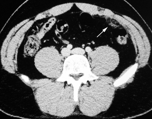
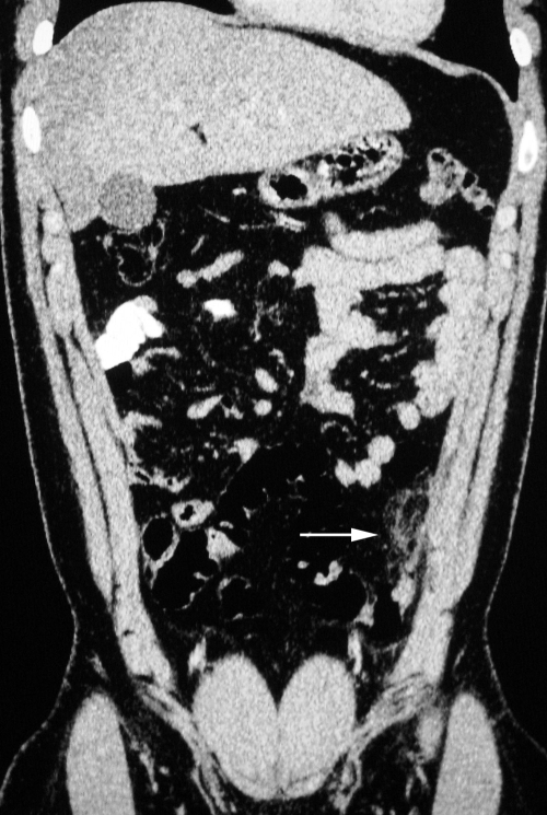
Abdominopelvic CT was performed after oral and intravenous administration of contrast material (figs 1 and 2). The CT scan showed thickening of visceral peritoneum and paracollic, attenuating strandings in the fat lateral to the descending colon. The descending colon was intrinsically normal. A diagnosis of appendagitis was made, and the patient was discharged from the emergency department with analgesia.
Figure 1 Axial view.
Figure 2 Coronal view: demonstrates thickening of visceral peritoneum and pericolonic fat strandings.
Discussion
Epiploic appendixes are small (0.5–5.0 cm long) pouches of peritoneum filled with fat and small vessels that protrude from the serosal surface of the colon. They occur in the rectosigmoid junction (57%), ileocecal region (26%), ascending colon (9%), transverse colon (6%) and descending colon (2%).3 Epiploic appendagitis may be primary or secondary. Primary epiploic appendagitis is caused by torsion or spontaneous venous thrombosis of the involved epiploic appendage. Secondary epiploic appendagitis is associated with inflammation of adjacent organs, such as diverticulitis, appendicitis or cholecystitis. Primary epiploic appendagitis occurs in the second to fifth decades of life without sexual predominance. The most common part of the colon affected by acute epiploic appendagitis in decreasing order of frequency is: the sigmoid colon, descending colon, cecum and ascending colon.4
Patients with epiploic appendagitis most commonly present with localised abdominal pain, more commonly on the left. The presenting clinical symptoms of epiploic appendagitis are non‐specific, leading to clinical misdiagnosis in most patients. Patients may present with localised abdominal pain of variable intensity and duration, rebound tenderness, an abdominal mass and mild fever. Nausea, vomiting and loss of appetite are rare symptoms. White blood cell count is normal or slightly elevated in most of the cases. The pain may be exacerbated by coughing, deep breathing or stretching because the infarcted appendage is adherent to the parietal peritoneum. Signs and symptoms are self‐limiting and rarely last more than 1 week.2,5 The non‐specific symptoms may mimic appendicitis, diverticulitis, omental infarction, pelvic inflammatory disease or a ruptured ovarian cyst.6
On CT, the lesion appears as a fatty mass, which is connected to the serosal surface of the colon and has slightly higher attenuation than peritoneal fat. All masses have periappendiceal fat stranding, and a few may have a central dot of high attenuation, possibly caused by a thrombosed vessel in the epiploic appendix or by the apposing surfaces of two adjacent appendixes.7 The CT changes of acute epiploic appendagitis completely resolved in all patients who underwent follow‐up CT 6 months after the acute presentation.4
Appendagitis is a self‐limiting disease and conservative treatment with analgesics is usually sufficient.8 Because these patients are conservatively managed, pathological confirmation of the disease is uncommon.9 The presumed diagnosis of this condition is primarily based on the CT features of inflammation centred over the epiploic appendage rather than the colon wall and lack of inflamed colonic diverticula.10
References
- 1.Pines B, Rabinovitch J, Biller S. Primary torsion and infarction of the appendices epiploicae. Arch Surg 194142775–787. [Google Scholar]
- 2.Molla E, Ripolles T, Martinez M.et al Primary epiploic appendagitis: US and CT findings. Eur Radiol 19988435–438. [DOI] [PubMed] [Google Scholar]
- 3.Thomas J, Rosato F, Patterson L. Epiploic appendagitis. Surg Gynecol Obstet 197413823–25. [PubMed] [Google Scholar]
- 4.Singh A, Gervais D, Hahn P.et al CT appearance of acute appendagitis. AJR 20041831303–1307. [DOI] [PubMed] [Google Scholar]
- 5.Rioux M, Langis P. Primary epiploic appendagitis: clinical, US, and CT findings in 14 cases. Radiology 1994191523–526. [DOI] [PubMed] [Google Scholar]
- 6.Boardman J, Kaplan K, Hollcraft C.et al Torsion of the epiploic appendage. AJR Am J Roentgenol 2003180748. [DOI] [PubMed] [Google Scholar]
- 7.Rao P, Wittenberg J, Lawrason J. Primary epiploic appendagitis: evolutionary changes in CT appearance. Radiology 1997204713–717. [DOI] [PubMed] [Google Scholar]
- 8.Rao P, Rhea J, Wittenberg J.et al Misdiagnosis of primary epiploic appendagitis. Am J Surg 199817681–85. [DOI] [PubMed] [Google Scholar]
- 9.Vazquez‐Frias J, Castaneda P, Valencia S. Laparoscopic diagnosis and treatment of an acute epiploic appendagitis with torsion and necrosis causing an acute abdomen. JSLS 20004247–250. [PMC free article] [PubMed] [Google Scholar]
- 10.Danielson K, Chernin M, Amberg J.et al Epiploic appendicitis: CT characteristics. J Comput Assist Tomogr 198610142–143. [DOI] [PubMed] [Google Scholar]


