Abstract
A 26‐year‐old man presented to the emergency department after a spontaneous 30 min bleed from his scrotal skin. He showed no other symptoms and denied any past medical history. He was exclusively sexually active, systemically well and haemodynamically stable. There were numerous (>50) 1–2 mm dark red, erythematous papules over the scrotum, sparing the shaft of penis, inner thigh and abdomen. A small area of blood marked the bleeding spot as a single papule. A diagnosis of angiokeratoma of the scrotum (Fordyce) was made and potential precipitants such as intra‐abdominal masses, urinary tract tumours, varicoceles, hernias and angiokeratoma corporis diffusum (Fabry syndrome) were excluded. He was discharged with dermatology follow‐up with a view to local laser treatment. The important differential diagnoses are angiokeratoma corporis diffusum and malignant melanoma (nodular type). In females, Fordyce angiokeratoma are distributed on labia majora.
A 26‐year‐old Portuguese man attended the emergency department complaining of scrotal skin bleeding. He described an episode of spontaneous bleeding while at work as a labourer, which stopped within 30 min. He could not identify a precipitating factor for the bleed. He was openly worried about the possibility of a sexually transmitted infection, but denied attendance at genitourinary medicine services for this problem. He denied any urethral discharge, dysuria or sexual dysfunction and was exclusively sexually active. He had no prior medical history and was not taking any regular medication.
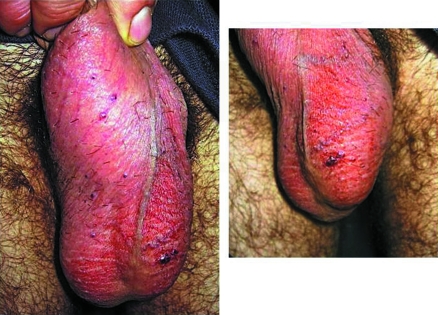
On examination the man was haemodynamically stable and systemically well. There were numerous (>50) 1–2mm dark red, erythematous papules over the scrotum (fig 1A), sparing the shaft of penis, inner thigh and abdomen. A small area of encrusted blood was visible on his left hemi‐scrotum, deemed to be the bleeding site (fig 1B). There was no active bleeding. There were no intra‐scrotal swellings palpable and no evidence of varicocele or epididymal pathology. His abdomen was soft and non tender, with no masses palpable. Urinalysis detected no abnormality.
Figure 1 1–to 2mm dark red, erythematous papules over the scrotum (A), encrusted blood on the left hemi‐scrotum, deemed to be the bleeding site (B) Informed consent was obtained for publication of this figure.
On further questioning, the man had noticed the papular lesions many years before, but they had remained asymptomatic. He was reassured that the lesions were benign and did not represent a sexually transmitted infection, and he was discharged. Dermatology outpatient review was organised to confirm the diagnosis of angiokeratoma of the scrotum (Fordyce).
Angiokeratoma are a group of eight clinically distinct vascular ectasias. They usually manifest as 1–6 mm red–blue, hyperkeratotic papules, occurring in isolation or groups, on the skin of the lower limbs, abdomen, trunk, tongue, scrotum, shaft of penis or labia majora.1 Accuracy on the prevalence of angiokeratoma is poor, as the lesions often go unnoticed, remaining asymptomatic with no systemic effects. However, some propose that the prevalence increases from 0.6% between the ages of 16–20 years, to 16.6% in the >70s. The lesions are most common in males, with Caucasian and Japanese populations predominantly affected.2
Fordyce angiokeratoma were first described in 1896, and refer to lesions occurring on the scrotum, shaft of penis, labia majora, inner thigh or lower abdomen. They range in size and can be solitary or in diffusely spread groups of <100. The pathophysiology remains uncertain.1 However, numerous reports and case reviews suggest that increased venous pressure proximal to the site is responsible.1,2,3 In men, varicoceles have been implicated as a common cause, although the data are variable.1,3 In women, increased venous pressure is noted during pregnancy, and in vulval varicosity, post‐partum and post‐hysterectomy. Urinary system tumours and hernias have also been suggested as causes.2 Patients most often present complaining of bleeding from the affected site, often following excoriation of the lesions or sexual intercourse. This can be substantial and difficult to stop. Some may not be aware of the lesions.
Although angiokeratoma of the scrotum is often a benign condition, it has the potential to cause considerable worry and distress to patients. In the emergency department it has an important differential diagnosis. Angiokeratoma corporis diffusum (Fabry syndrome) is an X linked in‐born error of metabolism characterised by recurrent fevers, chronic pain, peripheral oedema and multi‐system dysfunction and ultimately failure. It usually presents in late childhood and often causes death secondary to renal failure by age 39 years. Malignant melanoma (nodular melanoma type) can have a similar appearance and distribution to angiokeratoma, appearing as dark dome shaped papules prone to spontaneous bleeding. Importantly, this sub‐type of melanoma does not exhibit the warning signs of asymmetry, irregular border, variegation in colour, large diameter and rapid change in radial growth pattern, which are usually associated with malignant melanoma. It is also important to consider melanocytic naevii and genital warts in the differential diagnosis.2,4
The initial management of suspected angiokeratoma is to control bleeding with direct pressure. Once achieved, despite the evidence remaining unclear, it is important to exclude raised intra‐abdominal pressure as a cause, especially if there are associated systemic features. A good history and clinical examination of the abdomen for intra‐abdominal masses may help to exclude urinary tract tumours and hernias. Urinalysis to exclude microscopic haematuria may also help. The scrotum should be examined for evidence of varicocele, often described as a scrotal swelling mimicking a bag of worms, most evident when the patent is stood upright. The remaining skin should also be carefully examined for other lesions that may be associated with angiokeratoma corporis diffusum, malignant melanoma and melanocytic naevii.
If the diagnosis remains in doubt then refer to a dermatology clinic for opinion and skin biopsy. In angiokeratoma of the scrotum, biopsy will show numerous dilated, thin‐walled vessels in the papillary dermis or superficial sub‐mucosa. It may also show epithelial hyperkeratosis. The lesions generally respond well to local laser treatment.
Footnotes
Competing interests: None.
Informed consent was obtained for publication of the figure.
References
- 1.Erkek E, Basar M M, Bagci Y.et al Fordyce angiokeratomas as clues to local venous hypertension. Arch Dermatol 20051411325–1326. [DOI] [PubMed] [Google Scholar]
- 2.Atherton D J, Moss C. Naevi and other developmental defects. In: Burns T, Breathnach S, Cox N, et al eds. Rook's textbook of dermatology. Oxford: Blackwell 200489–90.
- 3.Orvieto R, Alcalay J, Leibovitz I.et al Lack of association between varicocele and angiokeratoma of the scrotum (Fordyce). Mil Med 1994159523–524. [PubMed] [Google Scholar]
- 4.Schiller P, Itin P. Angiokeratomas: an update. Dermatology 1996193275–282. [DOI] [PubMed] [Google Scholar]


