Abstract
Background: The diagnosis of systemic amyloidosis is determined through histological material from biopsy of different parenchymal organs, which have high diagnostic and informative value, but hide a high risk of bleeding because of the accumulation of amyloid in the vessels' wall. The main methods are kidney, liver, gastro–intestinal tract biopsy and aspiration of subcutaneous fatty tissue. The sensitivity of trans–dermal core kidney biopsy (KB) is close to 100%. The rectal biopsy is positive in 73% of cases, the biopsy of bone marrow in bout 50% and the one of gingival mucosa in 40–46 % of cases. The biopsy of subcutaneous fatty tissue (BSFT) is a new, highly sensitive method with sensitivity 73% and specificity 90%, so that it can be used as a screening test in patients without any clinical symptom or organ dysfunction.
Patients and Methods: One hundred fifteen patients, 59 male and 56 female with an average age 49.7±15.93 years were included in the study divided in two groups. The first group consisted of patients with kidney biopsy proved amyloidosis compared to biopsy findings from other parenchymal organs. The second group consisted of patients suspected having amyloidosis who underwent biopsies from various tissues or organs except kidney biopsy because there was contraindication.
Results: One hundred fifteen biopsies of subcutaneous fatty tissue (SFT) were performed for the diagnosis of systemic amyloidosis. In order to compare the data from the BSFT to the other known and practiced till the moment methods BSFT was performed in 54 patients with proved amyloidosis by KB. In 51/54 the positive result for amyloid was confirmed. A comparison of the data in a sample of 20 patients, 11 female and 9 male, in 18/20 patients the result from BSFT is positive (90%). In coloring with Congo red are typed with KMnO4 19/54 patients, 12 female and 7 male, with average age 48.12 (SD ±13.21). In 14/19 the amyloidosis was typed as AA (74.2%) and 5/19 non – AA, probably AL (25.8%). To reveal the meaning of so called screening–biopsy of subcutaneous fatty tissue for excluding accompanying amyloidosis in patients with significant proteinuria and/or uremia, dysglobulinemia, laboratory constellations for nephritic syndrome in immune nephropathies and chronic infections (Chronic Obstructive Lung Disease, purulent infections) with contraindications for kidney biopsy 61 screening BSFT were performed, accumulation of amyloid was defined in 37. In all of the patients the result was verified also by biopsies of rectal, gingival and stomach mucosa.
Conclusion: The purposeful searching and proving of amyloid in subcutaneous fatty tissue of the abdominal wall is a new, highly sensitive method. The receiving of richer material from SFT in the method "biopsy" in stead of "aspiration", makes it more reliable for proving amyloid in the case that it exists. The method is enough informative for proving not only amyloidosis AL, but also for amyloidosis AA, in treating with KMnO4.
The biopsy of SFT in combination with biopsies from other mucosa can prove the accumulation of amyloid in contraindications for performing KB.
Keywords: systemic amyloidosis, histological diagnosis, aspiration of subcutaneous fatty tissue, biopsy of subcutaneous fatty tissue
Amyloidosis is a disease resulting from of extra cellular accumulation of insoluble proteins in different organs and in blood vessel wall. This disorder is systemic by nature. The name "amyloid" is given by Virhov in 1854 from the Greek word amylon or from the Latin one – amylum, because of the coloring of amyloid substance with iodine, similar to carbohydrates1,2. The diagnosis of systemic amyloidosis is determined through histological material from biopsy of different parenchymal organs, which have high diagnostic and informative value, but hide a high risk of bleeding because of the accumulation of amyloid in the vessel wall1–3. The main methods are a) kidney and liver biopsy with high diagnostic value but with risk of hemorrhage, b) gastro–intestinal tract biopsy (rectal, gastric, duodenal and jejunal mucosa), gingival biopsy, and c) aspiration of subcutaneous fatty tissue which is highly informative for AL (60–80%), with low risk of bleeding. The sensitivity of the transdermal kidney biopsy is close to 100%. The rectal biopsy is positive in 73% of the cases, the biopsy of bone marrow in about 50 % and the one of gingival mucosa in 40–46 % of cases4,5,7,9. The disease can be missed in cases when it is not deliberated searched and the basic coloring methods for proving the amyloid are not applied. The epidemiological data for the frequency of the disease (4% at kidney pathology and 5.4% at autopsy, showing a tendency to rise4,6,8,10.
The biopsy of subcutaneous fatty tissue (BSFT) is a new, highly sensitive method with sensitivity 73% and specificity 90%, so that it can be used as a screening test in patients without any clinical symptom or organ dysfunction5. It is enough informative in the diagnosis not only of amyloidosis AL, but of amyloidosis AA. In 70% of cases with AL or AA amyloidosis, the aspiration of abdominal subcutaneous fatty tissue (ASFT) is positive and the method is indicative in the diagnosis of amyloid L7–9. The big advantage of the method is the lack of contraindications and no risk of complications.
The BSFT as a method of receiving material for diagnosing systemic amyloidosis and defining the type of the amyloid is a modification of the method "aspiration".
Patients and Methods
One hundred fifteen patients, 59 male and 56 female, 49.7±15.93 years old were included in the study. They were divided into two groups. The first group consisted of 54 patients with kidney biopsy proved amyloidosis compared to findings from BSFT, and rectal, gingival, gastric or intestinal biopsy. The second group consisted of 61 patients with significant proteinuria and /or uremia, dysglobulinemia, laboratory constellation for nephritic syndrome in immune nephropathies and chronic infections and patients with contra–indications for performing punction kidney biopsy with signs of accumulation of amyloid.
BSFT begins by applying local anesthesia with 2% solution of Lidocain subcutaneously; then cutaneous section of 3–4 mm length follows, with the help of a scalpel. A part of subcutaneous fatty tissue is clipped with a Kocher and the material is separated with a scalpel. On the place of the biopsy sterile gauze is put on for 2 days. In most cases the biopsy is performed on the right side of the umbilicus on the abdominal wall but it can be accomplished on some other place depending on the availability of subcutaneous fatty tissue. The advantages of the method are expressed in the greater reliability because of the always richer material in the manipulation and the greater reliability in defining the different types of systemic amyloidosis.In some cases the material from the subcutaneous fatty tissue was treated with KMnO4 for typing the amyloid A.
The investigation was applied in patients with chronic glomerulonephritis (CGN), lupus nephropathy, diabetic nephropathy, chronic pyelonephritis and kidney tuberculosis; in patients with pulmonary infections, chronic cholecystitis, rheumatic arthritis; and in patients with neoplasia.
Results
One hundrend fifteen biopsies of subcutaneous fatty tissue were performed for the diagnosis of systemic amyloidosis from 2001 to 2004 in the Clinic of Nephrology of the Devision of Internal Diseases in the Medical University in Sofia. Fifty four of them were executed for evaluating the method "biopsy of subcutaneous fatty tissue" in already established amyloidosis with KB.
First task:
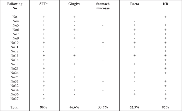
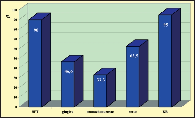
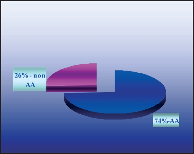
In order to compare the data from the biopsy of subcutaneous fatty tissue of the abdominal wall (BSFT) to the other known and practiced till the moment methods, BSFT was performed in 54 patients with proved, by KB, amyloidosis. In 51/54 the positive result for amyloid was confirmed. In Table 1 a comparison of the data in 20 patients, 11 female and 9 male, average age 46.42±13.94 is exposed. The female average age was 43.4±14.79 and 49.77±12.92 of male. In 18 patients the result from BSFT was positive (90%). The biopsy of the gingival mucosa gave a positive result in 7/15 cases (46.6%), from stomach or jejunal mucosa was positive in 3/9 patients (33.3%), while the biopsy from rectal mucosa gave a positive result in 10 /16 (62.5%), (Figure 1). In coloring with Congo red were typed with KMnO4 19/54 patients, 12 female and 7 male, with average age 48.12±13.21. In 14/19 the amyloidosis was typed as AA (74.2%) and 5/19 non – AA, probably AL (25.8%), (Figure 2).
Table 1: Comparison of data from biopsies from different tissues, for diagnosis of systemic amyloidosis in 20 patients.
SFT* subcutaneous fatty tissue
Figure 1: Graphics expression of the data from biopsies from different tissues for diagnosis of systemic amyloidosis in percentages.
Figure 2: Typing amyloid A after coloring with KMnO4.
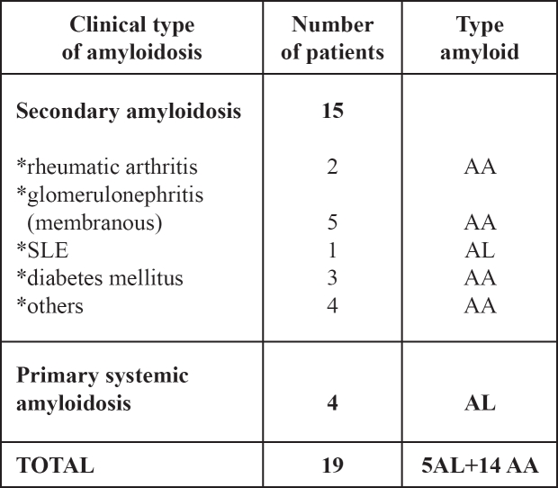
From the presented Tables and Figures it is clear that the modified method BSFT is reliable enough for typing amyloid in amyloidosis AA and amyloidosis AL. In all 19 patients the diagnosis was confirmed also by kidney biopsy and by biopsy from other mucosa. The amyloidosis in 15 patients was typed as AA. In 9 of them there was a combination of amyloidosis with other glomerulus pathology (5 with CGN, 1 with lupus nephritis, 3 with diabetic glomerulosclerosis), the rest were – 2 patients with rheumatoid arthritis and 4 with chronic infection. Four patients with amyloidosis as a single expression were typed as non–AA or AL amyloidosis.
Second task:
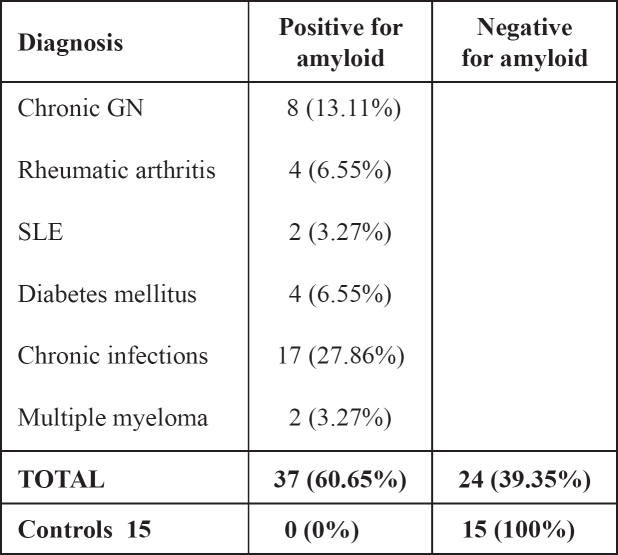
To reveal the meaning of so called screening–biopsy of subcutaneous fatty tissue for excluding accompanying amyloidosis in patients with significant proteinuria and/or uremia, dysglobulinemia, laboratory constellations for nephritic syndrome in immune nephropathies and chronic infections (Chronic Obstructive Lung Disease, purulent infections) with contraindications for kidney biopsy. Sixty one screening biopsies of subcutaneous fatty tissue were performed in patients with significant proteinuria and contraindications for KB, in patients with chronic pulmopathies, purulent chronic diseases, diabetes mellitus and high degree proteinuria, not responding to the prescription of the diabetes, immune diseases and laboratory constellations for nephrotic syndrome. From the 61 who underwent BSFT, accumulation of amyloid was defined in 37. In all of them the result was verified by biopsies of rectal, gingival and stomach mucosa (Table 3).
Table 3: Results from coloring with Congo red of material from subcutaneous fatty tissue purposeful searching of amyloid in 61 patients. Accumulation of amyloid was defined in 37 biopsies of SFT.
The accumulation of amyloid in the subcutaneous fatty tissue was defined semi–quantitatively in three–degree scale 1) Low (+), 2) Moderate (++), 3) High (+++).
Discussion
The BSFT, as a method of diagnosis in our patients, was modified and changed from the well known method – aspiration, because of insufficient material in most of the cases, the necessity of multiple aspirations (at least 3) from different places, especially in patients with thin abdominal wall.
The BSFT for diagnosis of systemic amyloidosis is a highly sensitive method. It is an easily performed method and the received material is considerably bigger in comparison with aspiration, and this is the reason why the method is more reliable and informative. Comparing the data from the various methods for the diagnosis of systemic amyloidosis, the BSFT is the second most informative method after KB (Table 1). The biopsy of subcutaneous fatty tissue in 90 % of the cases is indicative for diagnosis of systemic amyloidosis (Figure 1).
The modified method does not require any special preparations, materials and consummative, it is easily performed and can be applied in all conditions, including patients with disturbances of coagulation. The material, received by our method, is significantly richer in volume in comparison with the one of aspiration; the manipulation is single and in this connection is more reliable and informative. The method can be successfully used for screening, especially in patients with suspected having amyloidosis but with contraindications for KB.
From aur study we conclude that the purposeful searching and proving of amyloid in subcutaneous fatty tissue of the abdominal wall is a new, highly sensitive method. The extraction of richer material from subcutaneous fatty tissue in the method "biopsy" in stead of "aspiration", makes it more reliable for proving amyloid in the case that it exists. The method is enough informative for proving not only amyloidosis AL, but also for amyloidosis AA, in treating with KMnO4. The biopsy of subcutaneous fatty tissue of the abdominal wall does not possess any risk for the patient and is the second most informative method after KB. The biopsy of subcutaneous fatty tissue in combination with biopsies from other mucosa can prove the accumulation of amyloid in contraindications for performing KB. Searching amyloid in more than one tissue and using different methods excludes the possibility for false positive and false negative results and gives an idea for the heaviness and spread of the process.
Table 2: Distribution of different nosological groups of 19/54 patients with systemic amyloidosis in which BSFT is typed with KMnO4.
References
- 1.Westermark P, Stennkvist B. In: Amyloidosis. 3. Wegelius O, Paternack S, editors. New York: Academic press; 1976. 403 pp. [Google Scholar]
- 2.Westermark P, Benson L, Olofsson BO. Fine needle aspiration biopsy of abdominal subcutaneous fat tissue for the diagnosis and typing of amyloidosis. In: Glenner GG, Osserman EF, Benditt EP, Calkins E, Cohen AS, Zucker–Franklin D, editors. Amyloidosis. New York; London: Plenum Publishing Corporation; 1986. pp. 613–615. [Google Scholar]
- 3.Kyle RA. Multiple myeloma review of 869 cases. Mayo Clin Proc. 1975;50:29–40. [PubMed] [Google Scholar]
- 4.Barlogic B, Alexanian R, Jagannath S. Plasma cell dyscreasias. JAMA. 1992;268:2946–2951. [PubMed] [Google Scholar]
- 5.Gertz MA, Li CY, Shiracama T, Kyle RA. Utility of subcutaneous fat aspiration for the diagnosis of systemic amyloidosis (immunoglobulin light chain) Arch Intern Med. 1988;148:929–933. [PubMed] [Google Scholar]
- 6.Bogov B, Djerassi R, Kiperova B. Can abdominal ultrasonography suppose the histomorphological diagnosis of the diffuse renal diseases?". SonoAce International. 1997;4:15–19. [Google Scholar]
- 7.Duston MA, Skinner M, Meenan RF. Sensitivity, specificity, and predictive value of abdominal fat aspiration for the diagnosis of amyloidosis. Arthritis Rheum. 1989;32:82–5. doi: 10.1002/anr.1780320114. [DOI] [PubMed] [Google Scholar]
- 8.Sole Arques M, Campistol JM, Munoz–Gomez J. Abdominal fat aspiration biopsy in dialysis–related amyloidosis. Arch Intern Med. 1988;148:988. doi: 10.1001/archinte.1988.00380040228040. [DOI] [PubMed] [Google Scholar]
- 9.Van Gameren II, Hazenberg BP, Bijzet J, van Rijswijk MH. Diagnostic accuracy of subcutaneous abdominal fat tissue aspiration for detecting systemic amyloidosis and its utility in clinical practice. Arthritis Rheum. 2006;54:2015–2021. doi: 10.1002/art.21902. [DOI] [PubMed] [Google Scholar]
- 10.Haagsma EB, Van Gameren II, Bijzet J, Posthumus MD, Hazenberg BP. Familial amyloidotic polyneuropathy: longterm follow–up of abdominal fat tissue aspirate in patients with and without liver transplantation. Amyloid. 2007;14:221–226. doi: 10.1080/13506120701461368. [DOI] [PubMed] [Google Scholar]


