Abstract
Context:
Few researchers have examined shoulder strength in adolescent volleyball athletes despite increasing levels of participation in this age group.
Objective:
To compare medial and lateral isokinetic peak torque of the rotator cuff among skill levels and between athletes with and without a history of shoulder injury.
Design:
Cross-sectional design.
Setting:
The Human Performance Lab and Athletic Training Lab.
Patients or Other Participants:
Thirty-eight female adolescent club volleyball athletes from 10 to 15 years of age (mean = 13.02 ± 1.60 years).
Main Outcome Measure(s):
We measured concentric and eccentric peak torque of the medial and lateral rotators of the shoulder and calculated resultant cocking and spiking ratios based on peak torque values.
Results:
Athletes at higher skill levels had higher peak torque measurements in concentric and eccentric medial and lateral rotation compared with the athletes at lower skill levels. No differences in peak torque existed between participants with or without an injury history 6 months before the study. Strength ratios did not differ across skill levels, but previously injured participants produced lower eccentric medial rotation to concentric lateral rotation ratios compared with participants without a history of injury (P = .02). At the highest skill level, previously injured participants produced lower eccentric lateral rotation to concentric medial rotation ratios compared with participants without an injury history (P = .04).
Conclusions:
Differences in medial and lateral shoulder rotator strength ratios appear to be related more to injury prevalence than to absolute strength. Shoulder dysfunction related to strength ratio deficits also may exist in adolescent female volleyball athletes. Preventive shoulder strengthening programs focused on improving eccentric strength and correcting imbalances between medial and lateral rotators may be warranted for all female adolescent volleyball athletes.
Keywords: shoulder strength; strength ratios; glenohumeral joint, medial-lateral shoulder rotation
Key Points.
Rotator cuff strength was not predictive of injury history in our participants.
We found no differences in strength ratios among skill levels.
Participants with previous glenohumeral injuries had lower cocking ratios and nearly significant lower spiking ratios when compared with participants without a history of injury, indicating insufficient eccentric strength among those in the former group.
Rotator cuff strengthening, especially to improve eccentric function of the medial and lateral rotators, is indicated for injury prevention in early adolescent female volleyball players.
Among the most common injuries affecting volleyball athletes, glenohumeral joint injuries often contribute to long delays in return to training or competition.1–6 These injuries are often associated with frequent spiking and serving motions that require dynamic stabilization to maintain glenohumeral joint integrity.1,2,4–6 Musculoskeletal development of adolescent athletes is incomplete, so muscular imbalances may be present. Limitations in dynamic stabilization because of muscular imbalances may increase the likelihood of injury to the glenohumeral joint of adolescent club volleyball players. Unfortunately, research on the relationship between strength characteristics and glenohumeral joint injury in adolescent athletes is limited.7–9
Stability of the glenohumeral joint during the acceleration, deceleration, and follow-through phases of striking is provided by the rotator cuff muscles acting eccentrically to compress the humeral head.10 Active and passive mechanisms maintain dynamic stabilization and compression of the humeral head in the glenoid fossa during spiking and serving. As the upper extremity accelerates through its range of motion, the supraspinatus, infraspinatus, and teres minor muscles eccentrically resist translation of the humeral head and assist in deceleration of the moving limb.10–14 Consequently, rotator cuff weakness allows increased stress to be placed on the passive stabilizers of the shoulder, leading to detrimental translation of the humeral head. Conversely, laxity in the passive stabilizers increases the workload of the rotator cuff, leading to fatigue and malfunction of the dynamic stabilizers.15 Authors of previous studies16–19 have demonstrated the relationship between rotator cuff injuries and strength deficits in the glenohumeral and scapular muscles of injured participants; however, these studies17–19 primarily involved nonathletic adult participants.
Preventing shoulder injuries in adolescent populations through appropriate maintenance of rotator cuff strength is of particular concern because of musculoskeletal immaturity. The added demands of sport participation can lead to acute or overuse injuries in developing tissues14,20–22 and to long-term or permanent disability of the affected structures.20 In previous isokinetic studies involving volleyball athletes, investigators did not examine differences in rotator cuff strength between injured and uninjured shoulders in early adolescents.9,23–26 Therefore, the purpose of our retrospective study was to compare medial and lateral isokinetic peak torque of the rotator cuff among skill levels and between adolescent club volleyball athletes with and without a history of shoulder injury.
Methods
Participants
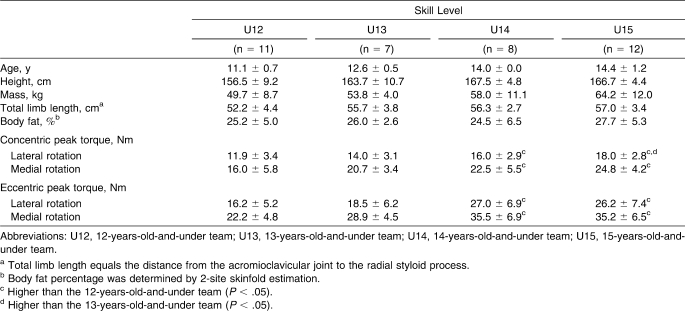
Participants were 38 highly trained, competitive, female club volleyball players ranging from 10 to 15 years of age (age = 13.0 ± 1.6 years). They completed a medical history questionnaire detailing injury history, pain, time lost because of glenohumeral joint injury, and pain in the striking arm within 6 months of study participation. No participant reported glenohumeral injury or shoulder pain at the time of testing, nor did anyone report experience with isokinetic testing. The coaching staff grouped participants on teams by skill level. Team age designations (12 years of age and under [U12], 13 years of age and under [U13], 14 years of age and under [U14], and 15 years of age and under [U15] teams) represented the maximum age allowed for team members, although younger, more skilled participants were eligible to compete on a higher age-limit team. Participant characteristics and groupings by skill levels are presented in Table 1.
Table 1.
Participant Characteristics and Concentric and Eccentric Peak Torque by Skill Level (Mean ± SD)
Questionnaire responses were used to divide participants into groups for initial analysis determined by previous glenohumeral joint injury (PI), or no history of injury (NI). Nine participants were placed in the PI group based on self-reported histories of glenohumeral joint injury that limited athletic participation (2 from U12, 2 from U14, and 5 from U15). The remaining 29 participants reported no previous pain or injury to the shoulder of the striking arm for at least 6 months before testing. Parents and participants provided informed consent, and the study was approved by the University of Hawai‘i Committee on Human Studies.
Procedures
All data were collected by the same Board-certified athletic trainer. All anthropometric and isokinetic peak torque data were collected during a single test session. Anthropometric measures included height, mass, body composition, and length of the striking arm. The striking arm was defined as the shoulder that the participant normally used for hitting or serving the volleyball. The athletic trainer estimated body composition via calf and triceps skinfolds using Lange skinfold calipers (Cambridge Scientific Industries Inc, Cambridge, MD).27
Using a Biodex System 3 dynamometer (Biodex Medical Systems Inc, Shirley, NY), the athletic trainer collected isokinetic medial and lateral shoulder rotation peak torque data in Newton · meters for the striking arm. Researchers have shown that this dynamometer is reliable in measuring glenohumeral medial and lateral rotation concentrically and eccentrically in neutral to slightly abducted positions.28,29 Using the Biodex system, Malerba et al30 reported that intraclass correlation coefficients (2,1) for medial and lateral rotation peak torque at 60°·s−1 in the scapular plane (45° abduction and 30° horizontal flexion) ranged from 0.82 to 0.86 and from 0.70 to 0.76, respectively, for concentric activity and from 0.70 to 0.90 and from 0.44 to 0.68, respectively, for eccentric activity. Although the testing position was not described, Toy and Rankin31 reported higher intraclass correlation coefficients when they used a Biodex system at a similar speed to measure glenohumeral medial and lateral rotation peak torque for both concentric (0.97, both) and eccentric (0.91 and 0.88–0.94, respectively) activity. Additionally, Hellwig and Perrin32 reported intraclass correlation coefficients of 0.80 for eccentric medial rotation (Eccmr) and 0.93 for eccentric lateral rotation (Ecclr) during isokinetic testing at 90° of abduction in the scapular plane using a KinCom system. Test-retest reliability was not assessed in our study.
The Biodex was calibrated after every fifth participant in our study. Peak torque data were collected at 60°·s−1 via 2 sets of 5 maximal repetitions of medial and lateral shoulder rotation; the first set was accomplished concentrically, and the second set was accomplished eccentrically. Sets were separated by a 5-minute rest period. Before collection of peak torque data, participants completed warm-up and familiarization trials consisting of submaximal efforts and a single maximal effort in concentric and eccentric modes. The decision to not use a counterbalanced design created the possibility of an order effect on peak torque generated concentrically versus eccentrically. However, we chose this predetermined order to provide the greatest safety for participants, most of whom had immature musculoskeletal development, by allowing them to increase their familiarity with maximal isokinetic testing before eccentric exposure. Additionally, we assumed that the limited number of repetitions performed combined with the 5-minute recovery period was adequate for complete replenishment of adenosine triphosphate via the adenosine triphosphate-phosphocreatine system, preventing an influence from the concentric trial on eccentric peak torque production.
Participants were tested in a seated modified neutral position of 90° of elbow flexion, 30° of glenohumeral joint flexion, and 30° of glenohumeral abduction with stabilization straps across the hips and upper body. Although testing in this position limited the ability to compare our values with those obtained by investigators using the 90° of glenohumeral abduction position,9,23–26 this position was chosen to increase the participants' comfort during the testing procedure33 because of the age of the participants and because of the reduced risk of anterior instability associated with the modified neutral position.34 Participants were given oral instructions regarding testing procedures and the importance of providing maximal effort during testing. To create a consistent testing environment, no oral or visual feedback or encouragement was given during the isokinetic testing protocol. All participants completed all phases of data collection without pain or discomfort in the shoulder.
Data Analysis
Isokinetic peak torque data were collected for each of the following measures: concentric medial rotation (Conmr), concentric lateral rotation (Conlr), Eccmr, and Ecclr. Based on peak torque values for each measure, the following functional ratios were derived for each participant in accordance with previous research: Conlr:Conmr, Ecclr:Eccmr, Ecclr:Conmr, and Eccmr:Conlr.7,8,24,35–43
All statistical analyses were performed with SAS (version 9.1.3; SAS Inc, Cary, NC) using a 2 (injury history) × 4 (skill level) research design. Data were analyzed via many multivariate analyses of covariance (MANCOVA) to control for interactions due to limb-length differences among participants. Initial analysis revealed no effect of the covariate limb length on any measure. Individual analysis of variance (ANOVA) and a priori Tukey HSD tests were used post hoc for each of the strength and ratio measures across all skill levels. The α level was set at .05. Because of the disproportionate rate of injury reported in the U15 skill level, additional analysis comparing PI and NI groups also was completed using only the data for the U15 skill level.
Results
Strength Measure and Ratio Differences Among Skill Levels
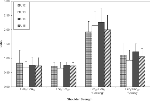
The ANOVA revealed differences among skill levels for Conmr, Conlr, Eccmr, and Ecclr. Post hoc Tukey HSD testing identified that the U15 and U14 teams were higher in all 4 strength measures compared with the U12 team (P = .005 and P = .03, respectively). Additionally, Conlr was higher in the U15 team than in the U13 team (P = .04; Table 1). No differences were found among skill levels for any of the 4 ratio measures, suggesting that peak torque for medial and lateral rotation increased at similar rates across skill levels. Reference values for shoulder strength ratios for each skill level are presented in Figure 1.
Strength Measure and Ratio Differences Between Injury History Groups
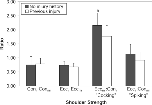
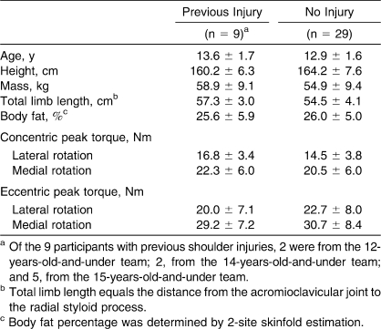
No differences were found between NI and PI groups for any of the 4 strength measures (Table 2). Concentric peak torque values were higher for the PI group than for the NI group for both lateral and medial rotation (P = .10 and P = .45, respectively). Eccentric peak torque values were higher for the NI group than for the PI group for both lateral and medial rotation (P = .37 and P = .63, respectively). However, differences existed between groups in the Eccmr:Conlr ratio, with PI participants producing lower Eccmr:Conlr peak torque ratios (1.77 ± 0.39) compared with NI participants (2.16 ± 0.44, P = .02). Although not different, the Ecclr:Conmr peak torque ratios for PI participants (0.92 ± 0.29) were lower at a level that approached significance (P = .08, power = .575) than these ratios for NI participants (1.14 ± 0.34). The Conlr:Conmr and Ecclr:Eccmr ratios for PI participants were not different from these ratios for NI participants (P = .79 and P = .31, respectively). Strength ratio data for NI and PI groups across skill levels are displayed in Figure 2.
Figure 2. Shoulder strength ratios by injury history across all skill levels (ratio + SD). Con indicates concentric; Ecc, eccentric; lr, lateral rotation; mr, medial rotation; a, difference between no injury and previous injury groups (P < .05).
Analysis of the U15 team revealed no differences for any of the 4 strength measures between the NI and PI groups. However, despite the small size of the sample groups when analyzing U15 alone (NI = 7, PI = 5), differences for both Eccmr and Ecclr peak torque between NI and PI groups approached significant levels (P = .07 and P = .08, respectively).
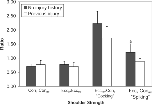
Analysis of strength ratio differences between the NI and PI groups for U15 alone produced similar results to those found when comparing participants at all skill levels. The Conlr:Conmr and Ecclr:Eccmr ratios for PI participants were not different from these ratios for NI participants (P = .37 and P = .45). Both Eccmr:Conlr and Ecclr:Conmr ratios were higher in the NI group than in the PI group when analyzing U15 alone (2.23 ± 0.43 versus 1.72 ± 0.40 and 1.21 ± 0.29 versus 0.88 ± 0.11, respectively). Interestingly, the difference in the Ecclr:Conmr ratio between the NI and PI groups, which only approached significance in the analysis including all participants (P = .08), was different when analyzing U15 alone (P = .04). Additionally, the difference in the Eccmr:Conlr ratio between the NI and PI groups was significant in the analysis including all participants (P = .02), but, when analyzing U15 alone, this difference only approached significance (P = .07). Figure 3 presents shoulder strength ratios for the NI and PI groups for U15 alone.
Figure 3. Shoulder strength ratios by injury history for the 15-years-old-and-under team (ratio + SD). Con indicates concentric; Ecc, eccentric; lr, lateral rotation; mr, medial rotation; a, difference between no injury and previous injury groups (P < .05).
Discussion
Conventional wisdom advocates the development of rotator cuff strength to prevent or alleviate shoulder injuries; however, we found that peak torque for both concentric and eccentric glenohumeral medial and lateral rotation was not different between the NI and PI groups. Although peak torque was not different between the NI and PI groups, it was higher for U14 and U15 participants than for U12 participants (Table 1). This was expected based on differences in physical development among skill levels related to muscle strength and sport-specific skills. Therefore, raw strength was not predictive of injury history in our participants. These findings have important clinical ramifications for identifying individuals at risk for developing shoulder injuries, because weakness of the rotator cuff muscles traditionally has been associated with the development of shoulder injuries.
Additionally, no differences in strength ratios were found among skill levels. These findings are similar to those of Ellenbecker and Roetert,7 who reported no differences in strength ratios of elite junior tennis athletes based on age. In our study, no differences in Conlr:Conmr peak torque ratios were found between NI and PI participants. These ratios previously have been reported to range from 0.57 to 1.19,* and Ellenbecker and Davies44 recommended Conlr:Conmr ratios of at least 2:3 to 3:4 (0.66–0.75) for the prevention of shoulder injuries. Interestingly, NI participants produced a mean Conlr:Conmr peak torque ratio of 0.75, while PI participants produced a mean Conlr:Conmr peak torque ratio of 0.79, exceeding the range that Ellenbecker and Davies44 recommended (Figure 2). Additionally, when analyzing U15 alone, NI participants produced a mean Conlr:Conmr peak torque ratio of 0.70, while PI participants produced a mean Conlr:Conmr peak torque ratio of 0.77, also exceeding the recommended44 range (Figure 3). Therefore, despite widespread use of Conlr:Conmr ratios for assessing and comparing shoulder strength profiles, our findings indicated that this ratio may have limited value for identifying adolescent girls at risk for shoulder injuries.
Investigators11,35,42 have suggested functional strength ratios as the most appropriate indicator for shoulder injury risk. Based on the function of rotator cuff muscles during serving and spiking motions, we calculated Eccmr:Conlr and Ecclr:Conmr ratios to represent the cocking and spiking phases, respectively. The PI participants produced lower Eccmr:Conlr (cocking) ratios (1.77 ± 0.39) than the NI participants (2.16 ± 0.44, P = .02). They also produced nearly significantly lower Ecclr:Conmr (spiking) ratios (0.92 ± 0.29) when compared with NI participants (1.14 ± 0.34, P = .08; Figure 2).
The significant difference in cocking ratio found between NI and PI groups despite insignificant differences in absolute strength measures may seem counterintuitive because ratios are derived directly from absolute strength measures. However, an examination of the individual relationships between the NI and PI groups for each of the 4 strength measures provides an explanation (P = .10 for lateral rotation and P = .45 for medial rotation). For the 2 concentric measures, peak torque of the PI group was higher, although not significantly, than that of the NI group. For the 2 eccentric measures, peak torque of the NI group was higher, although not significantly, than the PI group (P = .37 for lateral rotation and P = .63 for medial rotation). This combination of eccentric and concentric values resulted in a cocking ratio difference that reached significance despite the strength measures failing to reach significance.
The relationship between lower cocking ratio and injury history in our participants may appear difficult to explain when evaluating the ballistic nature of spiking compared with the cocking motion, which does not appear to subject the glenohumeral joint to the same degree of high-velocity rotational stress. However, the dynamic stability required to protect the glenohumeral joint from injury is the same for both motions. Imbalances between medial and lateral rotators may still allow excess joint translation, even during the cocking phase. Additionally, strength limitations of medial rotators acting eccentrically during the cocking phase may allow excess range of motion into lateral rotation, which is a position generally associated with increased risk of subacromial impingement of the supraspinatus and increased apprehension in individuals who tend to develop glenohumeral instability.46,47
The mean spiking ratios were 1.14 for NI participants and 0.92 for PI participants, which is a difference approaching significance (P = .08). Investigators11,35,42 have emphasized the importance of an adequate spiking ratio in maintaining shoulder stability in athletes whose sports involve repeated overhead motion. The trend toward significant differences that we observed between groups in spiking ratio supports the findings of Wang et al,26 who reported a greater risk of injury in adults with spiking ratios less than 1.0 combined with decreased glenohumeral flexibility. Conversely, Wang et al9 reported spiking ratios less than 1.0 among injured and uninjured junior volleyball athletes. They concluded that the risk factors for shoulder injury may be related to muscle imbalances less often in adolescents than in adults. However, these authors did not report differences in spiking ratios between injured and uninjured groups, so determining whether lower spiking ratios were associated with injury in their adolescent participants is difficult. Bak and Magnusson35 reported higher Ecclr:Conmr ratios in injured than in uninjured swimmers. However, injured participants in their study were tested while demonstrating a positive impingement sign and exhibited a decreased Conmr peak torque with nearly equal Ecclr peak torque relative to uninjured participants. Therefore, based on the results of our study and those reported by other authors, an adequate spiking ratio is clinically important because athletes with spiking ratios less than 1.0 appear to be at higher risk for shoulder injury.
Another mechanism may contribute to the relationship between spiking ratio and injury. During the cocking phase of spiking and serving motions, the combined action of Eccmr and Conlr positions the arm into 90° of elbow flexion and 90° of glenohumeral abduction with maximal lateral rotation. Placing the shoulder in this position may lead to suprascapular nerve compression by the supraspinatus and atrophy of the infraspinatus muscle. Although normally painless, this injury has been reported in volleyball athletes.48 However, this condition has generally not been reported in adolescent athletes48–51 and, therefore, was not assessed in our participants. It is possible that a deficiency, due to congenital anatomic differences or early sport participation, may lead to increased occurrence of this condition for volleyball athletes with a high training volume, resulting in decreased ability of the infraspinatus to provide eccentric dynamic stabilization during the acceleration phase of the spiking motion.
The lack of difference in the spiking ratio between groups (P = .08) may be due to the low number of participants who reported glenohumeral injury in the 6 months before the study (n = 9), resulting in relatively low statistical power (β = 0.575). This suggests that a larger sample size may have revealed a difference in the spiking ratio between PI and NI participants. The failure of this difference to rise to significant levels may also have been due to disproportionate numbers of PI participants in the highest skill-level group. Because higher skill-level groups generally produced higher peak torques compared with lower skill-level groups, the disproportionate number of PI participants in the U15 team may have attenuated strength differences between the NI and PI groups across all levels. It is likely that some PI participants on the U15 team produced peak torques equal to, or possibly greater than, those produced by NI participants on the U12 and U13 teams.
The disproportionate representation of PI participants from the U15 team led to subsequent analysis of differences between NI and PI participants in this skill level alone. Differences between NI and PI groups within U15 were similar but not identical to those found across skill levels. Although the cocking ratio was different between the NI and PI groups across all skill levels (P = .02), this ratio only approached significance for U15 alone (P = .07). Conversely, the difference in spiking ratio between NI and PI groups across all skill levels approached significance (P = .08), while it was significant only for the U15 team (P = .04) despite the small group size. Considering that the U15 team alone (n = 12) demonstrated differences in spiking ratios between the NI and PI groups (P = .04) and differences were found in cocking ratios across all skill levels between the NI and PI groups (n = 38, P = .02), we believe that these spiking and cocking ratios represent true differences between the NI and PI groups in our study. This conclusion is supported by the finding that U15 PI participants produced nearly significantly lower eccentric peak torque in both medial and lateral rotation compared with U15 NI participants (P = .07 and P = .08, respectively). Because eccentric strength is vitally important in maintaining high functional ratios, lower levels of eccentric strength would seem the most likely cause for a relationship between low functional ratios and injury history. Because decreasing concentric strength would not be a rational means of increasing cocking or spiking ratios, increasing eccentric strength should be the focus of protocols aimed at correcting ratio deficits.
In our study, cocking ratio deficits were greater in the PI group than in the NI group across all skill levels (P = .02) and among U15 participants alone, spiking ratio deficits were greater in the PI group than in the NI group (P = .04), indicating insufficient eccentric strength among those reporting a history of glenohumeral injury. Because of the retrospective design of this study, we could not determine if the strength ratio deficits led to the development of shoulder pain or if they resulted from previous shoulder injury. However, an association between injury status and functional strength ratios existed for our participants. Whether strength ratio deficits caused shoulder pain to develop or resulted from incomplete rehabilitation of previous injuries, they indicate the need for rotator cuff strengthening that emphasizes eccentric function among those with a history of shoulder injury. Based on our findings, differences in eccentric strength between medial and lateral rotators of the shoulder appear to relate more to injury prevalence than absolute strength does. Additionally, functional measures such as cocking and spiking ratios, appear more directly related to injury status compared with the traditionally used Conlr:Conmr ratio for adolescent female volleyball players.
Conclusions
The clinical relevance of our study is that rotator cuff strengthening, especially to improve eccentric function of medial and lateral rotators, is indicated for injury prevention in early adolescent girls. Furthermore, because nearly 25% of our participants reported glenohumeral injury severe enough to limit sport participation in the 6 months before the study, we believe that all female volleyball players at this age might benefit from a shoulder-strengthening program that focuses on eccentric strength deficits and correcting strength imbalances between medial and lateral rotators.
Figure 1. Shoulder strength ratios by skill level (ratio + SD). Con indicates concentric; Ecc, eccentric; lr, lateral rotation; mr, medial rotation; U12, 12-years-old-and-under team; U13, 13-years-old-and-under team; U14, 14-years-old-and-under team; U15, 15-years-old-and-under team.
Table 2.
Participant Characteristics and Concentric and Eccentric Peak Torque by Injury History (Mean ± SD)
Acknowledgments
We especially thank Andrea Harmon, MS, ATC, for her significant effort in acquisition of the data and Shuqiang Zhang, PhD, of the Department of Educational Psychology, University of Hawai‘i at Mānoa, for his assistance with the statistical analysis. We thank Mushtaq Ikramullah, MS, for his assistance with revision of the article. We also thank the coaches and members of the Jammers Volleyball Club, Honolulu, Hawaii, for their cooperation and participation in this study.
Footnotes
Christopher D. Stickley, PhD, ATC; Ronald K. Hetzler, PhD, FACSM; Bret G. Freemyer, MS, ATC; and Iris F. Kimura, PhD, PT, ATC, contributed to conception and design; acquisition and analysis and interpretation of the data; and drafting, critical revision, and final approval of the article.
*References 7-9, 23, 25, 26, 35, 36, 38-45.
References
- 1.Aagaard H, Jorgensen U. Injuries in elite volleyball. Scand J Med Sci Sports. 1996;6(4):228–232. doi: 10.1111/j.1600-0838.1996.tb00096.x. [DOI] [PubMed] [Google Scholar]
- 2.Aagaard H, Scavenius M, Jorgensen U. An epidemiological analysis of the injury pattern in indoor and in beach volleyball. Int J Sports Med. 1997;18(3):217–221. doi: 10.1055/s-2007-972623. [DOI] [PubMed] [Google Scholar]
- 3.Augustsson S.R, Augustsson J, Thomee R, Svantesson U. Injuries and preventive actions in elite Swedish volleyball. Scand J Med Sci Sports. 2006;16(6):433–440. doi: 10.1111/j.1600-0838.2005.00517.x. [DOI] [PubMed] [Google Scholar]
- 4.Bahr R, Reeser J.C. Injuries among world-class professional beach volleyball players: the Fédération Internationale de Volleyball beach volleyball injury study. Am J Sports Med. 2003;31(1):119–125. doi: 10.1177/03635465030310010401. [DOI] [PubMed] [Google Scholar]
- 5.Verhagen E.A, Van der Beek A.J, Bouter L.M, Bahr R.M, Van Mechelen W. A one season prospective cohort study of volleyball injuries. Br J Sports Med. 2004;38(4):477–481. doi: 10.1136/bjsm.2003.005785. [DOI] [PMC free article] [PubMed] [Google Scholar]
- 6.Wang H.K, Cochrane T. A descriptive epidemiological study of shoulder injury in top level English male volleyball players. Int J Sports Med. 2001;22(2):159–163. doi: 10.1055/s-2001-11346. [DOI] [PubMed] [Google Scholar]
- 7.Ellenbecker T, Roetert E.P. Age specific isokinetic glenohumeral internal and external rotation strength in elite junior tennis players. J Sci Med Sport. 2003;6(1):63–70. doi: 10.1016/s1440-2440(03)80009-9. [DOI] [PubMed] [Google Scholar]
- 8.Mulligan I.J, Biddington W.B, Barnhart B.D, Ellenbecker T.S. Isokinetic profile of shoulder internal and external rotators of high school aged baseball pitchers. J Strength Cond Res. 2004;18(4):861–866. doi: 10.1519/14633.1. [DOI] [PubMed] [Google Scholar]
- 9.Wang H.K, Juang L.G, Lin J.J, Wang T.G, Jan M.H. Isokinetic performance and shoulder mobility in Taiwanese elite junior volleyball players. Isokinet Exerc Sci. 2004;12(2):135–141. [Google Scholar]
- 10.Rokito A.S, Jobe F.W, Pink M.M, Perry J, Brault J. Electromyographic analysis of shoulder function during the volleyball serve and spike. J Shoulder Elbow Surg. 1998;7(3):256–263. doi: 10.1016/s1058-2746(98)90054-4. [DOI] [PubMed] [Google Scholar]
- 11.David G, Magarey M.E, Jones M.A, Dvir Z, Turker K.S, Sharpe M. EMG and strength correlates of selected shoulder muscles during rotations of the glenohumeral joint. Clin Biomech (Bristol, Avon) 2000;15(2):95–102. doi: 10.1016/s0268-0033(99)00052-2. [DOI] [PubMed] [Google Scholar]
- 12.Hess S.A. Functional stability of the glenohumeral joint. Man Ther. 2000;5(2):63–71. doi: 10.1054/math.2000.0241. [DOI] [PubMed] [Google Scholar]
- 13.Rizio L, Uribe J.W. Overuse injuries of the upper extremity in baseball. Clin Sports Med. 2001;20(3):453–468. doi: 10.1016/s0278-5919(05)70262-3. [DOI] [PubMed] [Google Scholar]
- 14.Wasserlauf B.L, Paletta G.A., Jr Shoulder disorders in the skeletally immature throwing athlete. Orthop Clin North Am. 2003;34(3):427–437. doi: 10.1016/s0030-5898(03)00032-4. [DOI] [PubMed] [Google Scholar]
- 15.Weldon E.J, III, Richardson A.B. Upper extremity overuse injuries in swimming: a discussion of swimmer's shoulder. Clin Sports Med. 2001;20(3):423–438. doi: 10.1016/s0278-5919(05)70260-x. [DOI] [PubMed] [Google Scholar]
- 16.Cools A.M, Witvrouw E.E, Mahieu N.N, Danneels L.A. Isokinetic scapular muscle performance in overhead athletes with and without impingement symptoms. J Athl Train. 2005;40(2):104–110. [PMC free article] [PubMed] [Google Scholar]
- 17.MacDermid J.C, Ramos J, Drosdowech D, Faber K, Patterson S. The impact of rotator cuff pathology on isometric and isokinetic strength, function, and quality of life. J Shoulder Elbow Surg. 2004;13(6):593–598. doi: 10.1016/j.jse.2004.03.009. [DOI] [PubMed] [Google Scholar]
- 18.McCabe R.A, Nicholas S.J, Montgomery K.D, Finneran J.J, McHugh M.P. The effect of rotator cuff tear size on shoulder strength and range of motion. J Orthop Sports Phys Ther. 2005;35(3):130–135. doi: 10.2519/jospt.2005.35.3.130. [DOI] [PubMed] [Google Scholar]
- 19.Tyler T.F, Nahow R.C, Nicholas S.J, McHugh M.P. Quantifying shoulder rotation weakness in patients with shoulder impingement. J Shoulder Elbow Surg. 2005;14(6):570–574. doi: 10.1016/j.jse.2005.03.003. [DOI] [PubMed] [Google Scholar]
- 20.Caine D, Caine C, Maffulli N. Incidence and distribution of pediatric sport-related injuries. Clin J Sport Med. 2006;16(6):500–513. doi: 10.1097/01.jsm.0000251181.36582.a0. [DOI] [PubMed] [Google Scholar]
- 21.Kocher M.S, Waters P.M, Micheli L.J. Upper extremity injuries in the paediatric athlete. Sports Med. 2000;30(2):117–135. doi: 10.2165/00007256-200030020-00005. [DOI] [PubMed] [Google Scholar]
- 22.Paterson P.D, Waters P.M. Shoulder injuries in the childhood athlete. Clin Sports Med. 2000;19(4):681–692. doi: 10.1016/s0278-5919(05)70232-5. [DOI] [PubMed] [Google Scholar]
- 23.Alfredson H, Pietila T, Lorentzon R. Concentric and eccentric shoulder and elbow muscle strength in female volleyball players and non-active females. Scand J Med Sci Sports. 1998;8(5, pt 1):265–270. doi: 10.1111/j.1600-0838.1998.tb00481.x. [DOI] [PubMed] [Google Scholar]
- 24.Forthomme B, Croisier J.L, Ciccarone G, Crielaard J.M, Cloes M. Factors correlated with volleyball spike velocity. Am J Sports Med. 2005;33(10):1513–1519. doi: 10.1177/0363546505274935. [DOI] [PubMed] [Google Scholar]
- 25.van Cingel R, Kleinrensink G, Stoeckart R, Aufdemkampe G, de Bie R, Kuipers H. Strength values of shoulder internal and external rotators in elite volleyball players. J Sport Rehabil. 2006;15(3):237–245. [Google Scholar]
- 26.Wang H.K, Macfarlane A, Cochrane T. Isokinetic performance and shoulder mobility in elite volleyball athletes from the United Kingdom. Br J Sports Med. 2000;34(1):39–43. doi: 10.1136/bjsm.34.1.39. [DOI] [PMC free article] [PubMed] [Google Scholar]
- 27.Slaughter M.H, Lohman T.G, Boileau R.A, et al. Skinfold equations for estimation of body fatness in children and youth. Hum Biol. 1988;60(5):709–723. [PubMed] [Google Scholar]
- 28.Frisiello S, Gazaille A, O'Halloran J, Palmer M.L, Waugh D. Test-retest reliability of eccentric peak torque values for shoulder medial and lateral rotation using the Biodex isokinetic dynamometer. J Orthop Sports Phys Ther. 1994;19(6):341–344. doi: 10.2519/jospt.1994.19.6.341. [DOI] [PubMed] [Google Scholar]
- 29.Meeteren J, Roebroeck M.E, Stam H.J. Test-retest reliability in isokinetic muscle strength measurements of the shoulder. J Rehabil Med. 2002;34(2):91–95. doi: 10.1080/165019702753557890. [DOI] [PubMed] [Google Scholar]
- 30.Malerba J.L, Adam M.L, Harris B.A, Krebs D.E. Reliability of dynamic and isometric testing of shoulder external and internal rotators. J Orthop Sports Phys Ther. 1993;18(4):543–552. doi: 10.2519/jospt.1993.18.4.543. [DOI] [PubMed] [Google Scholar]
- 31.Toy B.J, Rankin J.M. Concentric and eccentric glenohumeral joint peak torque reliability of the Biodex 2000 isokinetic dynamometer [abstract] J Athl Train. 1997;32(2 suppl):S–55. [Google Scholar]
- 32.Hellwig E.V, Perrin D.H. A comparison of two positions for assessing shoulder rotator peak torque: the traditional frontal plane versus the plane of the scapula. Isokinet Exerc Sci. 1991;1(4):202–206. [Google Scholar]
- 33.Perrin D.H. Isokinetic Exercise and Assessment. Vol. 1993. Champaign, IL: Human Kinetics Publishers; pp. 75–80. [Google Scholar]
- 34.Labriola J.E, Lee T.Q, Debski R.E, McMahon P.J. Stability and instability of the glenohumeral joint: the role of shoulder muscles. J Shoulder Elbow Surg. 2005;14(1 suppl S):32S–38S. doi: 10.1016/j.jse.2004.09.014. [DOI] [PubMed] [Google Scholar]
- 35.Bak K, Magnusson S.P. Shoulder strength and range of motion in symptomatic and pain-free elite swimmers. Am J Sports Med. 1997;25(4):454–459. doi: 10.1177/036354659702500407. [DOI] [PubMed] [Google Scholar]
- 36.Chandler T.J, Kibler W.B, Stracener E.C, Ziegler A.K, Pace B. Shoulder strength, power, and endurance in college tennis players. Am J Sports Med. 1992;20(4):455–458. doi: 10.1177/036354659202000416. [DOI] [PubMed] [Google Scholar]
- 37.Donatelli R, Ellenbecker T.S, Ekedahl S.R, Wilkes J.S, Kocher K, Adam J. Assessment of shoulder strength in professional baseball pitchers. J Orthop Sports Phys Ther. 2000;30(9):544–551. doi: 10.2519/jospt.2000.30.9.544. [DOI] [PubMed] [Google Scholar]
- 38.Ellenbecker T.S, Mattalino A.J. Concentric isokinetic shoulder internal and external rotation strength in professional baseball pitchers. J Orthop Sports Phys Ther. 1997;25(5):323–328. doi: 10.2519/jospt.1997.25.5.323. [DOI] [PubMed] [Google Scholar]
- 39.Ellenbecker T.S, Roetert E.P. Testing isokinetic muscular fatigue of shoulder internal and external rotation in elite junior tennis players. J Orthop Sports Phys Ther. 1999;29(5):275–281. doi: 10.2519/jospt.1999.29.5.275. [DOI] [PubMed] [Google Scholar]
- 40.Mikesky A.E, Edwards J.E, Wigglesworth J.K, Kunkel S. Eccentric and concentric strength of the shoulder and arm musculature in collegiate baseball pitchers. Am J Sports Med. 1995;23(5):638–642. doi: 10.1177/036354659502300520. [DOI] [PubMed] [Google Scholar]
- 41.Newsham K.R, Keith C.S, Saunders J.E, Goffinett A.S. Isokinetic profile of baseball pitchers' internal/external rotation 180, 300, 450 degrees·s−1. Med Sci Sports Exerc. 1998;30(10):1489–1495. doi: 10.1097/00005768-199810000-00004. [DOI] [PubMed] [Google Scholar]
- 42.Noffal G.J. Isokinetic eccentric-to-concentric strength ratios of the shoulder rotator muscles in throwers and nonthrowers. Am J Sports Med. 2003;31(4):537–541. doi: 10.1177/03635465030310041001. [DOI] [PubMed] [Google Scholar]
- 43.Sirota S.C, Malanga G.A, Eischen J.J, Laskowski E.R. An eccentric- and concentric-strength profile of shoulder external and internal rotator muscles in professional baseball pitchers. Am J Sports Med. 1997;25(1):59–64. doi: 10.1177/036354659702500111. [DOI] [PubMed] [Google Scholar]
- 44.Ellenbecker T.S, Davies G.J. The application of isokinetics in testing and rehabilitation of the shoulder complex. J Athl Train. 2000;35(3):338–350. [PMC free article] [PubMed] [Google Scholar]
- 45.Wilk K.E, Andrews J.R, Arrigo C.A, Keirns M.A, Erber D.J. The strength characteristics of internal and external rotator muscles in professional baseball pitchers. Am J Sports Med. 1993;21(1):61–66. doi: 10.1177/036354659302100111. [DOI] [PubMed] [Google Scholar]
- 46.Prentice W.E, Arnheim D.D. Arnheim's Principles of Athletic Training: A Competency-Based Approach. Vol. 2006. Boston, MA: McGraw-Hill; pp. 712–713. 12th ed. [Google Scholar]
- 47.Starkey C, Ryan J.L. Evaluation of Orthopedic and Athletic Injuries. Philadelphia, PA: FA Davis; 2002. p. xviii, 564. 2nd ed. [Google Scholar]
- 48.Sandow M.J, Ilic J. Suprascapular nerve rotator cuff compression syndrome in volleyball players. J Shoulder Elbow Surg. 1998;7(5):516–521. doi: 10.1016/s1058-2746(98)90205-1. [DOI] [PubMed] [Google Scholar]
- 49.Ferretti A, De Carli A, Fontana M. Injury of the suprascapular nerve at the spinoglenoid notch: the natural history of infraspinatus atrophy in volleyball players. Am J Sports Med. 1998;26(6):759–763. doi: 10.1177/03635465980260060401. [DOI] [PubMed] [Google Scholar]
- 50.Holzgraefe M, Kukowski B, Eggert S. Prevalence of latent and manifest suprascapular neuropathy in high-performance volleyball players. Br J Sports Med. 1994;28(3):177–179. doi: 10.1136/bjsm.28.3.177. [DOI] [PMC free article] [PubMed] [Google Scholar]
- 51.Witvrouw E, Cools A, Lysens R, et al. Suprascapular neuropathy in volleyball players. Br J Sports Med. 2000;34(3):174–180. doi: 10.1136/bjsm.34.3.174. [DOI] [PMC free article] [PubMed] [Google Scholar]


