Abstract
Tumors contain well-oxygenated (aerobic) and poorly oxygenated (hypoxic) regions, which were thought to utilize glucose for oxidative and glycolytic metabolism, respectively. In this issue of the JCI, Sonveaux et al. show that human cancer cells cultured under hypoxic conditions convert glucose to lactate and extrude it, whereas aerobic cancer cells take up lactate via monocarboxylate transporter 1 (MCT1) and utilize it for oxidative phosphorylation (see the related article beginning on page 3930). When MCT1 is inhibited, aerobic cancer cells take up glucose rather than lactate, and hypoxic cancer cells die due to glucose deprivation. Treatment of tumor-bearing mice with an inhibitor of MCT1 retarded tumor growth. MCT1 expression was detected exclusively in nonhypoxic regions of human cancer biopsy samples, and in combination, these data suggest that MCT1 inhibition holds potential as a novel cancer therapy.
The pioneering work of Peter Vaupel and his colleagues established that the partial pressure of oxygen (pO2) within human cancers is frequently much lower than that of the surrounding normal tissue and that intratumoral hypoxia is associated with an increased risk of local spread, metastasis, and patient mortality (1). Rakesh Jain’s laboratory demonstrated that in mouse tumor xenografts, the mean pO2 and pH declined as distance from the nearest blood vessel increased (2), reflecting the switch from oxidative to glycolytic metabolism that occurs in response to reduced O2 availability.
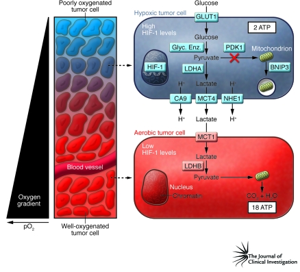
This metabolic reprogramming is orchestrated by HIF-1 through the transcriptional activation of key genes encoding metabolic enzymes, including: LDHA, encoding lactate dehydrogenase A, which converts pyruvate to lactate (3); PDK1, encoding pyruvate dehydrogenase kinase 1, which inactivates the enzyme responsible for conversion of pyruvate to acetyl-CoA, thereby shunting pyruvate away from the mitochondria (4, 5); and BNIP3, which encodes a member of the BCL2 family that triggers selective mitochondrial autophagy (6) (Figure 1). In addition, HIF-1 transactivates GLUT1 (7) — which encodes a glucose transporter that increases glucose uptake to compensate for the fact that, compared with oxidative phosphorylation, glycolysis generates approximately 19-fold less ATP per mole of glucose — and genes encoding the glycolytic enzymes that convert glucose to pyruvate (3). The extracellular acidosis associated with hypoxic tumor cells is due to both increased H+ production and increased H+ efflux through the HIF-1–mediated transactivation of: CA9, which encodes carbonic anhydrase IX (8); MCT4, which encodes monocarboxylate transporter 4 (9); and NHE1, which encodes sodium-hydrogen exchanger 1 (10).
Figure 1. Intratumoral hypoxia and metabolic symbiosis.
Tumors are characterized by gradients of O2 levels, based on the distance of tumor cells from a functional blood vessel. In the schematic, tumor cells surrounding the blood vessel are well oxygenated, whereas the tumor cells more distant from the vessel are poorly oxygenated and express high levels of HIF-1. HIF-1 induces the expression of proteins that increase: uptake of glucose (e.g., glucose transporter 1 [GLUT1]); conversion of glucose to pyruvate (e.g., glycolytic enzymes [Glyc. Enz.]); generation of lactate and H+ (e.g., LDHA); and efflux of these molecules out of the cell (e.g., carbonic anhydrase IX [CA9], sodium-hydrogen exchanger 1 [NHE1], MCT4). Two moles of lactate are produced for each mole of glucose consumed by the hypoxic cell. This increase in glycolytic metabolism is associated with reduced substrate delivery to the mitochondria (through the action of pyruvate dehydrogenase kinase 1 [PDK1]) and reduced mitochondrial mass (as a result of autophagy triggered by BNIP3). As described by Sonveaux et al. in their study in this issue of the JCI (11), aerobic tumor cells express proteins that allow them to take up lactate (e.g., MCT1) and use it (e.g., LDHB), in the presence of O2, as their principal substrate for mitochondrial oxidative phosphorylation. Note that hypoxic cells generate 2 mol of ATP and 2 mol of lactate per mol of glucose consumed, whereas aerobic cells generate 36 mol of ATP per 2 mol of lactate consumed. pO2, partial pressure of oxygen.
Metabolic symbiosis
In this issue of the JCI, the elegant article by Sonveaux, Dewhirst, et al. makes a major contribution to the field of cancer biology (11). The authors demonstrate the existence of a “metabolic symbiosis” between hypoxic and aerobic cancer cells, in which lactate produced by hypoxic cells is taken up by aerobic cells, which use it as their principal substrate for oxidative phosphorylation. As a result, the limited glucose available to the tumor is used most efficiently: hypoxic cells downregulate oxidative phosphorylation in order to maintain redox homeostasis (4, 6) and must consume large amounts of glucose to maintain energy homeostasis (12). At the molecular level, a key player in this symbiotic relationship is monocarboxylate transporter 1 (MCT1), which differs from MCT4 in two respects: the expression of MCT1 is hypoxia repressed rather than hypoxia induced, and it transports lactate into, rather than out of, cancer cells. O2-dependent expression of MCT1 allows aerobic cancer cells to efficiently take up lactate and, in concert with the O2-dependent expression of LDHB, to utilize lactate as an energy substrate, thereby freeing these cells from the need to take up large quantities of glucose (11). Thus, while Sonveaux et al. focused their attention on MCT1, it is important to note that O2 regulates the expression of many key enzymes in these metabolic pathways (Figure 1). It will be interesting to determine whether HIF-1 also controls MCT1 and LDHB expression, perhaps by inducing expression of a transcriptional repressor, as has been described for the O2-dependent regulation of adipogenesis (13).
Was there any precedent that should have alerted us to the existence of this symbiotic relationship between aerobic and hypoxic cancer cells (11)? Of course: the well-known recycling of lactate in exercising muscle. Just as tumors co-opt physiological mechanisms regulating vascularization, which are orchestrated by HIF-1 through transactivation of genes encoding VEGF, stromal-derived factor 1, and other angiogenic factors (14), so do they modulate metabolism through a program that functions to efficiently distribute glucose and lactate to fast-twitch (glycolytic) and slow-twitch (oxidative) muscle fibers.
The ability of tumor cells to adapt to regional variation (i.e., spatial heterogeneity) of oxygenation is remarkable. In another recent publication, Cárdenas-Navia, Dewhirst, and colleagues have also contributed to a large body of literature indicating that a cancer cell that is hypoxic at one moment may be aerobic an hour later and vice versa (15). In other words, there appears to be cyclic variation (i.e., temporal heterogeneity) in oxygenation. This, in turn, implies dynamic regulation of the metabolic symbiosis, such that cells may cycle between lactate-producing and lactate-consuming states.
Therapeutic implications
Hypoxic tumor cells are preferentially resistant to chemotherapy and radiation, thereby leading to treatment failure or disease relapse and, ultimately, to patient mortality (1). In this remarkable paper, Sonveaux et al. (11) demonstrate that treatment of tumor-bearing mice with an inhibitor of MCT1 provides a means to kill cancer cells. When MCT1 is inhibited, the metabolic symbiosis is disrupted: aerobic cells can no longer take up lactate, as demonstrated by the analysis of cultured human cancer cells. Instead, these cells increase their uptake of glucose and thereby deprive hypoxic cells of adequate glucose. Thus, MCT1 inhibition in aerobic cells leads to the death of hypoxic cells. Finally, the authors also show that the growth delay of mouse tumor xenografts that is induced by radiotherapy is increased when treatment is combined with MCT1 inhibition (11).
Recent studies reviewed here and elsewhere (16, 17) have advanced our understanding of the mechanisms whereby the metabolism of glucose, fatty acids, and amino acids is reprogrammed in human cancers. This metabolic reprogramming is a virtually universal characteristic of advanced metastatic disease. This is best illustrated by the observation that the most sensitive clinical test for detecting occult metastases is the uptake of [18F]fluorodeoxyglucose as determined by PET (18). Can a characteristic that is so useful for diagnosis also be exploited for therapy? Research that elucidated the mechanisms by which cancer cells induce blood vessel growth led to the development of angiogenesis inhibitors as anticancer agents over the last decade (19). Inhibitors of HIF-1, LDHA, PDK1, CA9, NHE1, and now MCT1 have shown anticancer effects in tumor xenograft models (11, 17, 20). It is likely that over the next decade, clinical translation of advances in our understanding of cancer metabolism will have an impact on cancer therapy. Furthermore, a treatment strategy in which tumors are deprived of both O2 (through angiogenesis inhibition) and their ability to adapt to hypoxia (through metabolic inhibition) may represent a formidable combination therapy.
Acknowledgments
Work in the author’s laboratory is supported by the American Diabetes Association; NIH grants R01-HL55338, P20-GM78494, P01-HL65608, and N01-HV28180; and the Johns Hopkins Institute for Cell Engineering.
Footnotes
Nonstandard abbreviations used: LDH, lactate dehydrogenase; MCT, monocarboxylate transporter.
Conflict of interest: The author has declared that no conflict of interest exists.
Citation for this article: J. Clin. Invest. 118:3835–3837 (2008). doi:10.1172/JCI37373.
See the related article beginning on page 3930.
References
- 1.Vaupel P., Mayer A. Hypoxia and cancer: significance and impact on clinical outcome. Cancer Metastasis Rev. 2007;26:225–239. doi: 10.1007/s10555-007-9055-1. [DOI] [PubMed] [Google Scholar]
- 2.Helmlinger G., Yuan F., Dellian M., Jain R.K. Interstitial pH and pO2 gradients in solid tumors in vivo: high-resolution measurements reveal a lack of correlation. Nat. Med. 1997;3:177–182. doi: 10.1038/nm0297-177. [DOI] [PubMed] [Google Scholar]
- 3.Semenza G.L., et al. Hypoxia response elements in the aldolase A, enolase 1, and lactate dehydrogenase A gene promoters contain essential binding sites for hypoxia-inducible factor 1. J. Biol. Chem. 1996;271:32529–32537. doi: 10.1074/jbc.271.51.32529. [DOI] [PubMed] [Google Scholar]
- 4.Kim J.W., Tchernyshyov I., Semenza G.L., Dang C.V. HIF-1-mediated expression of pyruvate dehydrogenase kinase: a metabolic switch required for cellular adaptation to hypoxia. Cell Metab. 2006;3:177–185. doi: 10.1016/j.cmet.2006.02.002. [DOI] [PubMed] [Google Scholar]
- 5.Papandreou I., Cairns R.A., Fontana L., Lim A.L., Denko N.C. HIF-1 mediates adaptation to hypoxia by actively downregulating mitochondrial oxygen consumption. Cell Metab. 2006;3:187–197. doi: 10.1016/j.cmet.2006.01.012. [DOI] [PubMed] [Google Scholar]
- 6.Zhang H., et al. Mitochondrial autophagy is a HIF-1-dependent adaptive metabolic response to hypoxia. J. Biol. Chem. 2008;283:10892–10903. doi: 10.1074/jbc.M800102200. [DOI] [PMC free article] [PubMed] [Google Scholar] [Retracted]
- 7.Ebert B.L., Firth J.D., Ratcliffe P.J. Hypoxia and mitochondrial inhibitors regulate expression of glucose transporter-1 via distinct cis-acting sequences. J. Biol. Chem. 1995;270:29083–29089. doi: 10.1074/jbc.270.49.29083. [DOI] [PubMed] [Google Scholar]
- 8.Wykoff C.C., et al. Hypoxia-inducible expression of tumor-associated carbonic anhydrases. Cancer Res. 2000;60:7075–7083. [PubMed] [Google Scholar]
- 9.Ullah M.S., Davies A.J., Halestrap A.P. The plasma membrane lactate transporter MCT4, but not MCT1, is up-regulated by hypoxia through a HIF-1alpha-dependent mechanism. J. Biol. Chem. 2006;281:9030–9037. doi: 10.1074/jbc.M511397200. [DOI] [PubMed] [Google Scholar]
- 10.Shimoda L.A., Fallon M., Pisarcik S., Wang J., Semenza G.L. HIF-1 regulates hypoxic induction of NHE1 expression and alkalinization of intracellular pH in pulmonary arterial myocytes. Am. J. Physiol. Lung Cell Mol. Physiol. 2006;291:L941–L949. doi: 10.1152/ajplung.00528.2005. [DOI] [PubMed] [Google Scholar]
- 11.Sonveaux P., et al. Targeting lactate-fueled respiration selectively kills hypoxic tumor cells in mice. J. Clin. Invest. . 2008;118:3930–3942. doi: 10.1172/JCI36843. [DOI] [PMC free article] [PubMed] [Google Scholar]
- 12.Seagroves T.N., et al. Transcription factor HIF-1 is a necessary mediator of the Pasteur effect in mammalian cells. Mol. Cell. Biol. 2001;21:3436–3444. doi: 10.1128/MCB.21.10.3436-3444.2001. [DOI] [PMC free article] [PubMed] [Google Scholar]
- 13.Yun Z., Maecker H.L., Johnson R.S., Giaccia A.J. Inhibition of PPAR gamma 2 gene expression by the HIF-1-regulated gene DEC1/Stra13: a mechanism for regulation of adipogenesis by hypoxia. Dev. Cell. 2002;2:331–341. doi: 10.1016/S1534-5807(02)00131-4. [DOI] [PubMed] [Google Scholar]
- 14.Liao D., Johnson R.S. Hypoxia: a key regulator of angiogenesis in cancer. Cancer Metastasis Rev. 2007;26:281–290. doi: 10.1007/s10555-007-9066-y. [DOI] [PubMed] [Google Scholar]
- 15.Cárdenas-Navia L.I., et al. The pervasive presence of fluctuating oxygenation in tumors. Cancer Res. 2008;68:5812–5819. doi: 10.1158/0008-5472.CAN-07-6387. [DOI] [PubMed] [Google Scholar]
- 16.Deberardinis R.J., Sayed N., Ditsworth D., Thompson C.B. Brick by brick: metabolism and tumor cell growth. Curr. Opin. Genet. Dev. . 2008;18:54–61. doi: 10.1016/j.gde.2008.02.003. [DOI] [PMC free article] [PubMed] [Google Scholar]
- 17.Kroemer G., Pouyssegur J. Tumor cell metabolism: cancer’s Achilles’ heel. Cancer Cell. 2008;13:472–482. doi: 10.1016/j.ccr.2008.05.005. [DOI] [PubMed] [Google Scholar]
- 18.Gatenby R.A., Gillies R.J. Why do cancers have high aerobic glycolysis? Nat. Rev. Cancer. 2004;4:891–899. doi: 10.1038/nrc1478. [DOI] [PubMed] [Google Scholar]
- 19.Kerbel R.S. Tumor angiogenesis. N. Engl. J. Med. . 2008;358:2039–2049. doi: 10.1056/NEJMra0706596. [DOI] [PMC free article] [PubMed] [Google Scholar]
- 20.Melillo G. Targeting hypoxia cell signaling for cancer therapy. Cancer Metastasis Rev. 2007;26:341–352. doi: 10.1007/s10555-007-9059-x. [DOI] [PubMed] [Google Scholar]


