Abstract
It is well established that ATP is co-secreted with insulin and zinc from pancreatic beta-cells (β-cells) in response to elevations in extracellular glucose concentration. Despite this knowledge, the physiological roles of extracellular secreted ATP and zinc are ill-defined. We hypothesized that secreted ATP and zinc are autocrine purinergic signaling molecules that activate P2X purinergic receptor (P2XR) channels expressed by β-cells to enhance glucose-stimulated insulin secretion (GSIS). To test this postulate, we performed ELISA assays for secreted insulin at fixed time points within a “real-time” assay and confirmed that the physiological insulin secretagogue glucose stimulates secretion of ATP and zinc into the extracellular milieu along with insulin from primary rat islets. Exogenous ATP and zinc alone or together also induced insulin secretion in this model system. Most importantly, the presence of an extracellular ATP scavenger, a zinc chelator, and P2 receptor antagonists attenuated GSIS. Furthermore, mRNA and protein were expressed in immortalized β-cells and primary islets for a unique subset of P2XR channel subtypes, P2X2, P2X3, P2X4, and P2X6, which are each gated by extracellular ATP and modulated positively by extracellular zinc. On the basis of these results, we propose that, within endocrine pancreatic islets, secreted ATP and zinc have profound autocrine regulatory influence on insulin secretion via ATP-gated and zinc-modulated P2XR channels.
Keywords: ATP, Zinc, P2X receptor channels, Insulin secretion, Islets, β-cells
Introduction
Glucose is a prominent physiological insulin secretagogue [1–5]. Elevations in extracellular glucose within the pancreatic islet microenvironment activate a series of events that culminate in secretion of insulin from β-cells [1–9]. These events are initiated by the influx of glucose into β-cells through glucose-specific transporters [1–5]. Elevated intracellular glucose is converted into intracellular ATP, thereby augmenting the ATP/ADP ratio. ADP bound to the K+ ATP channel maintains the membrane potential in a hyperpolarized state [1–5]. However, when the metabolism of the β-cell is accelerated, ATP displaces ADP from the K+ ATP channel [1–5]. As a result, this ATP-regulated channel is silenced, leading to depolarization of the membrane potential [1–5]. This depolarization opens voltage-dependent calcium (Ca2+) channels that facilitate Ca2+ entry from the extracellular environment [1–5]. Ca2+ entry triggered by this glucose- and voltage-dependent mechanism activates the insulin exocytotic pathway [1–5]. Fusion of insulin vesicles with the β-cell plasma membrane leads to the release of the vesicular contents−insulin, ATP, and zinc—into the microenvironment of the islet of Langerhans [1–5, 10–21].
Numerous studies have confirmed that ATP and zinc are secreted from β-cells of islets in response to glucose and other stimuli [10–21]. Furthermore, these studies have definitively shown that zinc is co-packaged and co-secreted with insulin [10–21]. However, it remains unclear whether the secreted ATP or zinc is derived solely from insulin secretory granules or if it emerges from secondary populations of vesicles and/or independent cellular pools of ATP and zinc [10–23]. In fact, studies conducted in multiple cell models and tissues have demonstrated that ATP is released physiologically via several pathways, including exocytosis from presynaptic terminals and diffusion through nucleotide transporters, anion channels, and large transmembrane pores (e.g., connexin hemi-channels and anion channels) [22, 23]. Regardless of the mode of ATP secretion into the islet microenvironment, extracellular ATP functions as a signaling molecule that evokes increases in insulin secretion by stimulating the influx of Ca2+ through P2 purinergic receptors located in the plasma membrane of β-cells [22–29]. P2 purinergic receptors are the sensors and intracellular transducers of extracellular purinergic signals [22–29]. This family of ATP-sensing membrane receptors functions physiologically to mobilize cytoplasmic Ca2+ and induce phospholipase C-mediated phospholipid turnover [22–29]. Furthermore, P2 receptors are subdivided into two classes: ATP-sensing G protein-coupled receptors (P2Y receptors) or ATP-gated cation channels (P2X receptor channels or P2XR channels or P2XRs), based on differences in molecular structure and signal transduction mechanisms [22–29]. Within the islet microenvironment, extracellular purinergic signaling molecules (nucleotides and nucleosides) have been postulated to have autocrine and paracrine functions [30–40]. Furthermore, numerous studies have demonstrated that purinergic molecules influence islet functions such as vascular tone and hormone secretion [30–40]. Specifically, these studies revealed that the activation of β-cell purinergic signaling cascades by exogenously applied ATP stimulated insulin secretion [30–40].
There are numerous immunohistochemical and pharmacological studies confirming the expression of P2Y purinergic receptor subtypes in islets and β-cells [30–40]. However, there is no complete molecular and biochemical characterization detailing the subtypes of P2XR channels expressed by pancreatic islets and β-cells. Furthermore, only a handful of studies suggest that P2XRs are expressed on pancreatic islet cells [38–40]. In studies performed on streptozocin-treated islets, the authors concluded that P2X7, an active player in apoptosis [41], was expressed by infiltrating neutrophils and on α-cells within islets [39, 40]. Another study provides pharmacological evidence of the expression of P2XR channels by pancreatic β-cells [38]. Notably, the activity of P2XR channels is modulated by extracellular divalent metals, such as zinc and cooper [42–44]. This fact is critical to our examination of ATP and zinc as autocrine extracellular signaling molecules and to the intriguing mixture of P2XR channel subtypes found. All expressed P2XR subtypes found in the study herein are opened (gated) by both ATP and zinc.
We believe that ATP and zinc are autocrine and paracrine factors that are secreted within the pancreatic islet microenvironment. Furthermore, we suspect that these extracellular signaling molecules have profound regulatory impact upon insulin secretion. We speculate that we have unveiled an underlying autocrine purinergic signaling loop that may govern minute-by-minute regulation of insulin secretion that is independent of a glucose stimulus. We also believe that this autocrine purinergic signaling loop may synergize with the glucose- and voltage-dependent insulin secretion pathway to fully stimulate insulin secretory granule exocytosis. Taken together, we conclude that P2XR channels govern insulin secretion triggered by the autocrine and paracrine co-ligands ATP and zinc, two ligands that potentiate glucose-stimulated insulin secretion by an autocrine mechanism.
Materials and methods
Rodent β-cell line culture
INS-1 cells, clone 832/13, [45] and βTC3 cells [46] were generous gifts from Dr. Christopher Newgard (Duke University) and Drs. Manju Surana and Norman Fleischer (Albert Einstein College of Medicine), respectively. These rodent insulinoma cell lines were grown in T-150 tissue culture flasks at 37°C and 5% CO2 in a humidified atmosphere. The β-cells were passaged every 5 days using 0.05% trypsin-EDTA. The complete culture medium was RPMI-1640 (Sigma; St. Louis, MO) supplemented with 5 mmol/l D-glucose, 10% fetal bovine serum, 100 U/ml penicillin, 100 μg/ml streptomycin, 10 mmol/l HEPES, 2 mmol/l L-glutamine, 1 mmol/l sodium pyruvate, and 50 μmol/l β-mercaptoethanol.
Rat primary islets of Langerhans isolation and culture
Islets of Langerhans were isolated from male Lewis rats in the Department of Surgery [47]. Briefly, a midline abdominal incision was made; the bile duct was cannulated and injected with cold rodent Liberase (Roche Diagnostics). Thereafter, pancreatic islets were isolated and purified using a standard liberase-based digestion method combined with a Ficoll gradient separation method. An aliquot of islets was counted as “islet equivalents” (IEQ) under a scaled microscope with the use of diphenylthiocarbazone (Sigma; St. Louis, MO) staining. One IEQ was the islet tissue mass equivalent to a spherical islet of 150 μm in diameter.
Additionally, purity was assessed by comparing the relative quantity of dithizone-stained (Sigma; St. Louis, MO) endocrine tissue with unstained exocrine tissue. Islet viability was assessed following staining with acridine orange (100 μg/ml) and ethidium bromide (100 μg/ml) and subjected to two-color fluorescence microscopy. Only islet isolations with >90% viability and >85% purity were used.
Islets were provided to our cell physiology laboratory unattached in a T-150 tissue culture flask. After isolation, islets were cultured in complete Miami Medium 1A (Cellgro/Mediatech; Herndon, VA) supplemented with L-glutamine, 100 U/ml of penicillin, 100 mg/ml of streptomycin, and 10% fetal bovine serum at 26°C and 5% CO2. One day after isolation, 200 IEQs were dispersed into 35-mm tissue culture dishes and then cultured at 37°C and 5% CO2 in a humidified atmosphere until they attached to the tissue culture dish.
Novel co-secretion bioassay for secreted ATP, zinc, and insulin from rat islets
Insulin, ATP, and zinc secretion was assayed in HBSS. Islets grown in 35-mm tissue culture dishes were washed in HBSS followed by an overnight pre-incubation in the same buffer at 37°C and 5% CO2 in a humidified atmosphere. At the time of the experiment, islets were placed on a 37°C slide warmer and washed in HBSS. After stimulating islets with 10 mmol/l glucose, samples of the supernatant were collected at appropriate timepoints (0, 5, 15, 30, and 45 s, and 1, 3, 5, and 30 min). These supernatant samples were separated into three parts, and secreted insulin was measured with a rat/mouse insulin ELISA kit (Millipore/Linco; St. Louis, MO), secreted zinc was detected with a Zinquin fluorescence indicator (Sigma; St. Louis, MO) and secreted ATP was analyzed by mixing with luciferin-luciferase detection reagent (Sigma; St. Louis, MO) [48]. Data are expressed as fold increase of post-stimulation response versus pre-stimulation.
For later experiments focusing on extracellular ATP and zinc signaling effects on insulin secretion, exogenous ATP (500 nmol/l) and/or zinc (20 μmol/l) were added in lieu of glucose as the stimulus and secreted insulin was assessed at 0, 5, 15, 30, and 45 s, and 1, 3, and 5 min via ELISA. In other experiments, glucose was the stimulus in the absence and presence of an ATP scavenger, zinc chelator, and P2 receptor antagonists.
Degenerate and specific RT-PCR to identify P2XR mRNA subtype expression in rodent primary islets and β-cell models
Total RNA was isolated from cells using PureLink Micro-to-Midi-Total RNA Purification System (Invitrogen; Carlsbad, CA). One microgram of total RNA was reverse transcribed to synthesize cDNA using iScript cDNA Synthesis Kit (Bio-Rad; Hercules, CA). The degenerate primers used for amplifying P2X1-P2X5 and P2X7 [49] were primer A: 5′-TTCACCMTYYTCATCAARAACAGCATC-3′ and primer B: 5′-TGGCAAAYCTGAAGTTGWAGCC-3′.
The reaction was run at a 52°C annealing temperature; this reaction generates a 330 base pair (bp) degenerate P2XR PCR product as a single band that contains multiple P2XR subtype sequences and spans multiple introns to insure that the product is not derived from genomic DNA.
The specific primers for amplifying P2X6 [50] were primer A: 5′-GCTCAGGCCAAGAACTTCAC-3′ and primer B: 5′-AAACCGGCCTCTCTATCCAC-3′.
The reaction was run at a 57.8°C annealing temperature; this reaction yields a specific P2X6 PCR product of 533 bp. The length of the PCR products was quantified by agarose gel electrophoresis and visualized by staining with ethidium bromide versus a 100-bp ladder.
Degenerate P2XR PCR products were gel-extracted and purified using a QIAquick gel extraction kit (Qiagen; Valencia, CA). The degenerate P2XR PCR products were ligated into the pGEM-T vector system (Promega; Madison, WI). Ligated P2XR PCR products were transformed into JM109 competent cells (Promega; Madison, WI). The transformed JM109 cells were plated on LB-agar plates containing ampicillin (100 μg/ml), 5-bromo-4-chloro-3-indolyl β-D-galactopyranoside (40 μg/ml), and isopropylthiogalactoside (100 μg/ml). White colonies were selected and cultured in LB medium containing ampicillin (100 μg/ml). Plasmids were isolated from selected JM109 cells using Perfectprep Plasmid Mini Kit (Eppendorf; Westbury, NY). Purified degenerate P2XR PCR product inserts were sequenced by a DNA sequencing core at the University of Alabama at Birmingham.
Western blot analysis to identify P2XR protein subtypes in rodent primary islets and β-cell models
Pancreatic islets and β-cell lines were lysed in a RIPA buffer containing 150 mM NaCl, 1% NP-40, 0.5% deoxycholate, 0.1% SDS, 50 mM pH 8.0 Tris-Cl, and a complete mini-protease inhibitor cocktail tablet (Roche Diagnostics; Mannheim, Germany). Proteins were loaded and run on a 4–12% Bis-Tris SDS-polyacrylamide gel (Invitrogen; Carlsbad, CA) and transferred to a polyvinylidene difluoride (PVDF) membrane (Bio Rad; Hercules, CA). Immunoblotting was performed with rabbit polyclonal antibodies against P2X2, P2X3, P2X4, and P2X6 receptors (Alomone Labs; Jerusalem, Israel) [49–52]. Reactivity was detected by horseradish peroxidase-labeled goat anti-rabbit secondary antibody (New England Biolabs; Ipswich, MA). Enhanced chemiluminescence was used to visualize the secondary antibody.
Immunofluorescence histochemistry to identify P2XR subtype expression in β-cells of islets of Langerhans of rats
Rat pancreatic tissues were fixed with 4% para-formaldehyde. Blocked tissue sections (5% BSA) were incubated in mouse monoclonal insulin antibody (1:100; Lab Vision; Fremont, CA) for 30 min. After washing, Alexa Fluor 488 goat anti-mouse IgG (1:100; Molecular Probes; Eugene, OK) was applied for 30 min. Sections were blocked again for 30 min and then incubated in rabbit polyclonal P2XR antibody (1:500; Alomone Labs; Jerusalem, Israel) [28] for 30 min. After washing, Alexa Fluor 594 goat anti-rabbit IgG (1:100; Molecular Probes; Eugene, OK) was applied for 30 min followed by 4,6-diamidino-2-phenylindole (DAPI; Invitrogen; Carlsbad, CA) for nuclear staining. Images were captured with an inverted Olympus IX70 microscope.
Rat/mouse insulin ELISA assay for secreted insulin
Insulin concentrations were determined from collected supernatant aliquots or samples from co-secretion bioassays by rat/mouse insulin ELISA (Linco Research; St. Charles, MO), according to the manufacturer’s protocol with no modifications.
Zinquin fluorescence assay for secreted zinc
An aliquot of the collected supernatant from co-secretion bioassays was mixed with Zinquin reagent (Sigma; St. Louis, MO) in a 96-well plate. After which, the plate was loaded into a fluorometer plate reader. Fluorescence was recorded at an excitation wavelength of 358 nm and emission wavelength of 497 nm. Data are expressed as fold increase of post-stimulation response versus pre-stimulation.
Luciferase-luciferin bioluminescence assay for secreted ATP
An aliquot of collected supernatant from co-secretion bioassays was mixed with lyophilized luciferase-luciferin reagent (Sigma; St. Louis, MO) in a cuvette. After which, the reaction was placed in a Turner TD 20/20 luminometer. Bioluminescence was recorded after a 15-s photon collection interval. Data are expressed as fold increase of post-stimulation response versus pre-stimulation.
Statistics
Results are expressed as means ± standard error of the mean (SEM). Statistics were performed using the software program Column Descriptive Statistics. Statistical significance was evaluated by t-test. P value of <0.05 was considered significant.
Materials
Analytical grade reagents and deionized water were used. D-glucose, adenosine 5′ triphosphate, zinc chloride, HEPES, diphenylthiocarbazone, dithizone pyridoxal-phosphate-6-azophenyl-2′,4′-disulfonate, diethylene triamine pentaacetic acid, apyrase, suramin, and bovine serum albumin were obtained from Sigma (St. Louis, MO). Sodium chloride, potassium chloride, potassium monophosphate, magnesium sulfate, calcium chloride, β-mercapoethanol, and sodium bicarbonate were purchased from Fisher Scientific. Fetal bovine serum, penicillin, streptomycin, L-glutamine, and sodium pyruvate were acquired from Invitrogen (Carlsbad, CA). All other sources for reagents, chemicals, drugs, or cell culture medium are identified above.
Results
Purinergic-signaling molecules ATP and zinc are secreted from isolated rat islets into the extracellular environment in response to the insulin secretagogue glucose
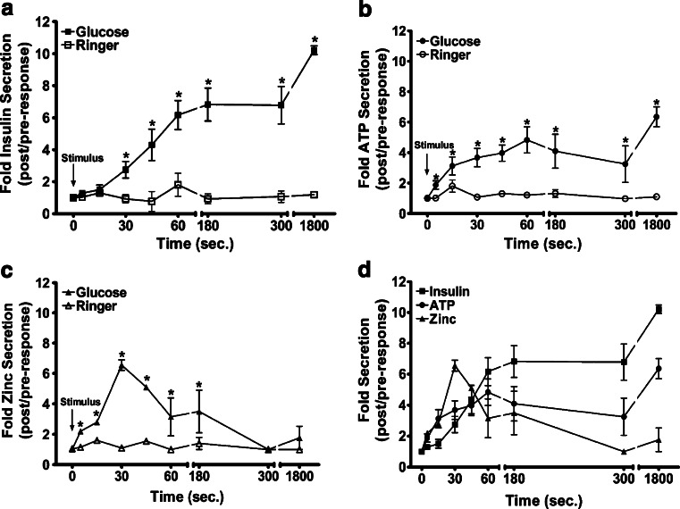
To address the hypothesis, the extracellular secretion of the purinergic-signaling molecules ATP and zinc was examined in parallel with insulin secretion. ATP and zinc were secreted from primary rat islets into the extracellular environment in response to the insulin secretagogue glucose (Fig. 1). Increasing the extracellular glucose concentration to 10 from 3 mmol/L elicited the immediate (0-60 s) secretion of insulin, ATP, and zinc (Fig. 1a-c). During this immediate phase of secretion, extracellular zinc peaked (6.6 ± 0.35 fold) and then declined (3.2 ± 1.3 fold), while insulin and ATP continued to accumulate (Fig. 1d). Between 180 and 300 s, extracellular insulin reached a steady state, but extracellular ATP and zinc declined (Fig. 1d). At later time points, all three molecules were increased at 1,800 s. During this phase of secretion, extracellular zinc increased to 1.8 ± 0.77 fold (Fig. 1c), insulin peaked at 9.5 ± 0.69 fold (Fig. 1a) and ATP peaked at 6.4 ± 0.65 fold (Fig. 1b). Taken together, these results demonstrate that the physiological insulin secretagogue glucose stimulates the extracellular secretion of the purinergic-signaling molecules ATP and zinc. Similar results with regard to insulin versus ATP secretion and insulin versus zinc have been described previously [10–21]; however, we contend that a “real-time” examination of secretion of these three critical mediators has not been assessed simultaneously in the literature.
Fig. 1.
The insulin secretagogue glucose stimulates the extracellular secretion of insulin, ATP, and zinc. Isolated rat primary islets were grown in adherent culture and were stimulated with 10 mmol/l glucose. Samples of the supernatant were collected at fixed time points, divided into three parts, and subsequently analyzed for the presence of secreted insulin, ATP, and zinc via rat/mouse insulin ELISA, luciferase-luciferin bioluminescence, and zinquin fluorescence, respectively. The results show that glucose stimulation elicited insulin, ATP, and zinc secretion. Values represent the means ± SE of three to five experiments. *P < 0.05 vs. pre-stimulation for all time points
Extracellular purinergic signaling molecules participate in glucose-stimulated insulin secretion (GSIS) from isolated rat islets
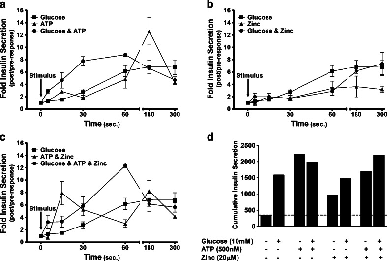
Initially, exogenous ATP and zinc were tested for their potency as insulin secretagogues in rat islets grown in primary culture. Results revealed that these molecules stimulated insulin secretion from primary rat islets (Fig. 2). Figure 2a shows that the insulin stimulatory effect of exogenous ATP was initially observed between 30 and 60 s. ATP-induced extracellular insulin peaked at 180 s to 12.7 ± 2.2 fold. Thereafter, extracellular insulin declined (4.3 ± 0.3 fold at 300 s) but remained elevated compared to baseline. Additionally, immediate and potentiated insulin secretion (5–60 s) was observed in response to the administration of exogenous ATP and glucose. ATP and glucose-induced extracellular insulin peaked to 8.8 ± 0.3 fold at 60 s; after which, extracellular insulin declined to 4.7 ± 0.5 fold over baseline. Figure 2b reveals that the application of exogenous zinc evoked insulin secretion. The zinc-induced increase in extracellular insulin was detected between 30 and 60 s and peaked to 3.7 ± 1.7 fold above baseline. Furthermore, zinc together with glucose stimulated an increase in extracellular insulin that began between 30 and 60 s and peaked at 300 s to 7.3 ± 1.9 fold. Since both P2XR channel ligands are secreted together, the collective effects of ATP and zinc on insulin secretion was also assessed. Figure 2c shows that ATP and zinc stimulated an increase in extracellular insulin that peaked initially to 7.9 ± 1.9 fold at 15 s. After a decline (15–60 s), extracellular insulin peaked again at 180 s to 8.3 ± 1.6 fold over baseline. The results also reveal that ATP, zinc, and glucose induced immediate and potentiated insulin secretion (5–15 s). Extracellular insulin peaked to 12.4 ± 0.4 fold at 60 s and then declined to 5.6 ± 0.8 fold at 300 s. Figure 2d shows the rank order of potency of the insulin secretagogue stimuli used in these studies: ringer (343) < zinc (957) < glucose + zinc (1,475) < ATP + zinc (1,691) ≤ glucose (1,780) < glucose + ATP (1,992) ≤ glucose + ATP + zinc (2,202) ≤ ATP (2,226). The results show that ATP and zinc are potent insulin secretagogues. However, ATP proved to be the most potent insulin secretagogue even over the physiological insulin secretagogue glucose. Additionally, ATP in conjunction with glucose did not display an additive or synergistic effect, thereby suggesting that these molecules function within the same insulin secretory pathway. Nonetheless, these findings suggest that extracellular purinergic signaling molecules stimulate insulin secretion from isolated rat islets.
Fig. 2.
Extracellular ATP and zinc stimulate insulin secretion from isolated rat islets. Isolated rat primary islets were stimulated with 500 nmol/l ATP ± 10 mmol/l glucose (a), 20 μmol/l zinc ± 10 mmol/l glucose (b), and 500 nmol/l ATP + 20 μmol/l zinc ± 10 mmol/l glucose (c). Samples of the supernatant were collected pre-stimulation and post-stimulation (5, 15, 30, 45, 60, 180, and 300 s) and subsequently analyzed for the presence of secreted insulin via a rat/mouse insulin ELISA assay kit. Cumulative insulin secretion was calculated for each trace (d). The results show that ATP and zinc evoked insulin secretion from rat primary islets. Values represent the mean ± SE of three to seven experiments. *P < 0.05 vs. pre-stimulation
To investigate a possible autocrine role for extracellular secreted purinergic signaling molecules in governing islet function, GSIS was evaluated in the presence of the extracellular ATP/ADP phosphatase apyrase and the zinc chelator DTPA. Figure 3 shows that glucose stimulated a 9.5 ± 0.69 fold increase in insulin secretion over basal secretion. However, in the presence of the extracellular ATP- and ADP-scavenging enzyme apyrase, GSIS was attenuated markedly from 9.5 ± 0.69 fold to 1.6 ± 0.4 to 2.2 ± 0.14 fold. Additionally, GSIS was lowered to 1.6 ± 0.4 to 3.6 ± 0.5 fold from 9.5 ± 0.69 fold in the presence of the extracellular zinc chelator DTPA. In the presence of both apyrase and DTPA, GSIS was also reduced but not eliminated fully (5.5 ± 1.3 to 5.7 ± 1.5 fold). It appeared from the results that apyrase and DTPA might be hampering each other’s inhibitory effect when added together in our experiments. The reasons for this are unclear. These results demonstrate that blocking endogenous extracellular ATP and zinc signaling attenuated GSIS. Taken together, these findings support the hypothesis that ATP and zinc are extracellular autocrine purinergic signaling molecules that participate in and potentiate physiological induction of insulin secretion by glucose.
Fig. 3.
Secreted ATP and zinc participate in insulin secretion from isolated rat islets. Glucose-stimulated insulin secretion in isolated rat islets was examined in the presence of an extracellular ATP scavenger (apyrase) and/or a zinc chelator (DTPA). The results show that the insulin secretory effects of glucose were attenuated in the presence of apyrase and DTPA. Values represent the means ± SE of 3–11 experiments. *P < 0.05 vs. glucose-stimulated insulin secretion response. Controls with apyrase alone and DTPA alone were without effect in the absence of glucose (data not shown)
Multiple ATP-gated and zinc-modulated P2X purinergic receptor channels are expressed by β-cells and primary islets
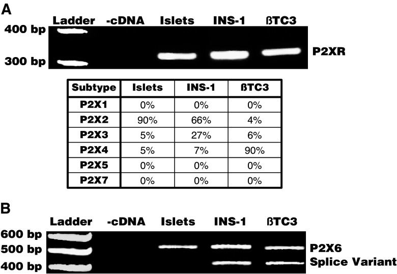
To test the hypothesis that P2XR channels may mediate extracellular ATP and zinc’s insulin secretagogue function, molecular, biochemical, and immunohistochemical studies were performed to investigate P2XR channel expression by normal primary pancreatic islet cultures and immortalized β-cell lines. As a first approach, RT-PCR was performed to detect mRNA expression of the P2XR channel subtypes in primary rat islets and in β-cell models from rat and mouse (INS-1 and βTC3, respectively). Figure 4 reveals that these insulin-secreting primary and immortalized model systems express mRNA for multiple P2XR channel subtypes. A degenerate P2XR PCR product of expected size, 330 bp, was amplified in primary rat islet cDNA samples and in the rat and mouse β-cell models (Fig. 4a). Sequencing of these degenerate P2XR PCR products revealed mRNA expression of P2X2, P2X3, and P2X4 subtypes in islets, INS-1, and βTC3 (see table within Fig. 4a). The only sequence that is not amplified by these degenerate PCR primers is P2X6 [49–52]. As such, specific primers for P2X6 were designed. Results from RT-PCR analysis revealed that P2X6 primers amplified a PCR product of the expected size, 533 bp (Fig. 4b) [50]. A single band was observed from islet cDNA, while two bands were detected in INS-1 and βTC3 β-cells. DNA sequencing confirmed that the larger band was P2X6 and that the smaller amplicon in cell lines was a splice variant that is likely generated due to immortalization (data not shown). PCR products were not observed in the cDNA negative controls (Fig. 4). Taken together, these data suggested that P2X2, P2X3, P2X4, and P2X6 are expressed within islets and β-cells.
Fig. 4.
P2XR channels are expressed at the mRNA level in rodent primary islets and β-cell models. Degenerate (a) and specific (b) RT-PCR was performed to detect P2XR channel mRNA expression in rat primary islets, a rat β-cell model (INS-1), and a mouse β-cell model (βTC3). a The results show that a P2XR PCR product of the expected size, 330bp, was detected in all rodent insulin-secreting model systems. Degenerate PCR primers recognize a consensus sequence of P2X1-P2X5 and P2X7 receptors. b The results also show that a specific P2X6 PCR product of the expected size, 550 bp, was detected in all insulin-secreting model systems. Additionally, a ∼450-bp P2X6 PCR product was unexpectedly detected, and we suspect it to be a splice variant
Western blot analysis using subtype-specific antibodies was performed to confirm the expression of the same P2XR channel subtypes at the protein level. Figure 5 shows representative immunoblots that confirm that multiple P2XR channel subtypes are expressed by primary islets and β-cell models derived from rodent endocrine pancreas. In all insulin-secreting model systems, two immunoreactive protein bands of ∼58 kD (unprocessed precursor isoform) and ∼64 kD (glycosylated isoform) were detected for P2X2 (Fig. 5a). The P2X3 antibody detected a ∼64-kD protein band (glycosylated isoform) in primary islets and both β-cell models (Fig. 5b). As shown in Fig. 5c, ∼45-kD (unprocessed precursor) and ∼60-kD (glycosylated isoform) immunoreactive bands were expressed in all endocrine pancreatic model systems when using the P2X4 antibody to immunoblot. Two immunoreactive isoforms of ∼48 kD (unprocessed precursor) and ∼68 kD (glycosylated isoform) were detected in primary islets and both β-cell models when using the P2X6 antibody to immunoblot (Fig. 5d). In the presence of respective blocking P2XR subtype peptides, antibodies for P2X2, P2X3, P2X4, and P2X6 did not detect protein (data not shown). Taken together, RT-PCR and Western blot analysis provide molecular and biochemical evidence to support the hypothesis that multiple P2XR channels, specifically P2X2, P2X3, P2X4, and P2X6, are expressed by β-cell lines and primary islets in vitro.
Fig. 5.
P2XR channels are expressed at the protein level in rodent primary islets and β-cell models. Western blot analysis was performed to confirm the mRNA-detected P2XR channel subtypes expressed by rat primary islets, INS-1, and βTC3. a The results show an unprocessed precursor isoform (∼58 kD) and a mature, glycosylated isoform (∼64 kD) of P2X2 were detected in islets, INS-1, and βTC3. b A mature, glycosylated isoform (∼64 kD) of P2X3 was detected in all insulin-secreting model systems. c Two mature, glycosylated isoforms (∼54 and ∼60 kD) of P2X4 were detected in all insulin-secreting model systems. d The unprocessed precursor (∼48 kD) and a mature, glycosylated isoform (∼68 kD) of P2X6 were detected in all insulin-secreting model systems
To ascertain whether β-cells, within islets of Langerhans, expressed P2XR channel subtypes, insulin (red) and P2XR channel (green) double-labeling immunofluorescence was performed on rat pancreatic tissue sections. Figure 6 demonstrates that pancreatic β-cells within islets of Langerhans express multiple ATP-gated and zinc-modulated P2XR channels. Results revealed that the majority of cells located within the islet core were immunoreactive for the β-cell marker insulin. Additionally, P2X2 (Fig. 6a), P2X3 (Fig. 6b), P2X4 (Fig. 6c), and P2X6 (Fig. 6d) channel subtypes were expressed by islets. Although more difficult to observe in the images because the plane of focus was set for the islet staining, acinar and duct cells of the pancreas were also positive for these P2XR channel subtypes as suggested from the other laboratories (Fig. 6) [24]. Figure 6 also revealed that insulin-positive cells within islets were immunoreactive for P2X2 (Fig. 6a, merge), P2X3 (Fig. 6b, merge), P2X4 (Fig. 6c, merge), and P2X6 (Fig. 6d, merge) subtypes. Similar results were obtained via immunostaining of isolated rat islets grown in attached primary culture (data not shown). These data demonstrate that pancreatic β-cells within islets of Langerhans express a unique subset of P2XR channels that are gated by extracellular ATP and modulated by extracellular zinc.
Fig. 6.
ATP-gated and zinc-modulated P2XR channel subtypes are expressed by β-cells within islets of Langerhans. Insulin (red) and P2XR channel (green) double-labeling immunofluorescence was performed to examine the expression of P2XR channel subtypes by β-cells within islets of Langerhans. The results show co-localization of anti-insulin staining with anti-P2X2 staining (a, merge), anti-P2X3 staining (b, merge), anti-P2X4 staining (c, merge), and anti-P2X6 staining (d, merge)
P2XR channels and P2Y G protein-coupled receptors participate in glucose-stimulated insulin secretion
To investigate a possible functional role for P2XR channels in β-cells, glucose-stimulated insulin secretion was evaluated in the presence of PPADS (P2XR antagonist) and suramin (P2XR and P2YR antagonist). Results demonstrate that P2XR channels participate in glucose-stimulated insulin secretion in isolated rat islets (Fig. 7). Figure 7 shows that glucose stimulated a 9.5 ± 0.69 fold increase in insulin secretion over basal secretion. However, in the presence of PPADS, GSIS was attenuated to between 6.6 ± 1.6 and 1.8 ± 0.26 fold compared to 9.5 ± 0.69 fold in the absence of the P2XR antagonist. Additionally, glucose only elicited between 4.9 ± 1.7 and 1.1 ± 0.08 fold increases in insulin secretion in the presence of suramin. The stronger inhibitory effect of suramin is likely due to antagonism of P2Y receptors as well as P2XRs. These results demonstrate that inhibition of P2XR channels and P2Y G protein-coupled receptors attenuates GSIS. This finding is consistent with the hypothesis that autocrine purinergic signaling by extracellular purinergic ligands, interacting with their specific P2 receptors, participates in and potentiates GSIS.
Fig. 7.
P2XR channels participate in glucose-stimulated insulin secretion. Glucose-stimulated insulin secretion in isolated rat islets was examined in the presence of a P2XR channels antagonist (PPADS) or a P2XR-channel and P2YR antagonist (suramin). The results show that the insulin secretory effects of glucose were attenuated in the presence of PPADS. Additionally, glucose-induced insulin secretion was further blunted in the presence of suramin. Values represent the means ± SE of three to seven experiments. *P < 0.05 vs. glucose-stimulated insulin secretion response. Controls with PPADS alone and suramin alone were without effect in the absence of glucose (data not shown)
Discussion
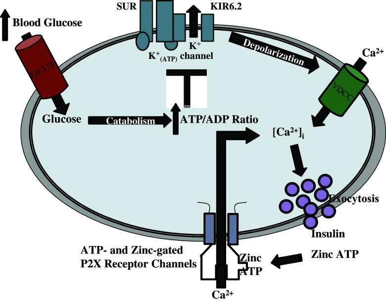
Our results support the view that the interstitium surrounding islets may provide an ideal microenvironment for purinergic signaling. To be more specific, we demonstrate that all components of the P2XR channel signaling pathway are present within the islet microenvironment, namely (1) extracellular secretion of P2XR channel ligands, ATP and zinc; (2) autocrine extracellular ATP and zinc signaling; (3) presence of ATP-gated and zinc-modulated P2XR channel on the β-cell surface within islets, and (4) intimate P2XR channel involvement in glucose-driven insulin secretion. On the basis of these results, we propose that autocrine P2XR channel purinergic signaling may play a profound underlying role in glucose-stimulated insulin secretion. This model is proposed based upon our work and the work of other laboratories (Fig. 8) and is discussed below.
Fig. 8.
Proposed model for an underlying P2XR-channel purinergic signaling loop within the glucose-stimulated insulin secretion coupling pathway. An increase in the extracellular glucose concentration leads to a series of events that result in the secretion of ATP and zinc into the islet microenvironment. Extracellular ATP and zinc bind to and activate a specific subtype of P2XR channels expressed by β-cells. The stimulation of these ATP-gated and zinc-modulated P2XR channels activates calcium influx from extracellular stores that synergizes with voltage-dependent calcium channel-driven calcium influx to enhance glucose-stimulated insulin secretion
It is well established that insulin secretagogues induce the release of insulin, ATP, and zinc into the microenvironment of the islet of Langerhans [1–5, 10–21]. Furthermore, studies have shown definitively that zinc is co-packaged and co-secreted with insulin [10–21]. We postulate that the reduced amount of extracellular zinc compared to insulin detected in co-secretion assays is likely due to the uptake of this biometal by cells of the islets via zinc influx transporters (ZIPs) as a possible recycling mechanism [10–21]. With regard to ATP, it remains unclear whether the secreted ATP is derived from insulin secretory granules, some secondary pool of vesicles, and/or independent cellular pools of ATP [22, 23]. Obermuller and co-workers have documented a difference between the timing of ATP and insulin secretion from β-cells [15]. This finding suggests a possible insulin vesicle-independent mechanism of ATP release. Furthermore, studies conducted in the central nervous system demonstrate that ATP can be released physiologically via several pathways, including exocytosis from neurotransmitter-containing vesicles and diffusion through nucleotide transporters, anion channels, and large transmembrane pores (e.g., connexin hemi-channels and anion channels) [22, 23]. We also speculate that the decline in ATP accumulation in our co-secretion assays is due to degradation by ecto-enzymes such as secreted or membrane-bound ATPases [22, 23]. It was interesting that there appeared to be an initial peak of zinc secretion that preceded that of insulin and ATP secretion. We speculate that there may also be efflux mechanisms for zinc and, possibly, ATP that may be independent of insulin secretion via exocytosis of insulin secretory granules. Taken together, our results confirm that ATP and zinc are secreted in a regulated manner into the islet microenvironment in parallel with insulin. Furthermore, our findings also hint at possible mechanisms by which extracellular purinergic signals are removed as well as the likely generation of adenosine in the islet microenvironment. To our knowledge, this report is the first to assess the co-release of insulin, zinc, and ATP over an extensive time course.
We provide new evidence on several levels that supports autocrine signaling roles for ATP and zinc in governing islet function in general and insulin secretion in particular. Although studies have previously documented exogenous ATP’s insulin secretagogue effects [32–38], no studies have examined the effects of exogenous zinc alone or the collective effects of exogenous ATP and zinc on insulin secretion. Thus, to our knowledge, we show for the first time that exogenous zinc is an insulin secretagogue in the absence and presence of ATP. Our findings also suggest that ATP and zinc may function within the same regulatory pathway governing insulin secretion. The most critical aspect of our work shows that endogenously secreted ATP and zinc play an integral autocrine regulatory role in governing glucose-stimulated insulin secretion. We postulated that the parallel secretion of ATP and zinc may trigger Ca2+ entry via P2XR channels to amplify the Ca2+ signal driven by voltage-activated Ca2+ entry channels (VDCCs) to trigger insulin secretion. This effect is not unlike the co-transmitter activity of ATP at glutamate-driven—and other stimulatory neurotransmitter—postsynaptic membranes to amplify the signal and trigger a more robust postsynaptic signal [53]. Taken together, these data suggest that autocrine ATP and zinc may collaborate to convey an underlying stimulatory signal within the “classical” glucose-stimulated insulin stimulus-secretion pathway.
We provide novel results examining the expression of P2XR channels on normal primary islets and immortalized β-cell lines. Molecular and biochemical examination of the P2XR channel subtypes reveal that multiple ATP-gated and zinc-modulated P2XR channels are expressed by β-cells in vitro and in vivo, namely P2X2, P2X3, P2X4, and P2X6. In our P2X6 RT-PCR studies, we speculate that the smaller band, ∼450 bp, is a P2X6 splice variant that has been reported in other cell systems [54]. We did not detect a different sized protein that correlates with this splice variant via immunoblotting. Alternative splicing has been described previously for P2X1, P2X2, P2X4, and P2X5 [55–59]. In regard to P2XR channel protein, all P2XR channel subtypes have multiple N-linked glycosylation sites [26, 28, 29]. We speculate that the detected P2XR channel protein bands with higher molecular weights represent the glycosylated forms of the proteins as we have demonstrated previously [49]. In support of our findings, previous pharmacological studies suggested that P2XR channels are expressed by β-cells [30–40]. Importantly, the particular P2XR channel subtypes found can heteromultimerize [60]. There is evidence in the literature for P2X2/P2X3, P2X2/P2X6, and P2X4/P2X6 heteromultimers. P2X6 expressed alone only confers functional Ca2+-permeable, non-selective cation currents upon complex glycosylation. More notably, P2X6 modulates the activity and the trafficking of other P2XR channels, most notably P2X2 and P2X4. Thus, we provide both molecular and biochemical evidence that demonstrates that a unique subset of P2XR channel subtypes, which are gated by extracellular ATP and positively modulated by extracellular zinc, are expressed by β-cells within islets in vitro and in vivo.
In closing, this work uncovers important roles for autocrine extracellular ATP and zinc signaling in the islet microenvironment via an intriguing subset of “fast-acting” P2X receptor Ca2+ entry channels expressed on normal β-cells within islets. We hypothesize that this purinergic signaling loop may normally underlie and/or amplify the glucose-driven stimulus-secretion coupling mechanism. Furthermore, we postulate similarly that extracellular ATP and zinc may also modulate insulin secretion on a minute-by-minute basis in the absence of the glucose stimulus. Finally, we propose that this subset of P2XR channels expressed on the β-cell surface may be therapeutic targets to augment and/or rescue insulin secretion in a glucose- and voltage-independent manner. Any approach to augment insulin secretion in an impaired, damaged, or transplanted β-cell or islet or in an implanted stem cell programmed to differentiate into an islet or β-cell would be welcomed to help control the multiple forms of diabetes mellitus.
Acknowledgements
We gratefully acknowledge the funding support of the Juvenile Diabetes Research Foundation (Innovation Grant 5-2007-262 to E.M.S.) and grants of our islet isolation collaborator, Dr. Juan Contreras, M.D. Dr. Contreras is director of the Southern Tissue Center, a funded provider of islets for the southeastern U/S. We thank Cheryl A. Smyth, MS, and Stacie Jenkins-Bryant, BA, colleagues of Dr. Contreras, who were very generous with their time, efforts, and education with regard to islet isolation. Dr. Contreras’s entire group was very generous with time and resources for this study. We also thank Dr. Susan Bellis, Ph.D., and Kristin Hennessy for assistance with and use of the fluorescence microscope. We thank Nai-Lin Cheng for help with molecular biology and biochemistry. We thank Dr. Lydia Aguilar-Bryan for tireless encouragement for entering the field of islet cell biology and diabetes. C.R.W. is supported by an NRSA Predoctoral Fellowship (5 F31 GM078758-02). We acknowledge previous support from the American Physiological Society (APS) in the form of an APS Porter Fellowship. C.R.W. is also currently a Fellow for the APS NIDDK K-12 Minority Outreach Program.
Abbreviations
- ATP
Adenosine 5′ triphosphate
- ADP
Adenosine 5′ diphosphate
- AMP
Adenosine 5′ monophosphate
- β-cells
Beta-cells
- Ca2+
Calcium
- [Ca2+]i
Intracellular calcium concentration
- DTPA
Diethylene triamine pentaacetic acid
- HBSS
HEPES balanced salt solution
- Islets
Islet of Langerhans
- K+ATP channel
Sulphonylurea receptor (SUR) and KIR 6.2 channel complex
- P2XR
P2X purinergic receptor channels
- P2YR
P2Y purinergic G protein-coupled receptors
- PPADS
Pyridoxal-phosphate-6-azophenyl-2′,4′-disulfonate
- VDCCs
Voltage-activated calcium channels
References
- 1.Foster DW, McGarry JD (2000) Glucose, lipid and protein metabolism. Griffin JE, Ojeda SR (eds) Textbook of endocrine physiology, 4th edn. Oxford University Press, New York, pp 393–420
- 2.Straub SG, Sharp GW (2002) Glucose-stimulated signaling pathways in biphasic insulin secretion. Diabetes Metab Res Rev 18:451–463 [DOI] [PubMed] [Google Scholar]
- 3.Straub SG, Sharp GW (2004) Hypothesis: one rate-limiting step controls the magnitude of both phases of glucose-stimulated insulin secretion. Am J Physiol Cell Physiol 287:C565–C571 [DOI] [PubMed] [Google Scholar]
- 4.Newgard CB, McGarry JD (1995) Metabolic coupling factors in pancreatic beta-cell signal transduction. Annu Rev Biochem 64:689–719 [DOI] [PubMed] [Google Scholar]
- 5.Bryan J, Crane A, Vila-Carriles WH, Babenko AP, Aguilar-Bryan L (2005) Insulin secretagogues, sulfonylurea receptors, and K(ATP) channels. Curr Pharm Des 11(21):2699–2676 [DOI] [PubMed] [Google Scholar]
- 6.Aponte G, Gross D, Yamada T (1985) Capillary orientation of rat pancreatic D-cell processes: evidence for endocrine release of somatostatin. Am J Physiol 249:G599–G606 [DOI] [PubMed] [Google Scholar]
- 7.Ballian N, Brunicardi FC (2007) Islet vasculature as a regulator of endocrine pancreas function. World J Surg 31:705–714 [DOI] [PubMed] [Google Scholar]
- 8.Bertrand G, Chapal J, Loubatieres-Mariani MM, Roye M (1987) Evidence for two different P2-purinoceptors on beta cell and pancreatic vascular bed. Br J Pharmacol 91:783–787 [DOI] [PMC free article] [PubMed] [Google Scholar]
- 9.Bratanova-Tochkova TK, Cheng H, Daniel S, Gunawardana S, Liu YJ, Mulvaney-Musa J, Schermerhorn T, Straub SG, Yajima H, Sharp GW (2002) Triggering and augmentation mechanisms, granule pools, and biphasic insulin secretion. Diabetes 51(Suppl 1):S83–S90 [DOI] [PubMed] [Google Scholar]
- 10.Chimienti F, Devergnas S, Favier A, Seve M (2004) Identification and cloning of a beta-cell-specific zinc transporter, ZnT-8, localized into insulin secretory granules. Diabetes 53:2330–2337 [DOI] [PubMed] [Google Scholar]
- 11.Chimienti F, Devergnas S, Pattou F, Schuit F, Garcia-Cuenca R, Vandewalle B, Kerr-Conte J, Van LL, Grunwald D, Favier A, Seve M (2006) In vivo expression and functional characterization of the zinc transporter ZnT8 in glucose-induced insulin secretion. J Cell Sci 119:4199–4206 [DOI] [PubMed] [Google Scholar]
- 12.Chimienti F, Favier A, Seve M (2005) ZnT-8, a pancreatic beta-cell-specific zinc transporter. Biometals 18:313–317 [DOI] [PubMed] [Google Scholar]
- 13.Zalewski PD, Millard SH, Forbes IJ, Kapaniris O, Slavotinek A, Betts WH, Ward AD, Lincoln SF, Mahadevan I (1994) Video image analysis of labile zinc in viable pancreatic islet cells using a specific fluorescent probe for zinc. J Histochem Cytochem 42:877–884 [DOI] [PubMed] [Google Scholar]
- 14.Hazama A, Hayashi S, Okada Y (1998) Cell surface measurements of ATP release from single pancreatic beta cells using a novel biosensor technique. Pflugers Arch 437:31–35 [DOI] [PubMed] [Google Scholar]
- 15.Obermuller S, Lindqvist A, Karanauskaite J, Galvanovskis J, Rorsman P, Barg S (2005) Selective nucleotide-release from dense-core granules in insulin-secreting cells. J Cell Sci 118:4271–4282 [DOI] [PubMed] [Google Scholar]
- 16.Hellman B, Dansk H, Grapengiesser E (2004) Pancreatic beta-cells communicate via intermittent release of ATP. Am J Physiol Endocrinol Metab 286:E759–E765 [DOI] [PubMed] [Google Scholar]
- 17.Leitner JW, Sussman KE, Vatter AE, Schneider FH (1975) Adenine nucleotides in the secretory granule fraction of rat islets. Endocrinology 96:662–677 [DOI] [PubMed] [Google Scholar]
- 18.Grapengiesser E, Dansk H, Hellman B (2004) Pulses of external ATP aid to the synchronization of pancreatic beta-cells by generating premature Ca(2+) oscillations. Biochem Pharmacol 68:667–674 [DOI] [PubMed] [Google Scholar]
- 19.Leitner JW, Sussman KE, Vatter AE, Schneider FH (1975) Adenine nucleotides in the secretory granule fraction of rat islets. Endocrinology 96:662–677 [DOI] [PubMed] [Google Scholar]
- 20.Qian WJ, Gee KR, Kennedy RT (2003) Imaging of Zn2+ release from pancreatic beta-cells at the level of single exocytotic events. Anal Chem 75:3468–3475 [DOI] [PubMed] [Google Scholar]
- 21.Qian WJ, Peters JL, Dahlgren GM, Gee KR, Kennedy RT (2004) Simultaneous monitoring of Zn2+ secretion and intracellular Ca2+ from islets and islet cells by fluorescence microscopy. Biotechniques 37:922–930 [DOI] [PubMed] [Google Scholar]
- 22.Schwiebert EM, Zsembery A (2003) Extracellular ATP as a signaling molecule for epithelial cells. Biochim Biophys Acta 1615:7–32 [DOI] [PubMed] [Google Scholar]
- 23.Schwiebert EM, Liang L, Cheng NL, Olteanu D, Richards-Williams C, Welty EA, Zsembery A (2005) Extracellular ATP- and zinc-gated P2X receptor calcium entry channels: physiological sensors and therapeutic targets. Purinergic Signal 1(4):299–310 [DOI] [PMC free article] [PubMed] [Google Scholar]
- 24.Novak I (2007) Purinergic receptors in the endocrine and exocrine pancreas. Purinergic Signal. doi:dx.doi.org/10.1007/s11302-007-9087-6 [DOI] [PMC free article] [PubMed]
- 25.Burnstock G (2007) Purine and pyrimidine receptors. Cell Mol Life Sci 64:1471–1483 [DOI] [PMC free article] [PubMed] [Google Scholar]
- 26.North RA (2002) Molecular physiology of P2X receptors. Physiol Rev 82:1013–1067 [DOI] [PubMed] [Google Scholar]
- 27.Ralevic V, Burnstock G (1998) Receptors for purines and pyrimidines. Pharmacol Rev 50:413–492 [PubMed] [Google Scholar]
- 28.Roberts JA, Vial C, Digby HR, Agboh KC, Wen H, Catterbury-Thomas A, Evans RJ (2006) Molecular properties of P2X receptors. Pflugers Arch 452:486–500 [DOI] [PubMed] [Google Scholar]
- 29.Khakh BS, North RA (2006) P2X receptors as cell-surface ATP sensors in health and disease. Nature 442:527–532 [DOI] [PubMed] [Google Scholar]
- 30.Novak I, Hede SE, Hansen MR (2008) Adenosine receptors in rat and human pancreatic ducts stimulate chloride transport. Pflugers Arch 456:437–447 [DOI] [PubMed] [Google Scholar]
- 31.Tuduri E, Filiputti E, Carneiro E, Quesada I (2008) Inhibition of Ca2+ signaling and glucagon secretion in mouse pancreatic {alpha}-cells by extracellular ATP and purinergic receptors. Am J Physiol Endocrinol Metab. doi:dx.doi.org/10.1152/10.1152/ajpendo.00641.2007 [DOI] [PubMed]
- 32.Bertrand G, Chapal J, Loubatieres-Mariani MM, Roye M (1987) Evidence for two different P2-purinoceptors on beta cell and pancreatic vascular bed. Br J Pharmacol 91:783–787 [DOI] [PMC free article] [PubMed] [Google Scholar]
- 33.Verspohl EJ, Johannwille B, Waheed A, Neye H (2002) Effect of purinergic agonists and antagonists on insulin secretion from INS-1 cells (insulinoma cell line) and rat pancreatic islets. Can J Physiol Pharmacol 80:562–568 [DOI] [PubMed] [Google Scholar]
- 34.Petit P, Hillaire-Buys D, Manteghetti M, Debrus S, Chapal J, Loubatieres-Mariani MM (1998) Evidence for two different types of P2 receptors stimulating insulin secretion from pancreatic B cell. Br J Pharmacol 125:1368–1374 [DOI] [PMC free article] [PubMed] [Google Scholar]
- 35.Petit P, Manteghetti M, Puech R, Loubatieres-Mariani MM (1987) ATP and phosphate-modified adenine nucleotide analogues. Effects on insulin secretion and calcium uptake. Biochem Pharmacol 36:377–380 [DOI] [PubMed] [Google Scholar]
- 36.Chapal J, Loubatieres-Mariani MM, Roye M (1981) Effect of adenosine and phosphated derivatives on insulin release from the newborn dog pancreas. J Physiol (Paris) 77:873–875 [PubMed] [Google Scholar]
- 37.Fernandez-Alvarez L, Hillaire-Buys D, Loubatieres-Mariani MM, Gomis R, Petit P (2001) P2 receptor agonists stimulate insulin release from human pancreatic islets. Pancreas 22:69–71 [DOI] [PubMed] [Google Scholar]
- 38.Farret A, Vignaud M, Dietz S, Vignon J, Petit P, Gross R (2004) P2Y purinergic potentiation of glucose-induced insulin secretion and pancreatic beta-cell metabolism. Diabetes 53(Suppl 3):S63–S66 [DOI] [PubMed] [Google Scholar]
- 39.Coutinho-Silva R, Parsons M, Robson T, Burnstock G (2001) Changes in expression of P2 receptors in rat and mouse pancreas during development and ageing. Cell Tissue Res 306:373–383 [DOI] [PubMed] [Google Scholar]
- 40.Coutinho-Silva R, Parsons M, Robson T, Lincoln J, Burnstock G (2003) P2X and P2Y purinoceptor expression in pancreas from streptozotocin-diabetic rats. Mol Cell Endocrinol 204:141–154 [DOI] [PubMed] [Google Scholar]
- 41.Liang L, Schwiebert EM (2005) Large pore formation uniquely associated with P2X7 purinergic receptor channels. Am J Physiol Cell Physiol 288(2):C240–C242 [DOI] [PubMed] [Google Scholar]
- 42.Coddou C, Morales B, Huidobro-Toro JP (2003) Neuromodulator role of zinc and copper during prolonged ATP applications to P2X4 purinoceptors. Eur J Pharmacol 472:49–56 [DOI] [PubMed] [Google Scholar]
- 43.Cuna-Castillo C, Morales B, Huidobro-Toro JP (2000) Zinc and copper modulate differentially the P2X4 receptor. J Neurochem 74:1529–1537 [DOI] [PubMed] [Google Scholar]
- 44.Xiong K, Peoples RW, Montgomery JP, Chiang Y, Stewart RR, Weight FF, Li C (1999) Differential modulation by copper and zinc of P2X2 and P2X4 receptor function. J Neurophysiol 81:2088–2094 [DOI] [PubMed] [Google Scholar]
- 45.Hohmeier HE, Mulder H, Chen G, Henkel-Rieger R, Prentki M, Newgard CB (2000) Isolation of INS-1-derived cell lines with robust ATP-sensitive K+ channel-dependent and -independent glucose-stimulated insulin secretion. Diabetes 49:424–430 [DOI] [PubMed] [Google Scholar]
- 46.D’Ambra R, Surana M, Efrat S, Starr RG, Fleischer N (1990) Regulation of insulin secretion from beta-cell lines derived from transgenic mice insulinomas resembles that of normal beta-cells. Endocrinology 126:2815–2822 [DOI] [PubMed] [Google Scholar]
- 47.Eckhoff DE, Eckstein C, Smyth CA, Vilatoba M, Bilbao G, Rahemtulla FG, Young CJ, Thompson J, Chaudry IH, Contreras JL (2004) Enhanced isolated pancreatic islet recovery and functionality in rats by 17beta-estradiol treatment of brain death donors. Surgery 136:336–345 [DOI] [PubMed] [Google Scholar]
- 48.Taylor AL, Kudlow BA, Marrs KL, Gruenert DC, Guggino WB, Schwiebert EM (1998) Bioluminescence detection of ATP release mechanisms in epithelia. Am J Physiol 275:C1391–C1406 [DOI] [PubMed] [Google Scholar]
- 49.Taylor AL, Schwiebert LM, Smith JJ, King C, Jones JR, Sorscher EJ, Schwiebert EM (1999) Epithelial P2X purinergic receptor channel expression and function. J Clin Invest 104:875–884 [DOI] [PMC free article] [PubMed] [Google Scholar]
- 50.Liang L, Zsembery A, Schwiebert EM (2005) RNA interference targeted to multiple P2X receptor subtypes attenuates zinc-induced calcium entry. Am J Physiol Cell Physiol 289:C388–C396 [DOI] [PubMed] [Google Scholar]
- 51.Schwiebert EM, Wallace DP, Braunstein GM, King SR, Peti-Peterdi J, Hanaoka K, Guggino WB, Guay-Woodford LM, Bell PD, Sullivan LP, Grantham JJ, Taylor AL (2002) Autocrine extracellular purinergic signaling in epithelial cells derived from polycystic kidneys. Am J Physiol Renal Physiol 282:F763–F775 [DOI] [PubMed] [Google Scholar]
- 52.Schwiebert LM, Rice WC, Kudlow BA, Taylor AL, Schwiebert EM (2002) Extracellular ATP signaling and P2X nucleotide receptors in monolayers of primary human vascular endothelial cells. Am J Physiol Cell Physiol 282:C289–C301 [DOI] [PubMed] [Google Scholar]
- 53.Burnstock G (2007) Physiology and pathophysiology of purinergic neurotransmission. Physiol Rev 87(2):659–797 [DOI] [PubMed] [Google Scholar]
- 54.da Silva RL, Resende RR, Ulrich H (2007) Alternative splicing of P2X6 receptors in developing mouse brain and during in vitro neuronal differentiation. Exp Physiol 92:139–145 [DOI] [PubMed] [Google Scholar]
- 55.Dhulipala PD, Wang YX, Kotlikoff MI (1998) The human P2X4 receptor gene is alternatively spliced. Gene 207:259–266 [DOI] [PubMed] [Google Scholar]
- 56.Le KT, Paquet M, Nouel D, Babinski K, Seguela P (1997) Primary structure and expression of a naturally truncated human P2X ATP receptor subunit from brain and immune system. FEBS Lett 418:195–199 [DOI] [PubMed] [Google Scholar]
- 57.Ohkubo T, Yamazaki J, Nakashima Y, Kitamura K (2000) Presence and possible role of the spliced isoform of the P2X1 receptor in rat vascular smooth muscle cells. Pflugers Arch 441:57–64 [DOI] [PubMed] [Google Scholar]
- 58.Simon J, Kidd EJ, Smith FM, Chessell IP, Murrell-Lagnado R, Humphrey PP, Barnard EA (1997) Localization and functional expression of splice variants of the P2X2 receptor. Mol Pharmacol 52:237–248 [DOI] [PubMed] [Google Scholar]
- 59.Worthington RA, Dutton JL, Poronnik P, Bennett MR, Barden JA (1999) Localisation of P2X receptors in human salivary gland epithelial cells and human embryonic kidney cells by sodium dodecyl sulfate-polyacrylamide gel electrophoresis/Western blotting and immunofluorescence. Electrophoresis 20:2065–2070 [DOI] [PubMed] [Google Scholar]
- 60.Stojilkovic SS, Tomic M, He ML, Yan Z, Koshimizu TA, Zemkova H (2005) Molecular dissection of purinergic P2X receptor channels. Ann NY Acad Sci 1048:116–130 [DOI] [PubMed] [Google Scholar]


