Summary
Brown fat can increase energy expenditure and protect against obesity through a specialized program of uncoupled respiration. We show here by in vivo fate mapping that brown but not white fat cells arise from precursors that express myf5, a gene previously thought to be expressed only in the myogenic lineage. Notably, the transcriptional regulator, PRDM16 controls a bidirectional cell fate switch between skeletal myoblasts and brown fat cells. Loss of PRDM16 from brown fat precursors causes a loss of brown fat characteristics and promotes muscle differentiation. Conversely, ectopic expression of PRDM16 in myoblasts induces their differentiation into brown fat cells. PRDM16 stimulates brown adipogenesis by binding to PPARγ and activating its transcriptional function. Finally, PRDM16-deficient brown fat displays an abnormal morphology, reduced thermogenic gene expression and elevated expression of muscle-specific genes. Taken together, these data indicate that PRDM16 specifies the brown fat lineage from a progenitor that expresses myoblast markers and is not involved in white adipogenesis.
Keywords: PRDM16, brown fat, skeletal muscle, myoblasts, UCP1, PGC-1α, PGC-1β, PPARα, PPARγ, obesity, thermogenesis, mitochondrial biogenesis
Introduction
The epidemic of obesity, closely associated with increases in diabetes, hypertension, hyperlipidemia, cancer and other disorders, has propelled a major interest in adipose cells and tissues. Adipose tissues contain two distinct types of fat cells, white and brown. White fat cells are specialized for the storage of chemical energy as triglycerides, while brown fat cells dissipate chemical energy in the form of heat1. Most fat depots can be characterized as either brown or white but some brown fat cells can also be found dispersed throughout white fat depots in rodents and humans2–4. Similarities in cell morphology, lipid metabolism and patterns of gene expression between the two fat cell types has led most investigators to assume that they share a common developmental origin5–7.
Until quite recently, brown adipose tissue (BAT) was thought to be of metabolic importance only in smaller mammals and infant humans. Recent studies using PET scanning, however, suggest that adult humans have several discrete areas of metabolically active BAT8. BAT may thus play a much more important role in human metabolism than was previously appreciated. The manipulation of fat stores is an obvious therapeutic dream, but disruption of the normal differentiation or development of white adipose tissues (WAT) causes ectopic lipid storage and severe pathology (lipodystrophy) in both humans and experimental animals. Promotion of increased BAT development in humans, however, offers the possibility of increasing energy expenditure without necessarily causing dysfunction in other tissues. Indeed, experimental increases in BAT in animals have been associated with a lean and healthy phenotype4, 9–11. By contrast, loss of BAT function is linked to obesity and metabolic disease12.
The ability to alter BAT development in a measured way depends upon a thorough understanding of the regulatory systems that control determination in this cell type. Several transcriptional suppressors of BAT development have been identified, including pRB, p107 and RIP14013–15. Positive transcriptional regulators include FOXC29 and PRDM1616, but only PRDM16 has been shown to determine brown fat cell fate in a cell-autonomous manner. This protein, which contains several zinc fingers and a PR domain, can turn on a full set of BAT-selective genes when expressed in WAT precursors in culture or in vivo, while turning off the expression of several white fat-enriched genes16, 17. These actions of PRDM16 are, at least partly, mediated by its interaction with other transcriptional co-regulators, PGC-1α and PGC-1β and do not require DNA binding of PRDM16. In this report, we show that PRDM16 can powerfully control a robust, bidirectional switch in cell fate between skeletal myoblasts and brown fat cells. Furthermore, lineage tracing studies in vivo show that depots of BAT but not WAT arise from myf5-expressing precursors that brown fat shares with skeletal muscle.
Results
Knockdown of PRDM16 in brown fat cells induces skeletal myogenesis
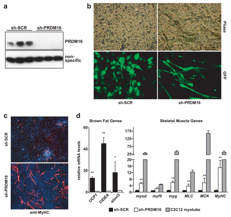
To examine in detail the cellular and molecular consequence of PRDM16 depletion, we used adenoviral vectors to express a sh-SCR (scrambled) control or a shRNA targeting PRDM16 in primary brown fat preadipocytes. PRDM16 protein was virtually undetectable in cultures expressing the sh-PRDM16 construct (Fig. 1a). By day 4 of adipogenic differentiation, cultures expressing the scrambled shRNA had undergone normal brown fat cell differentiation (Fig. 1b). In striking contrast, sh-PRDM16-expressing cultures contained long tube-like cells interspersed amongst fat cells that were strongly marked by GFP which was co-expressed from the adenoviral vector. In fact, the cells that adopted this morphology were those that had preferentially taken-up and expressed the sh-PRDM16 construct (Fig. 1b). Immunohistochemistry using a pan-skeletal myosin heavy chain (MyHC) antibody unequivocally identified these cells as skeletal myocytes (Fig. 1c). The myoctes were noted here but not previously16 because these studies achieved a more complete knock-down of PRDM16; and because the presence of the GFP gene in the adenoviral vector allowed a more focused examination of the transduced cells. Gene-expression analysis showed that knockdown of PRDM16 completely ablated the expression of brown fat cell-selective genes such as UCP1, CIDEA and elovl3/cig30 (Fig. 1d). Moreover, reduction of PRDM16 was associated with a large increase in the expression of myogenic genes, including: myod, myogenin (myg), myosin light chain (MLC), muscle creatine kinase (MCK) and MyHC. Knockdown of PRDM16 induced the expression of these genes to between 5–25% of their levels in C2C12 myotubes, a result generally consistent with the MyHC protein expression observed immunocytochemically in approximately 10–15% of the cells in these cultures. These results strongly suggest that PRDM16 functions in brown fat precursors to restrict skeletal muscle gene expression and development.
Fig. 1. Knockdown of PRDM16 in primary brown fat cells induces skeletal myogenesis.
(a) Western blot analysis for PRDM16 in primary brown fat cell cultures transduced with adenovirus expressing shRNA targeted to PRDM16 or a scrambled (SCR) control shRNA. (b) These cultures were visualized by phase contrast microscopy and by GFP fluorescence. (c) Immunocytochemistry for skeletal Myosin Heavy Chain (MyHC) expression. (d) Gene expression at day 4 of adipocyte differentiation including BAT-selective and skeletal muscle-specific genes (as indicated). C2C12 myotubes were also assayed for their expression of muscle-specific genes (n=3, error bars represent ± SD; *p<0.05, **p<0.01).
Brown fat and skeletal muscle but not white fat arise from myf5-expressing progenitors
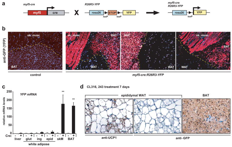
The induction of skeletal muscle in cultures of brown fat preadipocytes suggested a surprising developmental relationship between these two cell lineages. To directly address this question, we performed lineage tracing experiments in mice. Knock-in mice expressing cre recombinase from the regulatory elements of the skeletal muscle-specific myf5 gene18 were crossed with indicator mice that express YFP from the rosa26 gene locus (R26R3-YFP) in a cre-dependent manner. Myf5 is a myogenic regulatory factor that is expressed in committed skeletal myogenic precursors that were previously thought to give rise exclusively to skeletal muscle. Recombination at the rosa26 locus induced by cre recombinase is heritable and irreversible allowing the developmental progression of myf5-expressing descendents to be traced by YFP expression19 (Fig. 2a). Importantly, these myf5-cre knock-in mice have been successfully used to drive skeletal muscle-specific cre expression in other studies20–22.
Fig. 2. Brown fat and skeletal muscle arise from myf5-expressing precursors.
(a)myf5-cre mice were intercrossed with indicator mice that have a YFP gene integrated into the rosa26 locus downstream of a floxed transcriptional stop sequence (R26R3-YFP). Expression of Cre recombinase excises the stop sequence to irreversibly activate YFP expression. (b) Immunohistochemistry to detect YFP (GFP) expression in skeletal muscle (sk. musc), BAT and WAT from control (myf5-cre negative) and myf5-cre:R26R3-YFP mice. (c) Real-time PCR analysis of YFP mRNA levels in: liver; gluteal (glut), inguinal (ing) and epididymal (epid) WAT; skeletal muscle (skM) and BAT (n= 4/group; error bars are ± SEM; **p<0.01). (d) UCP1 and GFP expression in WAT and BAT from CL316, 243 treated myf5-cre:R26R3-YFP mice.
To examine the potential contribution of myf5-expressing progenitors to the adipose anlagen, immunohistochemistry with anti-GFP antibodies was used to localize expression of the YFP reporter gene in skeletal muscle, BAT and WAT from the interscapular region of 2–3 month old Myf5-Cre:R26R3-YFP mice. In myf5-cre negative (control) mice that harbor the R26R3-YFP reporter, YFP was not expressed in any tissues (Fig. 2b). However, in myf5-cre expressing mice, YFP was readily detected in skeletal muscle and BAT but not in WAT. YFP protein was also strongly expressed in the perirenal BAT (Figure S1-A) but not in any classic WAT depots (not shown). Moreover, real-time PCR showed that YFP mRNA was induced only in BAT depots and in skeletal muscle from myf5-cre expressing mice, and at comparable levels (Fig. 2c and Fig. S1-b). YFP mRNA expression was not activated in other tissues examined including the gluteal, inguinal and epididymal WAT depots (Fig. 2c and Fig. S1-b). These results indicate that myf5-expressing cells specifically gave rise to skeletal muscle and brown fat cells. Notably, myf5 mRNA itself was not detected in mature BAT (Fig. S1-c), indicating that it was transiently expressed at an earlier developmental stage.
In newborn mammals, BAT is apparent at distinct anatomical sites including the interscapular, perirenal and axillary depots. It is also well established that brown fat cells emerge in WAT in response to chronic cold exposure or prolonged β-adrenergic stimulation2, 4, 23. To determine whether these β-adrenergic induced brown fat cells in WAT are also derived from myf5-expressing precursors, we treated adult myf5-cre:R26R3-YFP mice with a selective β3-adrenergic agonist, CL 316, 243, for 7 days, and examined whether the newly formed brown fat cells expressed YFP. As shown in Fig. 2d, this treatment induced the development of numerous clusters of UCP1-expressing brown fat cells in epididymal WAT. However, these brown fat cells did not express the YFP reporter gene (Fig. 2d), whereas the interscapular brown fat cells from the same mice were readily marked by YFP expression (Fig. 2d). These results suggest that the brown fat cells that develop in WAT in response to β-adrenergic signaling have an independent developmental origin from the distinct and classic BAT depots that develop before birth.
PRDM16 induces brown adipogenesis in C2C12 and primary myogenic cells
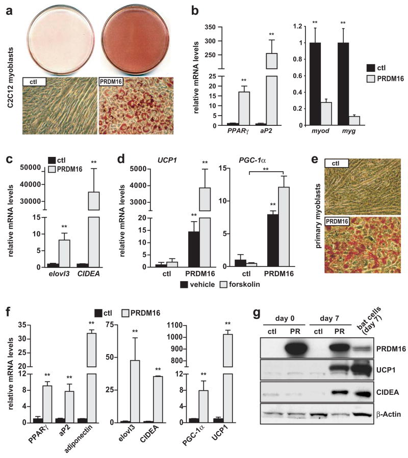
BAT depots arise from myf5-expressing cells during development. To investigate whether PRDM16 is sufficient to specify brown fat fate in skeletal myogenic progenitors, we ectopically expressed PRDM16 in the C2C12 myoblast cell line. First, C2C12 myoblasts were transduced with control or PRDM16-expressing retroviruses, and then exposed to pro-myogenic culture conditions to induce terminal myocyte differentiation. C2C12 cells expressing the control virus differentiated efficiently into multinucleated MyHC-expressing myotubes (Fig. S2a). PRDM16-expressing cells, however, failed to undergo myogenic differentiation (Fig. S2a). Gene expression analysis showed that PRDM16 blocked the induction of myotube-specific genes including: myod, myogenin (myg), mrf4, MyHC and MCK (Fig. S2b). Control and PRDM16-expressing C2C12 myoblasts were also stimulated to undergo adipogenesis using adipogenic inducers. Under these conditions, the control cultures differentiated into multinucleated skeletal myotubes, while the PRDM16-expressing cells uniformly differentiated into lipid filled adipocytes, as shown by Oil-Red-O staining (Fig. 3a). Gene expression studies showed that PRDM16 expression increased the mRNA levels of adipocyte-specific genes, including a 20-fold increase in PPARγ and 250-fold elevation of aP2/FABP4, along with decreased levels of myogenic genes such as myod and myg (Fig. 3b). Importantly, adipocytes induced by PRDM16 expression also expressed elevated levels of brown fat cell-specific genes such as elovl3 and CIDEA (more than 30,000-fold; Fig. 3c), as well as the thermogenic genes UCP1 and PGC-1α (Fig. 3d). Moreover, as in real brown fat cells, UCP1 and PGC-1α were further induced in PRDM16-derived adipocytes by elevating cAMP levels through forskolin treatment (Fig. 3d). These data demonstrate that PRDM16 expression alone is sufficient to drive brown adipocyte differentiation in committed and clonal skeletal myogenic cells.
Fig. 3. PRDM16 stimulates brown adipocyte differentiation in skeletal myoblasts.
(a–d) C2C12 myoblasts expressing retroviral PRDM16 or vector control (ctl) were stained with Oil-Red-O 6 days after inducing adipocyte differentiation (a); and analyzed by real-time PCR for their expression of markers specific to: adipocytes (PPARγ, aP2) (b, left); skeletal muscle (myod, myg) (b, right); BAT (elovl3, CIDEA) (c); and thermogenesis (UCP1, PGC-1α) (d). (e–g) ctl and PRDM16-expressing primary myoblasts were stained with Oil-Red-O 7 days after inducing adipocyte differentiation (e). Real-time PCR analysis of genes expressed selectively in adipocytes (PPARγ, aP2, adiponectin) (left); BAT (elovl3, CIDEA) and during thermogenesis (PGC-1α, UCP1). (g) Western blot analysis before (day 0) and after 7 days of differentiation. (n=4; error bars are ± SD; **p<0.05).
To further explore the adipogenic action of PRDM16, we ectopically expressed PRDM16 in primary mouse myoblasts isolated from postnatal skeletal muscle. As observed in C2C12 cells, control vector (ctl)–expressing cells differentiated into multinucleated myotubes in response to an adipogenic induction cocktail. By contrast, PRDM16-transduced myoblasts differentiated at near 100% efficiency into fat-storing adipocytes (Fig. 3e). Consistent with their morphological differentiation, PRDM16-transduced cultures expressed elevated mRNA levels of pan-adipocyte specific genes, such as PPARγ, aP2 and adiponectin (Fig. 3f). Moreover, adipocytes derived from PRDM16-expressing myogenic precursors strongly activated the expression of brown fat cell-specific genes including: elovl3, CIDEA, PGC-1α and a 1000-fold increase in UCP1 levels (Fig. 3f). By western blot analysis, PRDM16-derived adipocytes expressed the brown fat cell selective UCP1 and CIDEA proteins to levels comparable with that in bona fide brown fat cells (Fig. 3g). Altogether, these results demonstrate that PRDM16 activates a complete program of brown fat cell differentiation in muscle precursor cells.
PRDM16 binds to PPARγ and coactivates its transcriptional function
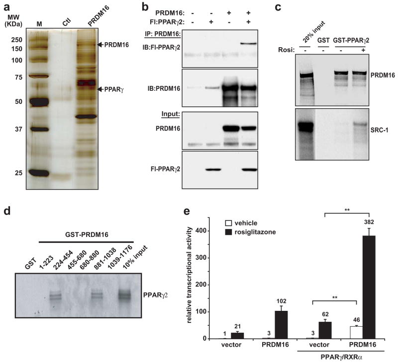
To explore mechanisms that could explain the pro-adipogenic actions of PRDM16, we used an unbiased approach to identify binding partners of PRDM16. To this end, the PRDM16 protein complex was immunopurified from fat cells and analyzed by mass spectrometry (Table S1). Of particular interest was the identification of PPARγ as a near-stoichiometric component of the PRDM16 complex (Fig. 4a). PPARγ was the only DNA-binding transcriptional component found in the PRDM16 complex. Independent co-expression assays in COS-7 cells again showed PPARγ in a complex with PRDM16 (Fig. 4b). This interaction is likely to be direct, since a full-length GST-PPARγ2 fusion protein purified from baculovirus infected insect cells bound to in vitro translated PRDM16 (Fig. 4c). PRDM16 and PPARγ2 interacted in a non-ligand dependent manner, since the binding was equivalent with or without the addition of PPARγ ligand, rosiglitazone, to the binding reaction. Steroid receptor coactivator-1 (SRC-1), by contrast, interacted with purified GST-PPARγ only in the presence of rosiglitazone (Fig. 4c). Domain mapping experiments using GST immunoaffinity assays identified two regions in PRDM16 that bound to PPARγ corresponding to zinc-finger-1 (ZF1) and zinc-finger-2 (ZF2) (Fig. 4d).
Fig. 4. PRDM16 binds and activates the transcriptional function of PPARγ.
(a) Components in the PRDM16 complex from fat cells were separated by SDS-PAGE and visualized by silver staining. (b) Immunoprecipitation of PRDM16 from COS-7 cells expressing exogenous PRDM16 and/or Flag-PPARγ2 followed by western blot analysis to detect PPARγ2. (c) GST alone or a GST fusion protein containing PPARγ2 was incubated with 35S-labeled PRDM16 or SRC-1 protein (+/− 1 μM rosiglitazone). (d) GST fusion proteins containing different regions of PRDM16 were incubated with 35S-labeled PPARγ2. (e) Transcriptional activity of a PPAR-driven reporter gene in response to PPARγ/RXRα and PRDM16 or vector expression in COS-7 cells (+/− 1 μM rosiglitazone) (n=3; error bars are ± SD; **p<0.05).
PPARγ is the master regulator of adipogenic differentiation being both absolutely necessary and sufficient to induce adipogenesis24–26. Furthermore, overexpression and activation of PPARγ was previously shown to promote adipogenic conversion of myoblasts27. We therefore investigated whether PRDM16 could enhance the transcriptional activity of PPARγ. As shown in Fig. 4e, the expression of PRDM16 stimulated the activity of a luciferase reporter gene controlled by three tandem repeats of a PPAR binding site (3 × DR1-Luciferase) by 15-fold. The reporter gene was further induced by PRDM16 in cells treated for 24 hours with 1 μM rosiglitazone. In PPARγ−/− cells, PRDM16 expression induced transcription of the reporter gene only in the presence of exogenously added PPARγ (Fig. S3). While the binding of PRDM16 to PPARγ was not ligand-dependent in vitro, the coactivation function of PRDM16 in cellular assays was augmented by ligand. We also examined whether PRDM16 could interact with PPARα, a related PPAR family member that is expressed at higher levels in brown versus white fat cells and has been implicated in UCP1 gene regulation28. In COS-7 cells, PRDM16 bound to PPARα with similar efficiency both in the absence and presence of the PPARα ligand, WY-14643 (Wy) (Fig. S4a). As observed with PPARγ, PRDM16 also activated the transcriptional function of PPARα in a ligand-dependent manner (Fig. S4b). These data indicate that PRDM16 can bind and coactivate the transcriptional function of both PPARα and PPARγ.
The strong coactivation of PPARγ transcriptional function by PRDM16 suggests that this is a key mechanism by which PRDM16 promotes adipogenesis. In this regard, PRDM16 is unable to promote adipogenesis in PPARγ-deficient fibroblasts (data not shown). Significantly, the adipogenic conversion of C2C12 (Fig. S5a) and primary myoblasts by PRDM16 was largely dependent on the presence of rosiglitazone, a highly specific agonist for PPARγ. In C2C12 cells, PRDM16 induced the expression of adipocyte-related genes like aP2 and adiponectin as well as brown fat cell-specific markers, UCP1 and CIDEA in a ligand-dependent manner (Fig. S5b). These results indicate that PPARγ activation is required for the adipogenic function of PRDM16.
We next examined whether expression of any potent coactivator of PPARγ could induce adipogenesis in C2C12 myoblasts. Interestingly, PGC-1β a strong activator of PPARα and PPARγ (29 and unpublished results) had no ability to promote adipogenesis or induce adipocyte-specific genes in C2C12 myoblasts (Fig. S6). It was however, able to induce several mitochondrial gene targets. These data suggest that the coactivation of PPARγ by PRDM16 is selectively potent in stimulating the adipocyte differentiation program in myogenic cells. Furthermore, PRDM16 was not pro-adipogenic in other non-myogenic cell types, including C3H10T1/2 cells, NIH-3T3 and several types of preadipocytes16. These data therefore suggest that the pro-adipogenic action of PRDM16 may be somewhat dependent on other factors present in myoblasts.
An important question arising from these studies was whether adipocytes derived from myoblasts through PPARγ function per se would always display a brown fat phenotype.
To address this question, we compared the adipogenic activity of ectopically expressed PRDM16 and PPARγ2 in C2C12 myoblasts. In these experiments, PRDM16 stimulated adipocyte development with equal, if not better, efficiency as compared to PPARγ2 (Fig. S7a). Interestingly, adipocytes formed from PRDM16-expressing cells had a uniform round shape; were smaller in size and contained numerous small lipid droplets. PPARγ-driven adipocytes were larger, irregularly shaped and contained large lipid droplets. Both PRDM16 and PPARγ2 induced the expression of pan-adipocyte genes such aP2 and adiponectin (Fig. S7b). However, only PRDM16 induced a complete brown fat differentiation program (Fig. S7b). These data demonstrate that PPARγ can convert myogenic cells into adipocytes but PRDM16 expression additionally commits cells to the brown fat fate.
Dysregulated gene expression in PRDM16-deficient brown fat
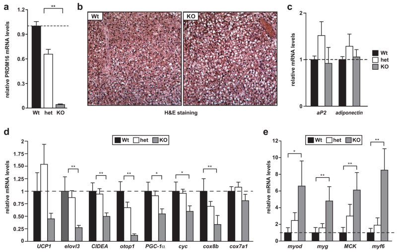
PRDM16 belongs to a family of 16 PR-domain containing proteins, some of which display an overlapping expression pattern with PRDM16. The effect of global PRDM16-deficiency on mouse and brown fat development has not been examined. Mice deficient for PRDM16 were created by targeted insertion of a gene-trap cassette into intron 2 of the gene. The creation and general phenotypic characterization of this mouse model will be described elsewhere. Notably, mice homozygous for the gene-trap insertion (PRDM16cGT/cGT) die at birth. We therefore analyzed the phenotype of putative BAT pads from wildtype (Wt), heterozygous carriers and homozygous PRDM16 mutant (knock-out, KO) late-stage embryos (E17). First, we established that PRDM16 mRNA was decreased in heterozygous BAT and virtually undetectable in the putative KO BAT (Fig. 5a). By hematoxylin and eosin (H&E) staining, the putative BAT from the KO animals had an unusual morphology, including substantially larger lipid stores (white areas in tissue) as compared to Wt BAT (Fig. 5b). No obvious muscle tissue was observed in these fat pads. Gene expression studies showed that Wt and KO BAT expressed similar levels of the pan-adipocyte markers aP2 and adiponectin (Fig. 5c). The PRDM16-deficient BAT, however, expressed significantly lower levels of essentially all BAT cell-selective and thermogenic genes (Fig. 5d). This included a 50% reduction of UCP1 mRNA levels, 75% reduction of elovl3, 50% reduction of CIDEA and a significant reduction of PGC-1α and mitochondrial target genes, cytochrome-c (cyc) and cox8b. Strikingly, the PRDM16-deficient BAT also exhibited large increases in the expression of skeletal myogenic genes including 4–6 fold elevated levels of myod, myogenin, MCK and myf6 (Fig. 5e). These data document a genetic requirement for PRDM16 in the appropriate development and gene expression program of BAT.
Fig. 5. Altered morphology and dysregulated gene expression in PRDM16-deficient brown fat.
(a)Real-time PCR analysis of PRDM16 mRNA levels in putative BAT depots from E17 wildtype (Wt), heterozygous (het) and PRDM16 knock-out (KO) mice. (b) Hematoxylin and Eosin (H&E) staining of representative sections of BAT from Wt and KO mice. (c–e) Wt, het and KO BAT were examined by real-time PCR for their expression of: general adipocyte markers (aP2 and adiponectin) (c); BAT-selective genes (d); and skeletal muscle-specific genes (e). (n= 7–11 mice per group; error bars represent ± SEM). (*p < 0.05; **p < 0.01).
Discussion
Most models of fat development have suggested that brown and white fat cells arise from a common adipogenic precursor cell5–7. Given the ability of PRDM16 to drive a complete brown fat gene program16; we had thus expected that brown fat cells lacking PRDM16 would adopt a white phenotype. However, very surprisingly, loss of PRDM16 from brown fat cells caused an increase in myogenic gene expression and bona fide skeletal muscle differentiation. Furthermore, this dependence on PRDM16 was bi-directional. Increased PRDM16 expression converted both immortalized and primary skeletal muscle myoblasts into brown fat cells.
By tracking the fate of cells that express the myoblast-specific myf5 gene, we provide evidence for a very close developmental relationship between skeletal muscle and BAT. Significantly, our analysis reveals that WAT and BAT depots arise from independent lineages and presumably have fundamentally distinct mechanisms of determination (Fig. S8). Cells with the morphology and molecular phenotype of brown fat can also be induced in WAT of rodents and humans by chronic cold exposure or by β-adrenergic stimulation2, 4, 23, 30. In our studies, the brown fat cells that emerged in the WAT of mice treated with a β3-adrenergic agonist were not derived from myf5-expressing precursors. Notably, genetic studies by Kozak and colleagues suggest that brown fat cell formation in WAT versus BAT is controlled by distinct mechanisms31, which is consistent with the view that these two types of brown fat cells have separate developmental origins. The identity of the cells in WAT that give rise to brown adipocytes remains unknown. It is still reasonable to speculate that a common white/brown fat progenitor exists in some/all WAT depots.
The genetic tracing experiments here do not determine whether a single Myf5+ cell gives rise to both BAT and skeletal muscle, or whether separate populations of Myf5+ progenitors exist. However, the interconversion between muscle and BAT induced through modulation of PRDM16 levels in culture suggest that bipotent muscle-brown fat precursors may give rise to both lineages (Fig. S8). Future studies addressing the exact nature of the muscle-brown fat precursor cell compartment(s) are needed. Interestingly, UCP1-expressing brown adipocytes have been localized in skeletal muscle and are correlated with resistance to obesity32. Our results suggest that myogenic precursor cells could be the source of these “ectopic” brown fat cells. In this regard, muscle stem cells called satellite cells can undergo adipogenesis under certain experimental conditions33, 34; though whether they assume a brown fat phenotype has not been examined.
Some recent experiments had hinted that BAT and muscle share important features. Timmons et al. showed that brown fat cell precursors express a wide array of muscle-related genes35. Lineage tracing experiments by Atit et al., revealed that embryonic BAT along with skin and muscle was derived from a population of Engrailed-1 (En1) expressing cells in the dermomyotome36. The contribution of En1-expressing cells to WAT depots was however not studied. En1 is expressed early in mouse development and marks a variety of other cell lineages including specific neuronal pools, thus a direct connection between muscle and BAT was not evident. The ontogenic relationship between BAT and skeletal muscle may explain why brown fat cells are specialized for lipid catabolism rather than storage much like oxidative muscle. Further, BAT and skeletal muscle are both highly responsive to sympathetic nerve activity and have the capacity to perform adaptive thermogenesis.
PRDM16 appears to function in brown fat cells via its ability to coregulate the activity of other transcription factors and coactivators (Fig. S8). The action of PRDM16 is almost certainly dependent on its interaction with PPARγ since PRDM16 stimulated adipogenesis in a PPARγ-ligand dependent manner, and PPARγ is known to be absolutely required for adipogenesis in cultured cells and in vivo24, 25. Our previous study indicated that an important contribution of the ability of PRDM16 to stimulate a brown fat phenotype in white fat precursors was through its association with PGC-1α and PGC-1β. It seems extremely likely that PRDM16 interacts with other factors, as yet undefined. At a mechanistic level, it remains to be determined how PRDM16 is able to stimulate the function of the PGC-1s and PPARs.
While the siRNA-mediated depletion of PRDM16 from cultured brown fat preadipocytes almost completely ablated their ability to differentiate into brown fat cells, chronic loss of PRDM16 in mice caused a significant but more modest reduction in brown fat cell character. Skeletal muscle gene expression was readily apparent in the KO tissue but no overt muscle fibers were observed. It is likely that one or more of the other 15 PR-domain containing family members can partially compensate for the loss of PRDM16 from BAT in vivo. It will be important to examine whether any of these family members has overlapping functions with PRDM16. The PRDM16-deficient mice die at birth, thereby precluding functional analyses of the mutant BAT in response to cold or its effect on energy balance. Tissue-selective ablation of PRDM16 will be required to address these issues.
The role of BAT in regulating energy balance and fighting obesity in rodents is well established. Humans have BAT and recent data suggests that it might play a more significant role in the physiology of adult humans than was previously recognized8. Since PRDM16 can function as a dominant regulator of the brown fat cell fate it will be important to investigate whether it can be used therapeutically. For example, it may be possible to use PRDM16 to drive the formation of brown-fat precursor type cells from myoblast or white fat precursors for autologous transplantation into fat tissues. Alternatively, identifying compounds that induce the expression of PRDM16 in white fat or muscle progenitors could be powerful in fighting obesity.
Methods Summary
DNA constructs
PRDM16 and PPARγ expression plasmids were described before 16, 37. Adenoviral shRNA constructs were made in the pAdtrack vector (Stratagene), using previously validated sequences16
Cell culture
Primary brown preadipocytes and myoblasts were prepared from P0–P4 Swiss-Webster mice as described before38, 39. Adipocyte differentiation was induced by treating cells for 48 hours in medium containing 10% FBS, 0.5 mM isobuylmethylxanthine, 125 nM indomethacin, 1 μM dexamethosone, 850 nM insulin, 1 nM T3 with or without 1 μM rosiglitazone (Alexis Biochemicals). After 48 hours, cells were switched to medium containing 10% FBS, 850 nM insulin, 1 nM T3 with or without 1 μM rosiglitazone.
Mice
For lineage tracing studies, heterozygous myf5-cre/+ mice18 were intercrossed with rosa26R3 (R26R3)-YFP mice19. PRDM16-deficient mice were created by homologous insertion of a gene-trap cassette and will be described elsewhere.
Immunohistochemistry
Paraffin embedded sections were subjected to citrate based antigen retrieval (Vector Labs) and incubated with antibodies against GFP or UCP1 (Abcam). Secondary detection was with anti-rabbit Alexa 647 (Invitrogen) or the Dako Envision+ system.
Binding studies
The PRDM16 transcriptional complex was immunopurified from adipocytes stably expressing Flag-tagged PRDM16 or a control vector. Gel-resolved proteins were excised and analyzed by MALDI-reTOF mass spectrometry40. Coexpression assays were performed in COS-7 cells to assess binding of PRDM16 with PPARα and PPARγ. Reporter gene assays were performed in COS-7 cells or PPARγ −/− fibroblasts using a luciferase reporter gene driven by 3 tandem copies of a PPARγ response element (3 × DR1-Luciferase)41.
Real-time PCR analysis
cDNA was prepared from total RNA using the ABI reverse transcription kit. Quantitative PCR reactions contained Sybr-Green fluorescent dye(ABI) Relative mRNA expression was determined by ΔΔ-Ct method using Tata-Binding Protein (TBP) levels as endogenous control.
Full Methods
Plasmids and viral vectors
Full-length mouse cDNA sequence for PGC-1β was cloned into pMSCV-puro retroviral vector (Stratagene). GST-PPARγ2 was cloned into pAcGHLT baculovirus expression plasmid (Pharmingen). Various fragments of PRDM16 were amplified by PCR and cloned into the EcoRI or BamH1/XhoI sites of the bacterial expression vector pGEX-4T-2 vector (GE healthcare).
Cell culture
COS-7, C2C12, and C3H-10T1/2 cells were obtained from the ATCC. Immortalized brown preadipocytes and PPARγ −/− fibroblasts were described elsewhere25, 42. For retrovirus production, phoenix packaging cells43 were transfected by calcium phosphate method as described16. For retroviral transduction, cells were treated overnight with viral supernatant supplemented with 8 μg/mL polybrene. For adenoviral infection of primary brown fat precursors, 70% confluent cell cultures were incubated with sh-PRDM16 or sh-SCR –expressing adenovirus (moi=100) overnight in complete growth medium. The medium was then replaced and cells were maintained in complete growth medium for an additional 24 hours before inducing adipogenic differentiation. Muscle differentiation was induced by culturing cells in 2% horse-serum/DMEM. To stimulate cAMP-induced thermogenesis in cells, 10 μM forskolin was added to cultures for 4 hours. Oil-Red-O staining was performed as described previously26. All chemicals were obtained from Sigma unless otherwise indicated.
Animals
All animal experiments were performed according to procedures approved by the Dana-Farber Cancer Institute’s Institutional Animal Care and Use Committee or in accordance with the University of Ottawa regulations for animal care and handling. CL316, 243, at 1 mg/kg was injected intraperitoneally into 8–10 week old male mice daily for 7 days (n=5 mice per genotype).
Immunostaining
Paraffin embedded sections of brown fat and white fat tissues were subjected to citrate based antigen retrieval (Vector Labs) and incubated with anti-rabbit GFP (Abcam) or anti-UCP1 (Abcam) antibodies for 30 min at room temperature. For fluorescent detection, sections were incubated with Alexa fluor 647 labeled anti-rabbit antibodies (Invitrogen) for 30 min and counterstained with 4′, 6-diamidino-2-phenylindole (DAPI). For horseradish-peroxidase-based detection, samples were processed using the Dako-Envision+ system and counterstained with hematoxylin (Vector Labs). Cell cultures were fixed in 4% paraformaldehyde, followed by permeabilization in 0.3% Triton-X 100. Fixed cells were incubated with anti-Myosin Heavy Chain (MF20, Developmental Studies Hybridoma Bank [DSHB], Iowa) or anti-Myogenin (F5D, DSHB) antibodies for 1 hour at room temperature. Secondary detection was performed with goat anti-mouse Alexa fluor 594 (Invitrogen), and nuclei were counterstained with DAPI. Samples were visualized using a Nikon Eclipse 80i upright microscope (Nikon Imaging Center, Harvard Medical School). Digital images were acquired by a Haramatsu orca 100 Cooled CCD camera and Metamorph image analysis software (Molecular Devices).
Binding studies
For affinity purification of the PRDM16 transcriptional complexes, PPARγ -deficient fibroblasts stably expressing PPARγ2, were infected with vectors expressing Flag-tagged PRDM16 or a control vector. These cells were grown to confluence and induced to differentiate for 4 days. Nuclei were isolated by homogenizing cells in hypotonic solution (10 mM HEPES pH 7.9, 10 mM KCl, 1.5 mM MgCl2). Nuclear proteins were extracted in hypertonic solution (20 mM HEPES pH 7.9, 400 mM NaCl, 1.5 mM MgCl2, 0.2 mM EDTA, 20% glycerol) and dialyzed against binding buffer (20 mM HEPES pH 7.9, 150 mM KCl, 0.2 mM EDTA, 20% glycerol). The PRDM16 complex was purified using Flag M2 agarose (Sigma), washed in the binding buffer (150 mM KCl), and eluted with 3× Flag peptide (Sigma). The eluted materials were separated by SDS-PAGE and visualized by silver staining or coomassie blue dye. Gel-resolved proteins were excised, digested with trypsin and analyzed by MALDI-reTOF mass spectrometry (UltraFlex TOF/TOF; BRUKER) as described40.
To examine the interaction of PRDM16 with PPARα and PPARγ2, COS-7 cells were co-transfected with expression plasmids for PRDM16 and either Flag-PPARα or Flag-PPARγ2. Whole-cell extracts were immunoprecipitated with anti-rabbit PRDM16 or anti-Flag M2 (Sigma) overnight at 4°C. Immunoprecipitates were washed 3x with washing buffer (20 mM Tris-HCl, 200 mM NaCl, 10% glycerol, 2 mM EDTA, 0.1% NP-40, 0.1 mM PMSF), separated by SDS-PAGE, transferred to PVDF membrane and blotted with anti-Flag and anti-PRDM16 antibodies. For GST immunoaffinity assays, GST-PRDM16 fusion proteins were purified from bacteria and GST-PPARγ2 (full-length) was purified from baculovirus infected SF9 cells on glutathione sepharose beads as described before44. 35S-labeled and in vitro translated PRDM16, SRC1 and PPARγ2 were prepared using the TNT Coupled Transcription/Translation System (Promega). Equal amounts of GST fusion proteins (2 μg) were incubated overnight at 4°C with in vitro translated proteins in binding buffer (20mM HEPES pH 7.7, 300 mM KCl, 2.5 mM MgCl2, 0.05% NP40, 1 mM DTT, 10% glycerol). The sepharose beads were washed 5× with binding buffer. Bound proteins were separated by SDS-PAGE and analyzed by autoradiography.
Reporter Gene Assays
COS-7 cells or PPARγ −/− fibroblasts were transfected with 50 ng PPARγ2 or PPARα and 50 ng RXRα or empty vector together with 250 ng PRDM16 expression plasmid or vector control and 100 ng of 3× DR1-Luciferase reporter in 12 well plates using Lipofectamine 2000 (Invitrogen). Cells were harvested 48 hours after transfection and assayed for luciferase activity using the Dual-Luciferase Reporter Assay System (Promega). Rosiglitazone or WY-14643 (1 μM) was added to some wells for 24 hours prior to harvesting cells. Firefly luciferase reporter gene measurements were normalized to Renilla luciferase activity.
Expression analysis and western blotting
Total RNA was isolated from cultured cells and tissue using TRIzol (Invitrogen) or RNeasy columns (Qiagen) according to manufacturer’s instructions. Real-time PCR oligo sequences are provided in Table S2. For western blot analysis, cells or tissues were lysed in RIPA buffer (0.5% NP-40, 0.1% SDS, 150 mM NaCl, 50 mM Tris-Cl [pH 7.5]). Proteins were separated by SDS-PAGE, transferred to PVDF membrane (Millipore) and probed with anti-UCP1 (Abcam), anti-PRDM16, anti-Cidea (Chemicon), and anti-β-Actin (Sigma) antibodies.
Supplementary Material
Table S1. Proteins identified by MALDI-TOF-MS/MS
Table S2. Primers used for real-time PCR analysis
Fig. S1. myf5-expressing cells give rise to brown fat depots and skeletal muscle (a)
Perirenal BAT from control (cre negative) and myf5-cre:R26R3-YFP mice were analyzed by immunohistochemistry to detect YFP (anti-GFP) and UCP1 protein. (b) Real-time PCR analysis of YFP gene activation in: heart, lung, spleen, kidney, interscapular BAT (IBAT), axial BAT (ABAT) and gastrocnemius muscle (Gast.) from myf5-cre:R26R3-YFP mice (c) Real time PCR analysis of PRDM16, UCP1, myf5 and cre recombinase expression in WAT, BAT and skeletal muscle (skM) (n=4–6 mice/group; error bars represent ± SEM). **p<0.01.
Fig. S2. PRDM16 expression blocks myogenic differentiation (a)
C2C12 myoblast cultures expressing retroviral PRDM16 or vector control (ctl) were induced to undergo muscle differentiation in 2% horse serum and analyzed by immunocytochemistry for expression of skeletal Myosin Heavy Chain (MyHC) protein. (b) Real-time PCR analysis of skeletal muscle-specific genes (n=3; error bars represent± SD; *p<0.05 **p<0.01).
Fig. S3. PRDM16 coactivates exogenous PPARγ in PPARγ-deficient cells
Transcriptional activity of a PPAR-driven reporter gene in response to PRDM16 or vector expression in PPARγ −/− cells with or without exogenous PPARγ/RXRα (+/− 1 μM rosiglitazone) (n=3; error bars represent ± SD; **p<0.05).
Fig. S4. PRDM16 binds and activates the transcriptional function of PPARα(a)
Flag-PPARα was immunoprecipitated from COS-7 cells expressing exogenous PRDM16 and/or Flag-PPARα followed by western blot analysis to detect PRDM16. The interaction was examined in the presence or absence of 1 μM of a specific PPARα ligand, WY-14643 (Wy). (b) Transcriptional activity of a PPAR-driven reporter gene (3× DR1) in response to PPARα/RXRα and PRDM16 or vector expression in COS-7 cells (+/− 1 μM Wy). (n=3; error bars represent ± SD; **p<0.05).
Fig. S5. PPARγ activation is required for the adipogenic action of PRDM16 (a)
Oil-Red-O staining of C2C12 myoblast cultures expressing retroviral PRDM16 or vector control (ctl) 6 days after inducing adipocyte differentiation in the presence or absence of 1 μM of the PPARγ-specific agonist, rosiglitazone. (b) Real-time PCR analysis of: PRDM16; adipocyte markers (PPARγ, aP2, Adiponectin); and BAT-selective genes (CIDEA, UCP1) (n=3; error bars represent ± SD; **p<0.05).
Fig. S6. PGC-1β expression does not stimulate adipogenesis in myoblasts (a)
Oil-Red-O staining of C2C12 myoblast cultures transduced with retroviral PRDM16, PGC-1β or vector control 7 days after inducing adipocyte differentiation. (b) Real-time PCR analysis of: adipocyte markers (left), BAT genes (UCP1, CIDEA); and mitochondrial genes (cyc, cox5b, cox4i1) (n=3, error bars are ± SD; *p<0.05 **p<0.01).
Fig. S7. PPARγ alone drives adipogenesis but does not induce the brown fat cell gene program in C2C12 myoblasts (a)
Oil-Red-O staining of C2C12 myoblast cultures expressing retroviral PRDM16, PPARγ2 or empty vector control (ctl) 7 days after induction of adipocyte differentiation. (b) Real-time PCR analysis of: adipocyte markers; BAT-selective genes; and skeletal muscle-specific genes (n=3 per group, error bars represent ± SD; *p<0.05 **p<0.01).
Fig. S8. Model of PRDM16 function in the specification of brown fat fate
Lineage tracing studies reveal that myf5-expressing precursors give rise to skeletal muscle and BAT but not WAT or any other tissues examined. PRDM16 expression in myoblasts induces BAT development and represses skeletal muscle differentiation. PRDM16 stimulates brown adipogenesis in myoblasts, at least in part, via binding and coactivating PPARγ. Together, these studies suggest that PRDM16 expression in bipotent muscle/brown fat progenitor cells determines brown fat identity. The mechanisms by which PRDM16 promotes brown fat determination and represses the muscle lineage in this upstream progenitor cell remain to be defined.
Supplementary Information is linked to the online version of the paper at www.nature.com/nature
Acknowledgments
We thank V. Seale and F. LeGrand for help with the lineage tracing studies and R. Gupta for discussions. We are grateful to P. Soriano for the myf5-cre mice and F. Constantini for the R26R3-YFP reporter mice. P.S. is supported by a fellowship from the American Heart Association. S.K.1 is supported by a fellowship from the Japan Society for the Promotion of Science. K.D. is supported by the Susan Komen Breast Cancer Foundation. This work is funded by an NIH grant to B.M.S and an NIH/NIAMS grant to M.A.R.
References
- 1.Cannon B, Nedergaard J. Brown adipose tissue: function and physiological significance. Physiol Rev. 2004;84:277–359. doi: 10.1152/physrev.00015.2003. [DOI] [PubMed] [Google Scholar]
- 2.Cousin B, et al. Occurrence of brown adipocytes in rat white adipose tissue: molecular and morphological characterization. J Cell Sci. 1992;103(Pt 4):931–42. doi: 10.1242/jcs.103.4.931. [DOI] [PubMed] [Google Scholar]
- 3.Garruti G, Ricquier D. Analysis of uncoupling protein and its mRNA in adipose tissue deposits of adult humans. Int J Obes Relat Metab Disord. 1992;16:383–90. [PubMed] [Google Scholar]
- 4.Guerra C, Koza RA, Yamashita H, Walsh K, Kozak LP. Emergence of brown adipocytes in white fat in mice is under genetic control. Effects on body weight and adiposity. J Clin Invest. 1998;102:412–20. doi: 10.1172/JCI3155. [DOI] [PMC free article] [PubMed] [Google Scholar]
- 5.Gesta S, Tseng YH, Kahn CR. Developmental origin of fat: tracking obesity to its source. Cell. 2007;131:242–56. doi: 10.1016/j.cell.2007.10.004. [DOI] [PubMed] [Google Scholar]
- 6.Hansen JB, Kristiansen K. Regulatory circuits controlling white versus brown adipocyte differentiation. Biochem J. 2006;398:153–68. doi: 10.1042/BJ20060402. [DOI] [PMC free article] [PubMed] [Google Scholar]
- 7.Rosen ED, Spiegelman BM. Molecular regulation of adipogenesis. Annu Rev Cell Dev Biol. 2000;16:145–71. doi: 10.1146/annurev.cellbio.16.1.145. [DOI] [PubMed] [Google Scholar]
- 8.Nedergaard J, Bengtsson T, Cannon B. Unexpected evidence for active brown adipose tissue in adult humans. Am J Physiol Endocrinol Metab. 2007;293:E444–52. doi: 10.1152/ajpendo.00691.2006. [DOI] [PubMed] [Google Scholar]
- 9.Cederberg A, et al. FOXC2 is a winged helix gene that counteracts obesity, hypertriglyceridemia, and diet-induced insulin resistance. Cell. 2001;106:563–73. doi: 10.1016/s0092-8674(01)00474-3. [DOI] [PubMed] [Google Scholar]
- 10.Ghorbani M, Claus TH, Himms-Hagen J. Hypertrophy of brown adipocytes in brown and white adipose tissues and reversal of diet-induced obesity in rats treated with a beta3-adrenoceptor agonist. Biochem Pharmacol. 1997;54:121–31. doi: 10.1016/s0006-2952(97)00162-7. [DOI] [PubMed] [Google Scholar]
- 11.Kopecky J, Clarke G, Enerback S, Spiegelman B, Kozak LP. Expression of the mitochondrial uncoupling protein gene from the aP2 gene promoter prevents genetic obesity. J Clin Invest. 1995;96:2914–23. doi: 10.1172/JCI118363. [DOI] [PMC free article] [PubMed] [Google Scholar]
- 12.Lowell BB, et al. Development of obesity in transgenic mice after genetic ablation of brown adipose tissue. Nature. 1993;366:740–2. doi: 10.1038/366740a0. [DOI] [PubMed] [Google Scholar]
- 13.Christian M, et al. RIP140-targeted repression of gene expression in adipocytes. Mol Cell Biol. 2005;25:9383–91. doi: 10.1128/MCB.25.21.9383-9391.2005. [DOI] [PMC free article] [PubMed] [Google Scholar]
- 14.Hansen JB, et al. Retinoblastoma protein functions as a molecular switch determining white versus brown adipocyte differentiation. Proc Natl Acad Sci U S A. 2004;101:4112–7. doi: 10.1073/pnas.0301964101. [DOI] [PMC free article] [PubMed] [Google Scholar]
- 15.Scime A, et al. Rb and p107 regulate preadipocyte differentiation into white versus brown fat through repression of PGC-1alpha. Cell Metab. 2005;2:283–95. doi: 10.1016/j.cmet.2005.10.002. [DOI] [PubMed] [Google Scholar]
- 16.Seale P, et al. Transcriptional control of brown fat determination by PRDM16. Cell Metab. 2007;6:38–54. doi: 10.1016/j.cmet.2007.06.001. [DOI] [PMC free article] [PubMed] [Google Scholar]
- 17.Kajimura S, et al. Regulation of the brown and white fat gene programs through a PRDM16/CtBP transcriptional complex. Genes Dev. 2008;22 doi: 10.1101/gad.1666108. [DOI] [PMC free article] [PubMed] [Google Scholar]
- 18.Tallquist MD, Weismann KE, Hellstrom M, Soriano P. Early myotome specification regulates PDGFA expression and axial skeleton development. Development. 2000;127:5059–70. doi: 10.1242/dev.127.23.5059. [DOI] [PubMed] [Google Scholar]
- 19.Srinivas S, et al. Cre reporter strains produced by targeted insertion of EYFP and ECFP into the ROSA26 locus. BMC Dev Biol. 2001;1:4. doi: 10.1186/1471-213X-1-4. [DOI] [PMC free article] [PubMed] [Google Scholar]
- 20.Huh MS, Parker MH, Scime A, Parks R, Rudnicki MA. Rb is required for progression through myogenic differentiation but not maintenance of terminal differentiation. J Cell Biol. 2004;166:865–76. doi: 10.1083/jcb.200403004. [DOI] [PMC free article] [PubMed] [Google Scholar]
- 21.Kuang S, Kuroda K, Le Grand F, Rudnicki MA. Asymmetric self-renewal and commitment of satellite stem cells in muscle. Cell. 2007;129:999–1010. doi: 10.1016/j.cell.2007.03.044. [DOI] [PMC free article] [PubMed] [Google Scholar]
- 22.Dong F, et al. Pitx2 promotes development of splanchnic mesoderm-derived branchiomeric muscle. Development. 2006;133:4891–9. doi: 10.1242/dev.02693. [DOI] [PubMed] [Google Scholar]
- 23.Himms-Hagen J, et al. Multilocular fat cells in WAT of CL-316243-treated rats derive directly from white adipocytes. Am J Physiol Cell Physiol. 2000;279:C670–81. doi: 10.1152/ajpcell.2000.279.3.C670. [DOI] [PubMed] [Google Scholar]
- 24.Barak Y, et al. PPAR gamma is required for placental, cardiac, and adipose tissue development. Mol Cell. 1999;4:585–95. doi: 10.1016/s1097-2765(00)80209-9. [DOI] [PubMed] [Google Scholar]
- 25.Rosen ED, et al. PPAR gamma is required for the differentiation of adipose tissue in vivo and in vitro. Mol Cell. 1999;4:611–7. doi: 10.1016/s1097-2765(00)80211-7. [DOI] [PubMed] [Google Scholar]
- 26.Tontonoz P, Hu E, Spiegelman BM. Stimulation of adipogenesis in fibroblasts by PPAR gamma 2, a lipid- activated transcription factor. Cell. 1994;79:1147–56. doi: 10.1016/0092-8674(94)90006-x. [DOI] [PubMed] [Google Scholar]
- 27.Hu E, Tontonoz P, Spiegelman BM. Transdifferentiation of myoblasts by the adipogenic transcription factors PPAR gamma and C/EBP alpha. Proc Natl Acad Sci U S A. 1995;92:9856–60. doi: 10.1073/pnas.92.21.9856. [DOI] [PMC free article] [PubMed] [Google Scholar]
- 28.Barbera MJ, et al. Peroxisome proliferator-activated receptor alpha activates transcription of the brown fat uncoupling protein-1 gene. A link between regulation of the thermogenic and lipid oxidation pathways in the brown fat cell. J Biol Chem. 2001;276:1486–93. doi: 10.1074/jbc.M006246200. [DOI] [PubMed] [Google Scholar]
- 29.Lin J, Puigserver P, Donovan J, Tarr P, Spiegelman BM. Peroxisome proliferator-activated receptor gamma coactivator 1beta (PGC-1beta ), a novel PGC-1-related transcription coactivator associated with host cell factor. J Biol Chem. 2002;277:1645–8. doi: 10.1074/jbc.C100631200. [DOI] [PubMed] [Google Scholar]
- 30.Huttunen P, Hirvonen J, Kinnula V. The occurrence of brown adipose tissue in outdoor workers. Eur J Appl Physiol Occup Physiol. 1981;46:339–45. doi: 10.1007/BF00422121. [DOI] [PubMed] [Google Scholar]
- 31.Xue B, et al. Genetic variability affects the development of brown adipocytes in white fat but not in interscapular brown fat. J Lipid Res. 2007;48:41–51. doi: 10.1194/jlr.M600287-JLR200. [DOI] [PubMed] [Google Scholar]
- 32.Almind K, Manieri M, Sivitz WI, Cinti S, Kahn CR. Ectopic brown adipose tissue in muscle provides a mechanism for differences in risk of metabolic syndrome in mice. Proc Natl Acad Sci U S A. 2007;104:2366–71. doi: 10.1073/pnas.0610416104. [DOI] [PMC free article] [PubMed] [Google Scholar]
- 33.Asakura A, Komaki M, Rudnicki M. Muscle satellite cells are multipotential stem cells that exhibit myogenic, osteogenic, and adipogenic differentiation. Differentiation. 2001;68:245–53. doi: 10.1046/j.1432-0436.2001.680412.x. [DOI] [PubMed] [Google Scholar]
- 34.Shefer G, Wleklinski-Lee M, Yablonka-Reuveni Z. Skeletal muscle satellite cells can spontaneously enter an alternative mesenchymal pathway. J Cell Sci. 2004;117:5393–404. doi: 10.1242/jcs.01419. [DOI] [PubMed] [Google Scholar]
- 35.Timmons JA, et al. Myogenic gene expression signature establishes that brown and white adipocytes originate from distinct cell lineages. Proc Natl Acad Sci U S A. 2007;104:4401–6. doi: 10.1073/pnas.0610615104. [DOI] [PMC free article] [PubMed] [Google Scholar]
- 36.Atit R, et al. Beta-catenin activation is necessary and sufficient to specify the dorsal dermal fate in the mouse. Dev Biol. 2006;296:164–76. doi: 10.1016/j.ydbio.2006.04.449. [DOI] [PubMed] [Google Scholar]
- 37.Mueller E, et al. Genetic analysis of adipogenesis through peroxisome proliferator-activated receptor gamma isoforms. J Biol Chem. 2002;277:41925–30. doi: 10.1074/jbc.M206950200. [DOI] [PubMed] [Google Scholar]
- 38.Megeney LA, Kablar B, Garrett K, Anderson JE, Rudnicki MA. MyoD is required for myogenic stem cell function in adult skeletal muscle. Genes Dev. 1996;10:1173–83. doi: 10.1101/gad.10.10.1173. [DOI] [PubMed] [Google Scholar]
- 39.Tseng YH, Kriauciunas KM, Kokkotou E, Kahn CR. Differential roles of insulin receptor substrates in brown adipocyte differentiation. Mol Cell Biol. 2004;24:1918–29. doi: 10.1128/MCB.24.5.1918-1929.2004. [DOI] [PMC free article] [PubMed] [Google Scholar]
- 40.Erdjument-Bromage H, et al. Examination of micro-tip reversed-phase liquid chromatographic extraction of peptide pools for mass spectrometric analysis. J Chromatogr A. 1998;826:167–81. doi: 10.1016/s0021-9673(98)00705-5. [DOI] [PubMed] [Google Scholar]
- 41.Forman BM, et al. 15-Deoxy-delta 12, 14-prostaglandin J2 is a ligand for the adipocyte determination factor PPAR gamma. Cell. 1995;83:803–12. doi: 10.1016/0092-8674(95)90193-0. [DOI] [PubMed] [Google Scholar]
- 42.Uldry M, et al. Complementary action of the PGC-1 coactivators in mitochondrial biogenesis and brown fat differentiation. Cell Metab. 2006;3:333–41. doi: 10.1016/j.cmet.2006.04.002. [DOI] [PubMed] [Google Scholar]
- 43.Kinsella TM, Nolan GP. Episomal vectors rapidly and stably produce high-titer recombinant retrovirus. Hum Gene Ther. 1996;7:1405–13. doi: 10.1089/hum.1996.7.12-1405. [DOI] [PubMed] [Google Scholar]
- 44.Drori S, et al. Hic-5 regulates an epithelial program mediated by PPARgamma. Genes Dev. 2005;19:362–75. doi: 10.1101/gad.1240705. [DOI] [PMC free article] [PubMed] [Google Scholar]
Associated Data
This section collects any data citations, data availability statements, or supplementary materials included in this article.
Supplementary Materials
Table S1. Proteins identified by MALDI-TOF-MS/MS
Table S2. Primers used for real-time PCR analysis
Fig. S1. myf5-expressing cells give rise to brown fat depots and skeletal muscle (a)
Perirenal BAT from control (cre negative) and myf5-cre:R26R3-YFP mice were analyzed by immunohistochemistry to detect YFP (anti-GFP) and UCP1 protein. (b) Real-time PCR analysis of YFP gene activation in: heart, lung, spleen, kidney, interscapular BAT (IBAT), axial BAT (ABAT) and gastrocnemius muscle (Gast.) from myf5-cre:R26R3-YFP mice (c) Real time PCR analysis of PRDM16, UCP1, myf5 and cre recombinase expression in WAT, BAT and skeletal muscle (skM) (n=4–6 mice/group; error bars represent ± SEM). **p<0.01.
Fig. S2. PRDM16 expression blocks myogenic differentiation (a)
C2C12 myoblast cultures expressing retroviral PRDM16 or vector control (ctl) were induced to undergo muscle differentiation in 2% horse serum and analyzed by immunocytochemistry for expression of skeletal Myosin Heavy Chain (MyHC) protein. (b) Real-time PCR analysis of skeletal muscle-specific genes (n=3; error bars represent± SD; *p<0.05 **p<0.01).
Fig. S3. PRDM16 coactivates exogenous PPARγ in PPARγ-deficient cells
Transcriptional activity of a PPAR-driven reporter gene in response to PRDM16 or vector expression in PPARγ −/− cells with or without exogenous PPARγ/RXRα (+/− 1 μM rosiglitazone) (n=3; error bars represent ± SD; **p<0.05).
Fig. S4. PRDM16 binds and activates the transcriptional function of PPARα(a)
Flag-PPARα was immunoprecipitated from COS-7 cells expressing exogenous PRDM16 and/or Flag-PPARα followed by western blot analysis to detect PRDM16. The interaction was examined in the presence or absence of 1 μM of a specific PPARα ligand, WY-14643 (Wy). (b) Transcriptional activity of a PPAR-driven reporter gene (3× DR1) in response to PPARα/RXRα and PRDM16 or vector expression in COS-7 cells (+/− 1 μM Wy). (n=3; error bars represent ± SD; **p<0.05).
Fig. S5. PPARγ activation is required for the adipogenic action of PRDM16 (a)
Oil-Red-O staining of C2C12 myoblast cultures expressing retroviral PRDM16 or vector control (ctl) 6 days after inducing adipocyte differentiation in the presence or absence of 1 μM of the PPARγ-specific agonist, rosiglitazone. (b) Real-time PCR analysis of: PRDM16; adipocyte markers (PPARγ, aP2, Adiponectin); and BAT-selective genes (CIDEA, UCP1) (n=3; error bars represent ± SD; **p<0.05).
Fig. S6. PGC-1β expression does not stimulate adipogenesis in myoblasts (a)
Oil-Red-O staining of C2C12 myoblast cultures transduced with retroviral PRDM16, PGC-1β or vector control 7 days after inducing adipocyte differentiation. (b) Real-time PCR analysis of: adipocyte markers (left), BAT genes (UCP1, CIDEA); and mitochondrial genes (cyc, cox5b, cox4i1) (n=3, error bars are ± SD; *p<0.05 **p<0.01).
Fig. S7. PPARγ alone drives adipogenesis but does not induce the brown fat cell gene program in C2C12 myoblasts (a)
Oil-Red-O staining of C2C12 myoblast cultures expressing retroviral PRDM16, PPARγ2 or empty vector control (ctl) 7 days after induction of adipocyte differentiation. (b) Real-time PCR analysis of: adipocyte markers; BAT-selective genes; and skeletal muscle-specific genes (n=3 per group, error bars represent ± SD; *p<0.05 **p<0.01).
Fig. S8. Model of PRDM16 function in the specification of brown fat fate
Lineage tracing studies reveal that myf5-expressing precursors give rise to skeletal muscle and BAT but not WAT or any other tissues examined. PRDM16 expression in myoblasts induces BAT development and represses skeletal muscle differentiation. PRDM16 stimulates brown adipogenesis in myoblasts, at least in part, via binding and coactivating PPARγ. Together, these studies suggest that PRDM16 expression in bipotent muscle/brown fat progenitor cells determines brown fat identity. The mechanisms by which PRDM16 promotes brown fat determination and represses the muscle lineage in this upstream progenitor cell remain to be defined.
Supplementary Information is linked to the online version of the paper at www.nature.com/nature


