Summary
Notch1-deficient epidermal keratinocytes become progressively hyperplastic, eventually producing tumors. In contrast, we observed that Notch1 deficient hair matrix keratinocytes have lower mitotic rates, resulting in smaller follicles with fewer cells. In addition, the ratio of melanocytes to keratinocytes is greatly reduced. Investigation into the underlying mechanistic basis for these phenotypes revealed that three pathways, not previously linked with Notch signaling, were significantly impacted: the KIT, TGFβ and IGF signaling pathways. The level of Kitl/Scf produced by follicular keratinocytes was reduced, resulting in a decline of resident melanocyte populations. TGFβ ligands were elevated in Notch1-deficient keratinocytes, which correlated with elevated expression of several targets, including the diffusible IGF antagonist IGFBP3 in the dermal papilla. Diffusible stromal targets remained elevated in the absence of epithelial TGFβ receptors, consistent with paracrine TGFβ actions. Overexpression of IGF1 in the keratinocyte reversed the phenotype, as expected if Notch1 loss altered the IGF/IGFBP equilibrium. Conversely, epidermal keratinocytes contained less stromal IGFBP4 and may thus be primed to experience an increase in IGF signaling as animals age. These results suggest that Notch1 participates in a bi-compartmental communication system that controls homeostasis, follicular proliferation rates and melanocyte population via modulation of KIT, TGFβ and IGF signaling.
Keywords: Hair follicle, Notch1, keratinocyte, Kitl/Scf, TGFβ, IGFBP
Introduction
During skin organogenesis, keratinocytes and the underlying mesenchymal cells engage in reciprocal, sequential communication events that result in formation of the hair follicles, the sebaceous gland, interfollicular epidermis with its region-specific thickness and the sweat glands (Hardy, 1992; Millar, 2002; Sengel, 1976). Migratory melanocytes, Merkel and Langerhans cells later join in to form a community of specialized cells that together form a unique constellation of physiological functions. Despite their shared origin, hair follicles and epidermal keratinocytes diverge to execute their unique differentiation programs and, in the adult, replenish themselves from distinct stem cell populations (Alonso and Fuchs, 2003; Ito et al., 2005). Epidermal keratinocytes restricted to the innermost (Basal) layer are the only ones to proliferate in the adult under normal circumstances. Asymmetric basal cell divisions result in one cell withdrawing from the cell cycle, commencing a terminal differentiation program and a journey outwards towards the skin surface (Lechler and Fuchs, 2005). Differentiation is manifest by unique gene expression profiles that distinguish the Spinous layer, Granular layer, and Cornified layer (Fuchs and Raghavan, 2002; Jamora and Fuchs, 2002).
In contrast, hair follicle keratinocytes surround a teardrop shaped core of mesenchymal cells (dermal papilla, DP) in a structure called a bulb, where only lineage-restricted stem cells maintain contact with the DP (Legue and Nicolas, 2005); the proliferation zone within the bulb contains many cells that are not in direct contact with a basement membrane. As matrix keratinocytes exit the cell cycle, they differentiate into several different cell layers (Hardy, 1992; Millar, 2002; Sengel, 1976). Starting in the third week of life, murine hair follicles start to cycle, i.e., they undergo a period of destruction, rest and regeneration which continues throughout adult life (Muller-Rover et al., 2001; Stenn and Paus, 2001). The phase in which the hair shaft is actively produced is called anagen, while the short destructive phase is called catagen. During catagen, the bottom two thirds of each hair follicle undergo apoptosis; only the upper third of the follicle, the stem cell niche (bulge) and DP, remain intact. Catagen lasts ~ 3 days, followed by a resting phase (telogen) of variable length. Subsequent entry into anagen is believed to begin when signals from DP activate stem cells in the bulge (Stenn and Paus, 2001).
Notch is a transmembrane receptor activated by transmembrane ligands on the neighboring cells (Kopan, 2002). Binding of Notch to its ligand leads to the cleavage of Notch and to the translocation of Notch intracellular domain (NICD) into the nucleus, where NICD binds to CSL (CBF1/RBPjk, Su(H), and Lag-1). CSL is a transcriptional repressor bound with other nuclear factors to the promoter region of particular genes. Association of NICD with CSL triggers an allosteric change that facilitates transient conversion of CSL from transcription repressor to transcription activator resulting in transcriptional activation of downstream targets (Fryer et al., 2004; Mumm and Kopan, 2000). Notch signaling promotes selection of the hair fate by activated bulge stem cells (Yamamoto et al., 2003) and plays an important role in maintenance of the inner root sheath fate (Pan et al., 2004). In the embryonic mouse epidermis, NICD is ubiquitous in supra-basal keratinocytes selecting the spinous fate (Blanpain et al., 2006; Lin and Kopan, 2003; Pan et al., 2004); after birth, NICD is detected transiently in a small fraction of spinous cell (YP and RK, unpublished observations). This pattern of Notch1 activation is consistent with its proposed role in suppressing proliferation and promoting differentiation via cell autonomous modulation of targets that may include Wnt, p21cip1, K1, K10, Hes1 and p63 (Blanpain et al., 2006; Devgan et al., 2005; Mammucari et al., 2005; Nguyen et al., 2006; Okuyama et al., 2004; Rangarajan et al., 2001). In addition, keratinocytes deficient in Notch1 are sensitive to chemical carcinogenesis, establishing Notch1 as a tumor suppressor in the epidermis (Nicolas et al., 2003). Although the role of Notch2 in the epidermis is thought to be minimal (Rangarajan et al., 2001), loss of Notch1 and 2, RBPjk or presenilin results in more severe epidermal phenotypes (Blanpain et al., 2006; Pan et al., 2004), indicating that Notch2 also contributes to epidermal differentiation.
The contribution of Notch signaling to follicular and epidermal homeostasis can result from both cell autonomous and cell non-autonomous effects. Both are consistent with the role of Notch as a transcriptional modulator: the former is built on the proven ability of Notch to modulate the transcription landscape within keratinocytes (Blanpain et al., 2006; Devgan et al., 2005; Nguyen et al., 2006), while the latter posits that among the transcripts altered by Notch, cell surface or secreted molecules would impact neighboring cells (Lin et al., 2000; Pan et al., 2004).
In this report, we demonstrate that Notch1 plays contrasting roles in keratinocyte proliferation of hair follicles and epidermis, attributed in large part to previously underappreciated cell non-autonomous signals. We provide evidence that TGFβ signaling is elevated and Kitl/Scf expression is reduced in Notch1-deficient hair matrix keratinocytes, correlating with reduced keratinocyte mitotic rates and melanocyte population, respectively. In addition, we demonstrate that the impact on the epithelial cell cycle is not dependent on autocrine TGFβ reception in the keratinocyte but on paracrine signaling to neighboring fibroblasts. The fibroblast, in turn, controls keratinocyte proliferation by modulation of IGF signaling: in the hair follicle, Insulin-like growth factor binding protein 3 (Igfbp3) levels were elevated in the dermal papilla (DP), while in the epidermis, a reduction in Igfbp4 production was detected. Increasing the IGF/IGFBP ratio by IGF1 overexpression in Notch1-deficient skin restored the cell numbers in the hair matrix. Thus, bi-compartmental signaling that involves diffusible epithelial TGFβ and stromal IGFBP acts downstream of Notch1. This paracrine loop could contribute to keratinocyte hyperproliferation in the epidermis, may explain why Notch1-deficient phenotypes differ in chimera vs. globally deleted skin, and may even facilitate the response of Notch1 deficient mice to chemical carcinogens in a manner analogous to the vicious cycle seen in bone metastasis.
Materials and methods
Production and Analysis of Single and Compound Mutant Mice
The generation of Tgfbr2flox,(Yang et al., 2004), Trp53 null (Jacks et al., 1994), Msx2-Cre (Sun et al., 2000), Ivl::IGF1 (Weger and Schlake, 2005a), and N1CKO (Msx2-Cre;Notchflox/flox) (Pan et al., 2004) mice were described previously.
N1IGF1
Ivl::IGF1 females were crossed with NICKO males, and the F1 Msx2-Cre/+;Notch1flox/+;Ivl::IGF1 male offspring were then crossed with F1 Notch1flox/+; Ivl::IGF1 females to produce Msx2-Cre/+;Notch1flox/flox;Ivl::IGF1 offspring.
N1P53DCKO
Female mice containing Trp53−/− were crossed with NICKO males, and the male Trp53−/− mice with Notch1flox/flox females. The F1 male offspring Msx2-Cre/+;Notch1flox/+;Trp53+/− were then crossed with F1 females Notch1flox/+;Trp53+/−. The F2 offspring, Msx2-Cre/+;Notch1flox/flox;Trp53+/− males and Notch1flox/flox;Trp53−/− females, were mated to produce the compound mice Msx2Cre/+;Notch1flox/flox;Trp53−/−, and their littermates were used for analysis.
N1TβR2CKO
Tgfbr2flox/flox males and females were similarly used to generate Msx2-Cre/+;Notch1flox/flox;Tgfbr2flox/flox for analysis.
PCR condition and protocols used for genotyping will be provided upon request.
Histology, In situ Hybridization (ISH), and Immunohistochemical Analysis
To obtain stereoscopic images, 9-day-old (P9) skins were torn as strips across the caudal-rostral axis with fine forceps in PBS and photographed with an Olympus SZH10 stereo dissecting microscope. For hematoxylin/eosin staining, ISH, and the immunohistochemical analyses, skins fixed in 4% paraformaldehyde in PBS were embedded in paraffin and sectioned at 5μm. ISH was performed as described (Pan et al., 2004) with probes prepared using cDNAs either generated by RT-PCR or from the following sources: Cdkn1a (IMAGE 3495942), Nrarp (IMAGE 6439916), and notch1 (Kopan and Weintraub, 1993). The primer sequences for RT-PCR are available upon request. For each reaction, a corresponding sense transcript was used as a negative control. Immunohistochemical analysis was performed as described previously (Lin et al., 2000; Pan et al., 2004). For BrdU staining, animals were intraperitoneally administered BrdU in 100μg/g body weight exactly 30 minutes before sacrifice. For the source and dilution condition for primary antibodies including AE13, BrdU, and Ki67, refer to previous reports (Lin and Kopan, 2003; Lin et al., 2000). Other antibodies used are Hes1 (rabbit, 1:1000; kindly provided by Dr. Sudo (Ito et al., 2000), Igfbp2 (goat, 1:200; R&D systems AF797), Igfbp3 (goat, 1:200; R&D systems AF775), and p73 (mouse, 1:1000; Labvision ER15). For β-galactosidase (rabbit, 1:1000, 5 Prime → 3 Prime, Inc) and Dct-1 (goat, 1:200, Santa Cruz sc-10451) antibody staining, skins were fixed in periodate-lysine-paraformaldehyde, cryoprotected with 30% sucrose in PBS overnight, embedded in OCT, and sectioned in 7μm thickness. Sections were counterstained with DAPI.
Cell counting and progenitor cell labeling index
For hair matrix cell counting, paraffin sections were stained with DAPI and photographed under both UV and visible light. The two pictures were overlapped and nuclei located below the first visible melanin granules were counted from each hair follicle. More than 20 hair follicles in proper longitudinal orientation per genotype were counted to calculate average and standard deviation. For progenitor cell labeling index, BrdU- and Ki67-positive cells were counted from more than 20 hair follicles per mice, three mice in each genotype. For calculating the melanocyte cell fraction, the number of Dct-1 positive cells was divided by the number of hair matrix cells in each follicle. The average percentage and standard deviation from more than 20 hair follicles per each genotype was calculated. In all cases, differences between means were determined by two-tailed Student’s t-test.
TUNEL (Terminal deoxynucleotide transferase dUTP-digoxigenin nick end labeling) staining
Fresh, unfixed frozen dorsal skins were embedded in OCT, and 10μm sections were prepared for TUNEL assay. TUNEL assay was performed as described (Lindner et al., 1997). The number of TUNEL-positive cells per follicle was counted from more than 60 longitudinally sectioned follicles with proper orientation from each individual, and 3 different individuals were used per each genotype (P9 anN1, emN1, wild-type and P16 wild-type). Statistical significance was measured by two-tailed Student’s t-test.
Isolation of hair bulbs and epidermis, total RNA extraction, and microarray
For hair bulb isolation, freshly obtained P9 skin samples were immediately immersed in RNAlater (Ambion, TX) and individual hair follicles were collected using a fine tungsten needle and precisely cut with a 26G syringe needle (Becton Dickinson and Company, NJ) in the place two times higher than the distance between the tip of the bulb and the onset of pigmentation. For epidermis preparation, a patch of P9 dorsal skin was harvested and flash-frozen on dry ice, and epidermis was isolated by scraping it off the skin using a cold scalpel. Isolated hair bulbs and epidermis samples were then subjected to total RNA isolation using QIAGEN RNeasy Micro Kit (QIAGEN Sciences, MD) according to the manufacturer’s protocol. For hair bulb microarray analyses, 50–100ng of hair bulb total RNA collected from two mice of each genotype (emN1, anN1, and wild-type) was subjected to double-round amplification/labeling procedures. The cRNA products were then hybridized to Affymetrix MOE430v2 chips. All protocols were conducted at the Washington University Genechip Facility as described in the Affymetrix manual. Affymetrix microarray suite 5.0 and dChip software (http://www.dchip.org) were used for comparisons and GO (Gene Ontology)-based classification. Ingenuity (https://analysis.ingenuity.com) and David2 (http://niaid.abcc.ncifcrf.gov/) were used for pathway analyses.
cDNA preparation and Quantitative RT-PCR
Total RNA from hair bulbs and epidermis were used for cDNA preparation using QIAGEN Sensiscript RT Kit (QIAGEN Sciences, MD) according to the manufacturer’s protocol. The cDNA was amplified with SYBR-Green PCR Master Mix (Applied Biosystems, CA) and gene-specific exon-exon junction spanning primers in ABI7700 sequence detection system (Applied Biosystems, CA) or in MyIQ iCycler (Bio-rad, CA). Reaction was performed in 95°c for 10 minutes followed by 40 cycles of 95 for 15 seconds, 55°c for 45 seconds, and 72°c for 30 seconds. Normalizations across samples were performed using hypoxanthine guanine phosphoribosyl transferase 1 (hprt1) or glyceraldehyde-3-phosphate dehydrogenase (gapdh) primers. The differences between samples and controls were calculated using the Comparative CT (2−ΔΔCT) Method. The histograms in Figures show mean ± s.e.m. of two or three independent experiments for hair follicles and five for epidermal samples, each of which was performed in triplicate. All qRT-PCR primers were tested for the presence of nonspecific primer dimers and for linear amplification. The identity of the qRTPCR amplicons was validated by DNA sequencing or restriction enzyme digestion of the amplicon (cyclinD1). Primer sequences are available upon request.
Immunoblotting
For immunoblotting, frozen epidermal samples as described above were immediately lysed in NP40 lysis buffer (1% NP-40, 150mM NaCl, 10% glycerol, and 1mM EDTA, and 20mM Tris, pH 8.0) with protease and phosphatase inhibitors; lysate was combined with 2x Laemmli buffer for SDS-PAGE. Western blot transfer was done as previously described. The following primary antibodies were used: a-tubulin (mouse, 1:1000; Sigma-Aldrich B-5-2), CyclinD1 (rabbit, 1:1500; Santa Cruz sc-718), IGFBP4 (rabbit, 1:5000; kindly provided by Dr. Chernausek, Cincinnati Children’s Hospital), p15 (rabbit, 1:200; Santa Cruz sc-612), p21 (mouse, 1:100; Santa Cruz sc-6246) and anti-TGFβ2 (rabbit, 1:200; Santa Cruz sc-90). Western blot quantification was performed using Quantity One software (Bio-Rad Laboratories, CA). The histograms in Figures show mean ± s.e.m. of two or three independent epidermal lysates per genotype.
Results and Discussion
The bulbs of Notch1-deficient hair follicles contain fewer keratinocytes
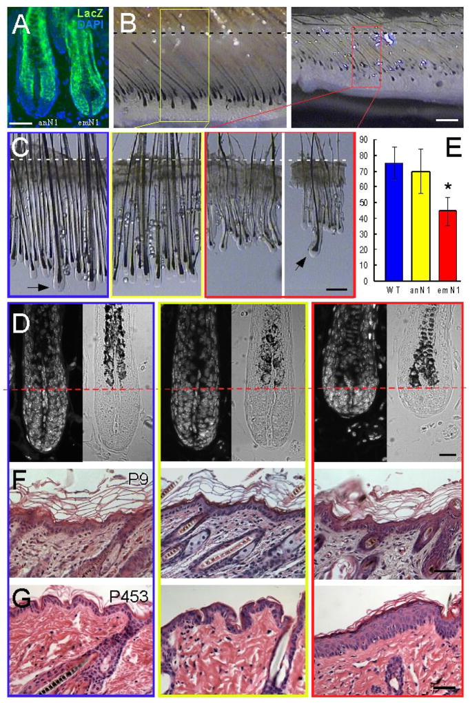
Conditional deletion of Notch1 in Msx2Cre; Notch1flox/flox (N1CKO) mice produced two different patterns of gene loss in hair follicles, previously referred to as anagen-deleted and embryo-deleted (Fig. 1A and (Pan et al., 2004)). As the names imply, Embryo deleted (emN1) epidermis and follicles are derived from cells experiencing a pulse of Cre activity at E9.5 and thus lack Notch1 from that point onwards. Anagen deleted (anN1) follicles express Cre recombinase in the mid-bulb region a few days after the start of the growth phase (anagen); Notch1 is thus only removed in the upper bulb (Fig. 1A). We observed that emN1 follicles are substantially shorter than anN1 or wild type follicles (Figs 1B and C). Because anN1 follicles are wild type in appearance, we concluded that Notch1 function is required in the proximal, actively proliferating matrix during hair morphogenesis (Figs 1B and C; (Pan et al., 2004)).
Figure 1. Reduced cell numbers in hair follicles while epidermal keratinocytes expand in response to Notch1 loss.
(A) LacZ immunohistochemistry on Msx2Cre;Notch1flox/flox;Rosa26R/+ skin. Note that LacZ staining displays the area of Notch1 deletion. Scale Bar 50μM.
(B) Stereoscopic side view of the anagen-deleted (anN1) (left) and embryo-deleted (emN1) (right) skin at P9. Scale Bar 300μM.
(C) Stereoscopic view magnified from (B) after dissection. Wild type is in blue box, anN1 in yellow, and emN1 in red. Arrows indicate guard hairs. Scale Bar 200μM.
(D) Histological view of (B) and (C). Box color indicates the genotype as in (C). Nuclei staining with DAPI. Scale Bar 20μM.
(E) Average number of hair matrix cells (below a red line in D) per hair follicle. (* p < 0.00001). Error Bars represent s.d.
(F–G) H&E staining of P9 (F) and P453 (G) skin. Note the epidermal hyperplasia in old emN1 skin (G). Scale Bar 40μM.
Closer inspection of the emN1 hair bulbs revealed that the portion below the first visible melanin granules (marked by a dashed red line in Fig. 1D) was shorter than wild type or anN1 follicles at postnatal day 9 (Fig. 1D). Cell size in all genotypes was not altered, suggesting that the PI3K (Ciofani and Zuniga-Pflucker, 2005; Luo et al., 2005) and Myc pathways impacted in Notch1-deficient thymocytes (Weng et al., 2006) are intact in keratinocytes. Instead, fewer cells were detected in longitudinal sections of Notch1-deficient follicles: emN1 hair follicles had only 44.69 ± 8.92 cells, whereas wild type (75.32 ± 10.30) and anN1 (69.86 ± 14.17) hair follicles had nearly twice as many cells (Fig. 1E). Notch1-deficient epidermis did not display any observable hyperplasia at this age, but the epidermis of older mice did (Figs 1F and G; (Nicolas et al., 2003; Rangarajan et al., 2001)). We thus conclude that in contrast to the epidermis, where the number of Notch1-deficient epidermal keratinocytes increase as mice age (Nicolas et al., 2003; Rangarajan et al., 2001), fewer Notch1-deficient keratinocytes formed the hair bulb. Therefore, Notch1 loss leads to context-dependent impact on related keratinocyte population.
Notch1-deficient hair matrix cells have decreased mitotic rates
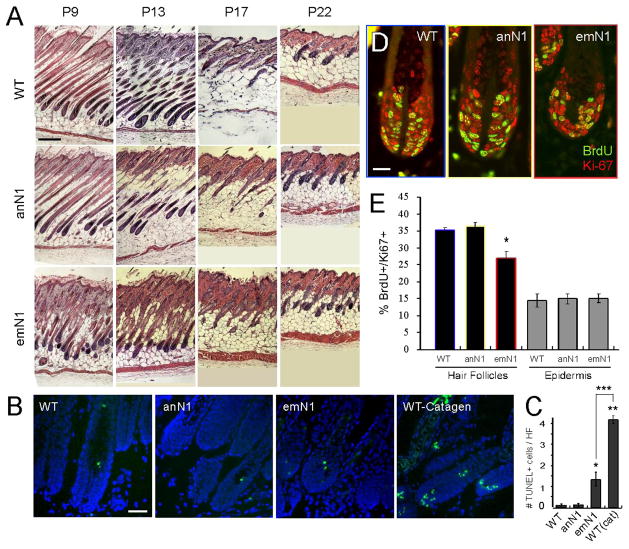
During catagen, hair bulbs involute and regress quickly (during three to four days) by a combination of cell cycle arrest and apoptotic cell death (Dry, 1926; Lindner et al., 1997; Muller-Rover et al., 2001; Weedon and Strutton, 1981). One trivial explanation for reduced keratinocyte numbers in emN1 bulbs is accelerated catagen onset, observed in mice with global epithelial deletion of Notch1 (K14-Cre; Notch1flox/flox and K5-CreERT; Notch1flox/flox (Vauclair et al., 2005)). To ask if reduced cell numbers in emN1 hair bulbs reflected catagen onset at P9, we examined emN1 skin from P9, P13, P17, and P22 (Fig. 2A). If catagen had started at P9, follicles would have lost their lower two thirds by P13. Instead, characteristic follicular catagen destruction was detected in all genotypes at P17, indicating that Notch1 deficient skin in chimeric mice did not accelerate entry into catagen (Fig. 2A).
Figure 2. Notch1 loss causes decreased mitotic rates and a slight increase in apoptosis, but catagen was not accelerated.
(A) H&E staining of anN1, emN1, and wild type at P9, 13, 17, and 22. Note that hair cycling was not altered in Notch1-deficient hair follicles. Scale Bar 200μM.
(B) TUNEL staining of emN1, anN1, and wild type hair follicles at P9 and P16.
(C) Average number of TUNEL-positive cells per hair follicle in each genotype shown in (B). (* p < 0.05 for both WT vs emN1 and anN1 vs emN1; ** p < 0.05 for both WT vs WT(cat) and anN1 vs WT(cat); *** p < 0.005).
(D) Immunofluorescence staining of BrdU (green) and Ki67 (red). Scale Bar 20μM
(E) Progenitor labeling index of anN1, emN1, and wild type hair follicles and epidermis. (* p <0.01 for anN1 vs emN1 and WT vs emN1). Error Bars represent s.d.
Hair bulbs including >2 TUNEL-positive cells are morphologically identical to follicles in anagen, but have been classified as being in catagen I (Muller-Rover et al., 2001). Early catagen II follicles contain, on average, 4 or more TUNEL-positive cells ((Lindner et al., 1997), Figs 2B and C). While we observed a few Notch1-deficient follicles containing >2 TUNEL positive cells, more than 40% of emN1 hair follicles lacked TUNEL-positive cells (data not shown). Moreover, the average number of TUNEL positive cells was significantly lower than the 4 cells seen in P16 (early catagen, 1.34 in emN1 at P9 vs 4.18 TUNEL-positive cells/HF p< 0.005; Fig. 2C). Given that no further morphological changes were detected until after P13, we concluded that increased apoptotic cell death did not indicate catagen onset nor could it explain the reduction in cell number seen in TUNEL-negative hair follicles.
Another possible explanation for the lower cell numbers in emN1 follicles is a reduced proliferation rate. To examine mitotic rates at P9, we identified mitotic cells with Ki67 (red in Fig. 2D) and BrdU-incorporating S-phase cells (green in Fig. 2D) following a 30-minute BrdU pulse-label (Chenn and Walsh, 2002). The ratio of S-phase cells in the cycling population (BrdU+/Ki67+) was tabulated in hair follicles and the epidermis of mutant and wild type mice. Interestingly, emN1 hair follicles displayed a reduced progenitor-labeling index (27.07±1.99%) compared with anN1 (36.26±1.33%) and wild type (35.28±0.71%) follicles (Fig. 2E), which correlated with the cell numbers in these follicles. In contrast, P9 epidermal keratinocytes did not display any difference in the progenitor-labeling index (Fig. 2E). Collectively, it is evident that delayed cell cycle, with a small contribution from increased apoptotic cell death, accounts for the lower cell numbers within Notch1-deficient hair bulbs and that the proliferation defect in epidermal keratinocytes is progressive, evident only as the animals age.
Notch1-deficient hair follicles contain elevated levels of transcripts encoding cytostatic and proapoptotic proteins
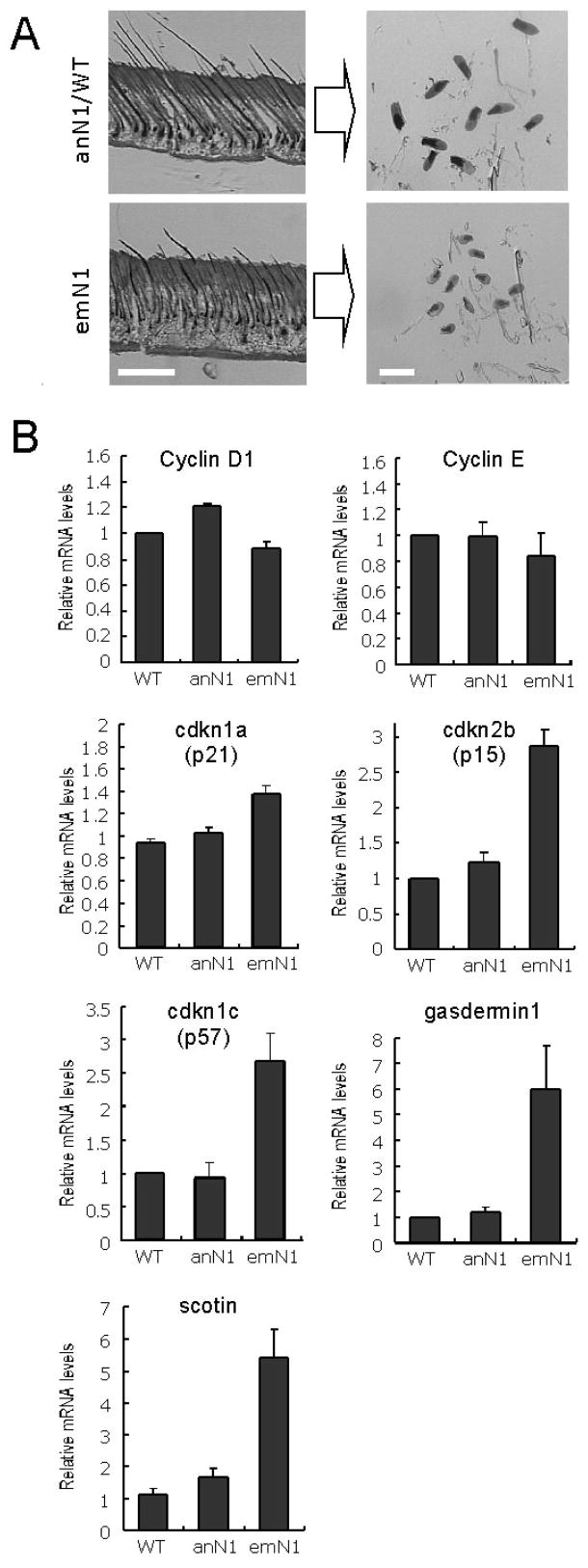
The length of the G1 phase in mammalian cells can vary, while the length of S and G2+M phases remain relatively unchanged (Chenn and Walsh, 2002; DiSalvo et al., 1995; Malkinson and Keane, 1978). If the reduced mitotic rate in emN1 is attributable to changes in G1 length or the G1/S checkpoint, either the expression/stability of G1 cyclins has decreased or the abundance of Cyclin kinase inhibitors (CKIs) has increased (Mainprize et al., 2001; Massague, 2004; Sherr and Roberts, 1999). To address the molecular mechanism underlying the lowered mitotic index and increased apoptosis seen in Notch1-deficient hair follicles, we mechanically micro-dissected hair bulbs of emN1, anN1, and wild type mice (~100 follicles/mouse; two animals per genotype, Fig. 3A). Isolated total RNA was subjected to microarray analysis, and genes altered in emN1 matrix but not in anN1 matrix were subjected first to GO-based classification. We identified 47 genes altered in Notch1-deficient hair matrix that are involved in cell growth, cycle, proliferation, or programmed cell death (Table S1; see Material and methods for details).
Figure 3. Reduced mitotic rates in hair follicles correlates with elevated expression of cytostatic and proapoptotic genes.
(A) Microdissection of hair bulbs from anN1, emN1, and wild type follicles. Scale Bars (left) 100μM; (right) 200μM.
(B) qRT-PCR of G1 cyclins, CKIs, gasdermin1, and Scotin using total RNA isolated from micro-dissected samples shown in (A). Error bars represent s.e.m.
The microarray analysis and qRT-PCR revealed that transcription of the cyclin kinase inhibitors Cdkn1a (p21Cip1), Cdkn2b (p15INK4b) and Cdkn1c (p57Kip2) was increased upon Notch1 loss, while mRNA expression of G1 cyclins, cyclin D1 and cyclin E did not change (Table S1 and Fig. 3B). In addition, the expression of gsdm1 (gasdermin 1), so named due to its presence in skin and the upper gastrointestinal tract (Lunny et al., 2005; Saeki et al., 2000), was significantly increased (Table S1 and Fig. 3B). gsdm1 is absent from proliferating keratinocytes and from several gastric cancer cell lines, leading to the hypothesis that it promotes cell cycle arrest (Saeki et al., 2000). Moreover, the mRNA level of a proapoptotic gene, Scotin, was also significantly induced upon Notch1 loss (Fig. 3B), consistent with the observed increase in apoptosis (Fig. 2C). These data are consistent with the hypothesis that loss of Notch1 affected the length of the G1 phase via increased cytostatic genes and a slight increase of apoptosis through elevated levels of proapoptotic genes in follicular keratinocytes. However, these changes clearly fell short from full induction of catagen.
The impact of Notch1 loss is not mediated via p53 family members
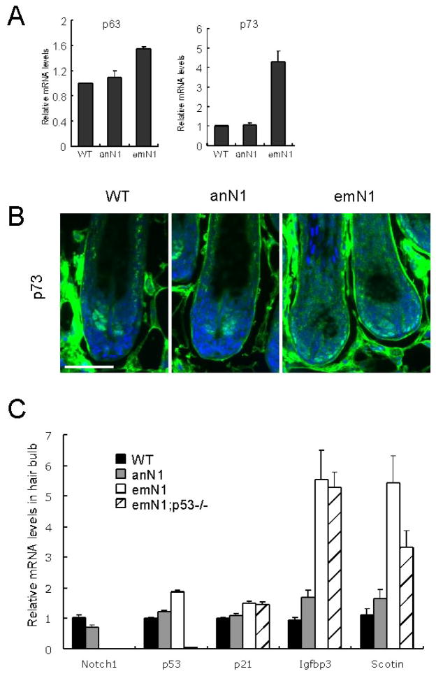
The cytostatic and proapoptotic transcripts elevated in Notch1-deficient follicles include shared p53 family targets Cdkn1a, Scotin (Fig. 3B; (Harms and Chen, 2005; Rossi et al., 2004; Terrinoni et al., 2004)) and Igfbp3 (Fig. 5A, described in detail below). Members of the p53 family are known regulators of G1/S arrest and apoptosis (Bartek and Lukas, 2001; Massague, 2004; Moll and Slade, 2004); Notch loss can lead to a reduction in p53 activity (Huang et al., 2004; Yang et al., 2004). In the epidermis, however, loss of Notch1 results in elevated p63 levels (Nguyen et al., 2006), which in turn contributes to proliferation at the expense of differentiation.
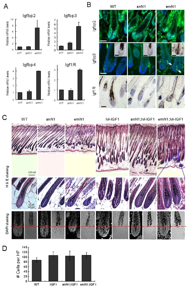
Figure 5. Altered IGF/IGFBP equilibrium in the hair matrix of Notch1-deficient hair follicles.
(A) qRT-PCR of IGF signaling components. Error bars represent s.e.m. (B) Immunohistochemistry and in situ hybridization of IGF signaling components on hair follicles. DAPI was used for nuclear staining in IGFBP2 and 3 antibody staining. Arrows indicate DP-specific IGFBP3 immunoreactivity. Insets, mRNA in situ hybridization of the genes specified. Note the mRNA and protein expression of Igfbp3 in dermal papilla, where Notch1 is not expressed. Scale Bars 40μM. (C) Histological comparisons of the hair follicles from the genotypes specified. Note that the matrix size is restored in emN1;Ivl-IGF1 follicles. Scale Bars 200μM (skin H&E staining); 50μM (follicular H&E and DAPI staining) (D) Average number of hair matrix cells (below a red line in C) per hair follicle. Error Bars represent s.d.
Trp63 (p63) transcripts were not significantly altered in Notch1-deficient follicles (Fig 4A). In contrast, transcripts of Trp73 (p73) were greatly elevated (Fig. 4A). p73 expression was restricted to the subset of lineage-restricted stem cells adjacent to the DP (Fig. 4B). In the absence of Notch1, p73 expression extended to additional IRS and ORS cells (Fig. 4B), indicating that Notch1 or its targets may negatively regulate p73 expression. This expression pattern, however, did not overlap with the expression domain of any shared p53 family targets (Figs 4B, 5B, and 8B). Moreover, p73-positive cells are actively cycling, as evident from BrdU incorporation studies (compare Fig. 2D with 4B).
Figure 4. p53 family members do not contribute to Notch1-deficient follicular phenotype.
(A) qRT-PCR of p63 and p73. (B) Immunohistochemistry of p73 in hair follicles of anN1, emN1, and wild type littermates. Scale Bars 50μM. (C) qRT-PCR with Notch1, Trp53 (p53), and p53 target genes (Cdkn1a, Scotin, and Igfbp3) using hair bulb total RNA from the genotypes specified (emN1;p53−/− indicates Msx2Cre;Notch1flox/flox;p53−/−). Note the sustained increase of Scotin and Igfbp3 in the absence of p53. Error bars represent s.e.m.
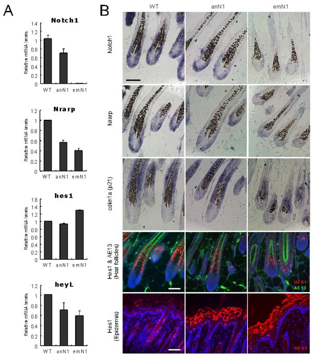
Figure 8. Monitoring epidermal Notch targets in the hair follicles.
(A) qRT-PCR for Notch1, Nrarp, Hes1, and Heyl. Note that Hes1 expression was sustained in emN1 hair follicles. Error bars represent s.e.m.
(B) mRNA (Notch1, Nrarp, and Cdkn1a) and protein (Hes1) expression in skin. Hes1 (red) expression is found in differentiating hair matrix and epidermal suprabasal cells. AE13 is a marker for follicular cortex and cuticle. Blue is nuclear staining by DAPI. Scale bars 40μM.
To explore if changes in p53 accumulation contributed to the Notch1 phenotype, we crossed our N1CKO with p53-deficient mice (Msx2-Cre;Notch1flox/flox;p53−/−). Skin morphology and histology of compound homozygous mice were indistinguishable from Notch1-deficient hair follicles (Fig. S1). Moreover, the mRNAs of putative targets (Igfbp3, cdkn1a, and Scotin) did not change in compound Msx2-Cre;Notch1flox/flox;p53−/− hair follicles (Fig. 4C). Therefore, we conclude that none of the p53 members contribute significantly to the Notch1 phenotype in the hair follicle.
Altered IGF signaling in Notch1-deficient hair follicles
As mentioned above, transcripts for IGF signaling modulators (Igfbp2, 3, and 4) are elevated in Notch1-deficient hair bulbs (Table S1 and Fig. 5A). Importantly, diffusible IGF binding proteins (IGFBPs) antagonize the mitogenic (Baserga et al., 1997; Firth and Baxter, 2002) and antiapoptotic (Butt et al., 1999; Resnicoff and Baserga, 1998) activity of IGF1 by sequestering it from its receptors (Edmondson et al., 2003; Firth and Baxter, 2002). IGF signaling promotes cell proliferation by reducing the length of G1 phase (Edmondson et al., 2003), the inverse of the phenotype we observed in Notch1-deficient hair follicles (Fig. 2). Pathway analysis of transcripts altered in emN1 relative to wild type follicles confirmed that the vast majority of possible protein networks were anchored around IGFBPs (data not shown).
We therefore attempted to confirm, at the tissue level, the observed changes in IGFBP expression. IGFBP2 expression was detected in the nuclei of hair matrix cells, and no change in its distribution was evident at the protein level in Notch1-deficient hair follicles (Fig. 5B). Strikingly, while IGFBP3 mRNA and protein are undetectable in wild type hair follicles, their expression was evident in DP fibroblasts of Notch1-deficient hair follicles, not keratinocytes (Fig. 5B). However, the expression levels fell far below those seen during catagen (see below and Supplemental Figure S5).
IGFBP3 is a diffusible molecule and its ectopic expression in hair follicles decreased hair length and the volume of the bulb (Weger and Schlake, 2005b), a phenotype strikingly similar to Notch1-deficient hair follicles (Figs 1 and 2). Conversely, ectopic expression of IGF1 promotes hair growth and follicular elongation (Su et al., 1999a; Su et al., 1999b; Weger and Schlake, 2005a). Importantly, mice overexpressing both IGF1 and IGFBP3 are normal (Weger and Schlake, 2005a), indicating that IGF/IGFBP3 balance is an important regulator of follicular keratinocyte proliferation.
To determine if IGF1 overexpression (and thus a higher IGF/IGFBP ratio) can rescue Notch1-deficient hair follicles, we generated Msx2-Cre;Notch1flox/flox;Ivl::IGF1 mice (Weger and Schlake, 2005a) and analyzed the morphology of hair follicles at P9. The cell numbers in Notch1-deficient, IGF1-expressing hair follicles increased (Figure 5C and D), indicating that equilibrium between IGF ligand and IGFBPs was indeed a contributing factor to the phenotype of Notch1-deficient follicles.
We reported that a change in protein expression in one compartment (Notch1-loss in keratinocytes) led to a change in another (IGFBP3 gain in DP). Dermal fibroblasts and DP do not contain Notch1 mRNA or protein (Figs. 8 and S6), have not experienced Msx2-Cre activity (as evident from lack of LacZ expression in Msx2-Cre; Rosa26R skin; Fig. 1A and data not shown), and are separated from Notch1 expressing cells by a basement-membrane and a layer of keratinocytes lacking Notch1 proteins. Therefore, a longer G1 in Notch1-deficient matrix keartinocytes is brought about either by a cell-autonomous mechanism (elevated expression of Igfbp2, cytostatic and proapoptotic genes), a cell non-autonomous mechanism (elevation of Igfbp3 in the DP), or a combination of both. We again turned to the array information in search for molecules that can participate in bi-compartmental communication.
Bi-compartmental signals that impact melanocyte populations are not likely to contribute to the follicular phenotype
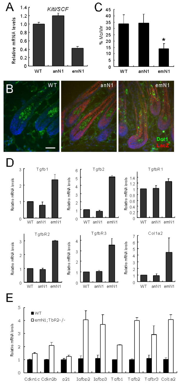
In silico comparison of proximal transcripts from Notch1-deficient and wild type hair bulbs revealed that expression of the diffusible factor Kitl/SCF was significantly down regulated in Notch1-deficient hair bulbs (Table S1 and Fig. 6A), suggesting that Notch1 is a positive regulator of this melanocyte survival factor in hair matrix cells. Further analysis uncovered reduction in multiple melanocyte-signature transcripts (Table S2, (Rendl et al., 2005)). To ascertain if this reflected a reduced transcriptional output from the same number of cells or change in the number of melanocytes residing within the bulb, Dct1-positive melanocytes were counted (Figs 6B and 6C). Strikingly, a significant reduction in melanocyte number was observed in Notch1-deficient hair bulbs (Figs 6B and 6C), even after correcting for keratinocyte numbers (emN1 bulbs contain fewer cells, Fig. 1E). Notch-mediated activation of Hes1 contributes autonomously to melanoblast survival (Moriyama et al., 2006); however, melanocytes in N1CKO skin did not experience a Cre-mediated gene deletion (Fig. S4) and express Hes1 (Fig. 8B). Therefore, proliferation and survival of follicular melanocytes, which requires activated c-Kit receptors, depends on a ligand produced by keratinocytes (Botchkareva et al., 2001; Ito et al., 1999) under the control of Notch1.
Figure 6. Notch1-loss alters the expression of the diffusible ligands Kitl and TGFβ.
(A) qRT-PCR of Kitl using hair follicle total RNA from genotypes specified (p < 0.0001 in both WT vs emN1 and anN1 vs emN1). (B) Immunohistochemical staining of wild type and Msx2Cre;Notch1flox/flox;Rosa26R/+ with Dct-1 (green) and LacZ (red). Nuclei were stained with DAPI. Note that melanocytes (Dct-1-positive) do not show LacZ staining. Scale bar 50 μM. (C) The ratio of melanocytes (Dct-1 positive) to the matrix cells in each follicle was obtained from different genotypes specified. Note the reduced number of melanocytes in Notch1-deficient hair follicles (p < 0.0001 in both WT vs emN1 and anN1 vs emN1). (D) qRT-PCR of TGFβ signaling components and its target (Col1a2). (E) qRT-PCR of the genes specified using Notch1- and Tgfbr2-deficient hair bulbs. Error bars represent s.e.m. (A, D, and E) and s.d. (C)
Notch signaling has been shown to control the proliferation and differentiation of neighboring cells (Baker and Schubiger, 1996; Buckles et al., 2001; Giraldez and Cohen, 2003; Lin and Kopan, 2003; Lin et al., 2000; Pan et al., 2004). Although several diffusible molecules, including Wingless and Unpaired, have been identified as mediators of such cell non-autonomous Notch functions in Drosophila (Giraldez and Cohen, 2003; Vaccari and Bilder, 2005), KITL is the first mammalian factor mediating bi-compartmental, Notch1-dependent signals that regulate proliferation or survival of a neighboring cell population (melanocytes within the hair follicles). Ascertaining whether Kitl is a direct or indirect target of Notch signaling will be addressed elsewhere. Nonetheless, reduced autocrine Kitl is not sufficient to explain the Notch1-deficient follicular phenotypes because animals treated with ACK2 (anti c-Kit antibody) or animals with mutations that reduced KITL/SCF levels do not display any phenotypes reminiscent of Notch1 loss in their hair follicles (Besmer et al., 1993; Botchkareva et al., 2001; Lecoin et al., 1995; Okura et al., 1995).
Changes in epithelial TGFβ ligands lie downstream of Notch1 and may contribute to the skin phenotype
Another signaling pathway shown to induce both IGFBPs and CKIs, and thus capable of promoting G1 arrest, was altered in N1CKO skin: the TGFβ signaling pathway (Firth and Baxter, 2002; Jetten et al., 1986; Moustakas et al., 2002). Transcription of Tgfb1 and Tgfb2, the receptor Tgfbr2 (itself a target of TGFβ signaling (Cui et al., 1995)), the co-receptor Tgfbr3 (Brown et al., 1999), as well as the TGFβ target Col1a2 (Inagaki et al., 1994) and Thbs1, were all elevated in the emN1 hair bulb (Table S1 and Fig. 6D). The ligands TGFβ1 and TGFβ2 and the receptors TGFβRI and TGFβRII are all expressed in the hair follicle (Fig. S2 and (Paus et al., 1997)). These receptors are also expressed in DP (Fig. S2), and Col1a2 is specifically expressed in dermal fibroblasts (Zheng et al., 2002), indicating that TGFβ signals are received in fibroblasts. Because TGFβ-mediated signaling can act both as an autocrine and a paracrine signal, this pathway is capable of activating transcription of Igfbp2 in keratinocytes (Moustakas et al., 2002; Scandura et al., 2004), as well as that of Igfbp3 in neighboring cells (Guo et al., 1995; Martin and Baxter, 1991).
To ask if autocrine reception of TGFβ plays a role in hair matrix homeostasis, we removed the essential type II receptor Tgfbr2 in all Notch1-deficient keratinocytes by generating Msx2Cre;Tgfbr2flox/flox (TβR2CKO) and Msx2Cre;Notch1flox/flox;Tgfbr2flox/flox (N1TβR2CKO) mice. Only dermal fibroblasts and DP cells in these compound mice retain the ability to respond to TGFβ ligands, yet the skin of TβR2CKO mice is indistinguishable from wild type (data not shown). Importantly, hair follicles from N1TβR2CKO and N1CKO mice were morphologically identical, and qRT-PCR analysis confirmed that the expression of Tgfb1, Tgfb2, Tgfbr3, Igfbp2, Igfbp3, Col1a2, and CKIs remained elevated in TβR2CKO emN1 hair bulbs (Fig. 6E).
These observations indicate that (1) TGFβRII-mediated TGFβ signals do not regulate the proliferation/apoptosis of hair matrix keratinocytes cell-autonomously, and (2) elevated Igfbp2 or CKI mRNA levels are not a consequence of autocrine TGFβ signaling in hair matrix. Collectively, the results in this and the previous sections are consistent with the hypothesis that Notch1 regulates hair matrix proliferation both autonomously, by preventing expression of proapoptotic and cytostatic genes (including gsdm1), and non-autonomously, by establishing a bi-compartmental feed-back loop: loss of Notch1 in transit amplifying keratinocytes induced TGFβ ligands, which are received in DP cells. Diffusible IGFBP3, produced exclusively in DP cells as a possible consequence of TGFβ signaling, could inhibit follicular IGF signaling to suppress the proliferation of follicular keratinocytes.
Entry into catagen was accelerated in 90% of follicles when Notch1 is removed over the entire skin surface (K14-Cre and K5-CreERT, (Vauclair et al., 2005)). However, catagen was not accelerated in Notch1-deficient hair follicles when they reside in a heterogeneous environment with anN1 follicles present. Instead, synchronicity is maintained in the chimeric skin with Notch1-deficient follicles entering catagen when their wild type neighbors do. Until this report, no mechanistic connection was made between Notch1 and catagen induction that could explain either phenomenon.
While several pro-catagen transcripts were elevated when Notch1 was lost, these changes were not sufficient to permit catagen onset in our chimeric mice. For example, detection of Igfbp3 mRNA in N1CKO skin by ISH required extended incubation time in NBT reagent (48 hours) compared to signal development in early catagen follicles (3 hours; Supplemental Figure S5). If catagen onset relied only on follicle-intrinsic mechanisms, it would be expected that Notch1-deficient follicles would enter catagen early, even if embedded in a heterogeneous environment. It is important to consider that while our mice have a chimeric skin, most individual follicles are not salt-and-paper mosaics; they either express Notch1 or they do not (Figs 1A, 8B, and S6). This unequivocal finding eliminates one trivial explanation (rescue by wild type matrix cells) for the milder phenotype we see in Notch1-deficient follicles.
Several other possible explanations for this observation exist. First, the timing of Notch1 deletion in these experimental paradigms differs: dorsal ectodermal Msx2-Cre expression (Pan et al., 2004) precedes dorsal Keratin 14 expression (Byrne et al., 1994); the earliest catagen was induced by the postnatal Cre-mediated deletion (by K5-CreERT in newborn mice; (Vauclair et al., 2005)). Perhaps early deletion allowed for compensation by an intrinsic follicular factor. Alternatively, accelerated catagen may be due in part to Cre mediated toxicity (Loonstra et al., 2001; Schmidt et al., 2000), which may be more severe in later-deleting follicles. However, we cannot rule out a third possibility: the presence of wild type epidermis and follicles in Msx2-Cre lines ameliorate the impact of Notch1 loss by generation of systemic, diffusible factor(s) that can entrain the cycling of Notch1-deficient follicles (Stenn and Paus, 2001) and prevent them from entering into catagen.
It is important to note that transgenic mice expressing activated Notch1 enter catagen as their wild type littermates (Lin et al., 2000), indicating that Notch1 do not antagonize catagen-inducing factors such as FGF5.
Differential response in the stroma may contribute to the differential impact of Notch1 loss on hair follicle and epidermal keratinocytes
In Notch1-deficient skin, the proliferation of epidermal keratinocytes increases progressively until a hyperplastic skin phenotype is evident, whereas follicular keratinocytes are hypoplastic, resulting in a shorter hair shaft (Fig. 1). To gain further insight into the basis of the differential response in these related keratinocyte populations, we asked if any proteins whose expression was altered in Notch1-deficient hair follicles were also affected in the epidermis. IGFBP2, p15INK4b and TGFβ2 protein levels were similarly regulated in the Notch1-deficient epidermis and hair follicles at P9 (Figs 3B and 7). The mRNA levels of Nrarp and Heyl, known Notch1 targets (Krebs et al., 2001; Lamar et al., 2001), were reduced in the epidermis and in the hair follicle (Figs 7C and 8A). In addition, Nrarp and Notch1 transcripts overlap in the hair follicle (Fig. 8B).
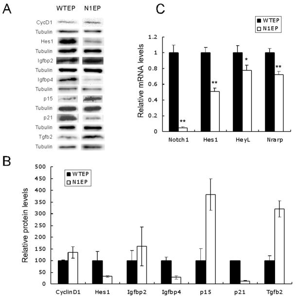
Figure 7. Monitoring the epidermal response of genes altered in hair follicles.
(A) Western blot analysis with P9 epidermal cell lysates from embryo-deleted NICKO (N1EP) and its wild type littermate (WTEP). (B) Quantification of the western data from (A). (C) qRT-PCR of Notch1 and its targets, Hes1, HeyL, and Nrarp, using P9 epidermal RNA preparation. Error bars represent s.e.m. * p = 0.05; ** p < 0.001
We confirmed previous reports that epidermal Hes1 and p21Cip1 protein levels were reduced in Notch1-deficient epidermal keratinocytes (Fig. 7A). In contrast, mRNA expression of Hes1 and p21Cip1 was sustained in Notch1-deficient hair follicles (Figs 3B and 8). Moreover, Hes1 protein, unarguably the best-studied Notch target, did not accumulate where Notch1 mRNA (Fig. 8B) or protein (Fig. S6) were present. Instead, it overlapped with the expression domain of Notch2 and Notch3 (see (Pan et al., 2004)). In Notch1-deficient hair follicles, p21Cip1 mRNA expression was sustained and its expression overlapped well with Hes1. This observation supports an indirect p21Cip1 activation mechanism by Hes1 through Calcineurin/NFAT (Mammucari et al., 2005). Therefore, we conclude that p21Cip1 may be a target of Hes1 in both the epidermis and the hair follicle, and that Hes1 acts as a Notch1 target only in the epidermal keratinocytes; in the hair follicle, its expression may be regulated by other Notch paralogs (Notch2 or Notch3). These data collectively agree with the common hypothesis that cellular context affects usage of targets, which contribute to the distinct proliferative behavior of hair follicles and epidermal keratinocytes in response to Notch1 loss.
The second striking molecular difference between epidermis and hair follicles is the behavior of two IGF binding proteins: IGFBP3 and IGFBP4. In Notch1-deficient hair follicles, induction of IGFBP3 in DP, presumably by reception of TGFβ in wild type DP cells, underlie the follicular phenotype in N1CKO animals. In contrast, the levels of IGBP4 were significantly down regulated (Fig. 7A and B) in Notch1-deficient epidermis, opposite to the trend seen in the hair follicles (Fig. 5A). Igfbp4 mRNA accumulates in dermis (Batch et al., 1994) and overexpression of this diffusible protein can decrease the growth of prostate cancer in vivo (Durai et al., 2006). Therefore, we posit that the differential modulation of IGF signaling that could result from changes in dermal IGFBP levels may underlie the difference in the response of epidermis and hair follicles to Notch1 loss. Thus, the contribution of Notch signaling to differential follicular and epidermal homeostasis results from both cell autonomous (differential target selection) and cell non-autonomous effects (differential stromal response to the same diffusible factors), the later mediated possibly by TGFβ.
TGFβ signaling, which is negatively regulated by Notch1 in both keratinocyte populations observed here (Figs 6D and 7), has been shown to cooperate with Notch1 in other systems (Blokzijl et al., 2003; Zavadil et al., 2004). However, to our knowledge, this is the first report to place Notch1 upstream of TGFβ signaling in any cell type. While at this time we can only speculate how TGFβ ligands responded to Notch1 loss, the broad spectrum of co-regulated TGFβ response genes confirm that this pathway is perturbed in response to Notch1 loss. The published observations we summarized and the data presented in this manuscript suggest that prior to overt changes in Notch1-deficient epidermal keratinocytes, a dialog between stromal fibroblast and keratinocyte, mediated in part via TGFβ signaling, is established to control keratinocyte proliferation. It was reported that TGFβ expression in basal epidermal cells leads to epidermal hyperplasia, most likely due to stromal involvement: epidermal overexpression is accompanied by changes in dermal compartments, which include increased angiogenesis, inflammatory responses, and dermal hyperplasia (Li et al., 2004; Liu et al., 2001), all seen in Notch1 deficient skin (data not shown). Although TGFβ1 is the major ligand in epidermal keratinocytes (Li et al., 2006), TGFβ2 can also promote keratinocyte proliferation (Foitzik et al., 1999). In this scenario, the elevated expression of p15INK4b in Notch1-deficient epidermal keratinocytes may initially keep proliferation in check but eventually may become overridden by the strong mitogenic effect of TGFβ-induced inflammatory and/or fibroblast responses (Li et al., 2004), which include the paracrine impact on stromal IGFBPs.
The role of cellular context in interpreting Notch1 signals is not unique to keratinocytes: an opposite impact of Notch1 on cell proliferation has also been demonstrated in lymphocytes, where Notch1 suppresses proliferation and induces apoptosis in B cells (Morimura et al., 2000) while promoting proliferation and blocking apoptosis or differentiation in T cells (Weng et al., 2004; Weng et al., 2003). Like P9 skin keratinocytes, B and T cells are derived from a common lymphoid progenitor (CLP). At P9, hair follicular and epidermal keratinocytes are both derived from a common pool of progenitors, yet they contain different Notch targets, creating a different molecular context. Unlike lymphocytes, the differences in proliferative behavior of keratinocytes may reflect autonomous differences (e.g. p21 and Hes1), as well as differential stromal responses to common targets (e.g. TGFβ2).
Conclusions
We demonstrate that Notch1 contributes to an elaborate network of signaling pathways that monitors and controls proliferation in skin. The response of wild type stromal cells to signals produced by mutant epithelial neighbors critically contributes to cancer (Krtolica and Campisi, 2002; Krtolica and Campisi, 2003). The observations summarized above suggest that ligand reception in the stroma may contribute to the tumor suppressor Notch1 phenotype, similar to the ‘viscous cycle,’ prompting bone metastasis in several solid cancers (Mundy, 2002; Roodman, 2004) and transformation of cells adjacent to Notch-deficient clones in Drosophila (Moberg et al., 2005; Thompson et al., 2005; Vaccari and Bilder, 2005). In a broader context, the role of Notch signaling in tumor suppression may reflect both an autonomous function promoting differentiation of keratinocytes and a non-autonomous function in a monitoring network, which responds by modulating keratinocyte proliferation.
Supplementary Material
Acknowledgments
The authors wish to thank Drs. Anne Bowcock, Tatiana Efimova, Jeffrey Gordon, Meei-hua Lin, Liang Ma, and the members of the Kopan laboratory for careful reading and numerous consultations throughout the course of this work. We wish to thank Dr. Gail Martin for the Msx2-Cre mice. JL and RK were supported in part by Washington University and by grants from the NIH-NIGMS (GM55479-10) and NIHD HD044056-01.
References
- Alonso L, Fuchs E. Stem cells in the skin: waste not, Wnt not. Genes & Development. 2003;17:1189–200. doi: 10.1101/gad.1086903. [DOI] [PubMed] [Google Scholar]
- Bartek J, Lukas J. Mammalian G1- and S-phase checkpoints in response to DNA damage. Current Opinion in Cell Biology. 2001;13:738–47. doi: 10.1016/s0955-0674(00)00280-5. [DOI] [PubMed] [Google Scholar]
- Baserga R, Hongo A, Rubini M, Prisco M, Valentinis B. The IGF-I receptor in cell growth, transformation and apoptosis. Biochimica et Biophysica Acta. 1997;1332:F105–26. doi: 10.1016/s0304-419x(97)00007-3. [DOI] [PubMed] [Google Scholar]
- Batch JA, Mercuri FA, Edmondson SR, Werther GA. Localization of messenger ribonucleic acid for insulin-like growth factor-binding proteins in human skin by in situ hybridization. Journal of Clinical Endocrinology & Metabolism. 1994;79:1444–9. doi: 10.1210/jcem.79.5.7525637. [DOI] [PubMed] [Google Scholar]
- Besmer P, Manova K, Duttlinger R, Huang EJ, Packer A, Gyssler C, Bachvarova RF. The kit-ligand (steel factor) and its receptor c-kit/W: pleiotropic roles in gametogenesis and melanogenesis. Development Supplement. 1993:125–37. [PubMed] [Google Scholar]
- Blanpain C, Lowry WE, Pasolli HA, Fuchs E. Canonical notch signaling functions as a commitment switch in the epidermal lineage. Genes Dev. 2006;20:3022–35. doi: 10.1101/gad.1477606. [DOI] [PMC free article] [PubMed] [Google Scholar]
- Blokzijl A, Dahlqvist C, Reissmann E, Falk A, Moliner A, Lendahl U, Ibanez CF. Cross-talk between the Notch and TGF-beta signaling pathways mediated by interaction of the Notch intracellular domain with Smad3. Journal of Cell Biology. 2003;163:723–8. doi: 10.1083/jcb.200305112. [DOI] [PMC free article] [PubMed] [Google Scholar]
- Botchkareva NV, Khlgatian M, Longley BJ, Botchkarev VA, Gilchrest BA. SCF/c-kit signaling is required for cyclic regeneration of the hair pigmentation unit. FASEB Journal. 2001;15:645–58. doi: 10.1096/fj.00-0368com. [DOI] [PubMed] [Google Scholar]
- Brown CB, Boyer AS, Runyan RB, Barnett JV. Requirement of type III TGF-beta receptor for endocardial cell transformation in the heart. Science. 1999;283:2080–2. doi: 10.1126/science.283.5410.2080. [DOI] [PubMed] [Google Scholar]
- Butt AJ, Firth SM, Baxter RC. The IGF axis and programmed cell death. Immunology & Cell Biology. 1999;77:256–62. doi: 10.1046/j.1440-1711.1999.00822.x. [DOI] [PubMed] [Google Scholar]
- Byrne C, Tainsky M, Fuchs E. Programming gene expression in developing epidermis. Development. 1994;120:2369–83. doi: 10.1242/dev.120.9.2369. [DOI] [PubMed] [Google Scholar]
- Chenn A, Walsh CA. Regulation of cerebral cortical size by control of cell cycle exit in neural precursors.[see comment] Science. 2002;297:365–9. doi: 10.1126/science.1074192. [DOI] [PubMed] [Google Scholar]
- Ciofani M, Zuniga-Pflucker JC. Notch promotes survival of pre-T cells at the beta-selection checkpoint by regulating cellular metabolism. Nat Immunol. 2005 doi: 10.1038/ni1234. [DOI] [PubMed] [Google Scholar]
- Cui W, Fowlis DJ, Cousins FM, Duffie E, Bryson S, Balmain A, Akhurst RJ. Concerted action of TGF-beta 1 and its type II receptor in control of epidermal homeostasis in transgenic mice. Genes & Development. 1995;9:945–55. doi: 10.1101/gad.9.8.945. [DOI] [PubMed] [Google Scholar]
- Devgan V, Mammucari C, Millar SE, Brisken C, Dotto GP. p21WAF1/Cip1 is a negative transcriptional regulator of Wnt4 expression downstream of Notch1 activation. Genes Dev. 2005;19:1485–1495. doi: 10.1101/gad.341405. [DOI] [PMC free article] [PubMed] [Google Scholar]
- DiSalvo CV, Zhang D, Jacobberger JW. Regulation of NIH-3T3 cell G1 phase transit by serum during exponential growth. Cell Proliferation. 1995;28:511–24. doi: 10.1111/j.1365-2184.1995.tb00089.x. [DOI] [PubMed] [Google Scholar]
- Dry FW. The coat of the mouse (Mus musculus) Jour Genetics. 1926;16:287–340. [Google Scholar]
- Durai R, Davies M, Yang W, Yang SY, Seifalian A, Goldspink G, Winslet M. Biology of insulin-like growth factor binding protein-4 and its role in cancer (review) International Journal of Oncology. 2006;28:1317–25. [PubMed] [Google Scholar]
- Edmondson SR, Thumiger SP, Werther GA, Wraight CJ. Epidermal homeostasis: the role of the growth hormone and insulin-like growth factor systems. Endocrine Reviews. 2003;24:737–64. doi: 10.1210/er.2002-0021. [DOI] [PubMed] [Google Scholar]
- Firth SM, Baxter RC. Cellular actions of the insulin-like growth factor binding proteins. Endocrine Reviews. 2002;23:824–54. doi: 10.1210/er.2001-0033. [DOI] [PubMed] [Google Scholar]
- Foitzik K, Paus R, Doetschman T, Dotto GP. The TGF-beta 2 isoform is both a required and sufficient inducer of murine hair follicle morphogenesis. Developmental Biology. 1999;212:278–289. doi: 10.1006/dbio.1999.9325. [DOI] [PubMed] [Google Scholar]
- Fryer CJ, White JB, Jones KA. Mastermind Recruits CycC:CDK8 to Phosphorylate the Notch ICD and Coordinate Activation with Turnover. Mol Cell. 2004;16:509–20. doi: 10.1016/j.molcel.2004.10.014. [DOI] [PubMed] [Google Scholar]
- Fuchs E, Raghavan S. Getting under the skin of epidermal morphogenesis [Review] Nature Reviews Genetics. 2002;3:199–209. doi: 10.1038/nrg758. [DOI] [PubMed] [Google Scholar]
- Giraldez AJ, Cohen SM. Wingless and Notch signaling provide cell survival cues and control cell proliferation during wing development. Development. 2003;130:6533–43. doi: 10.1242/dev.00904. [DOI] [PubMed] [Google Scholar]
- Guo YS, Townsend CM, Jr, Jin GF, Beauchamp RD, Thompson JC. Differential regulation by TGF-beta 1 and insulin of insulin-like growth factor binding protein-2 in IEC-6 cells. Am J Physiol Endocrinol Metab. 1995;268:E1199–1204. doi: 10.1152/ajpendo.1995.268.6.E1199. [DOI] [PubMed] [Google Scholar]
- Hardy MH. The secret life of the hair follicle. Trends in Genetics. 1992;8:55–61. doi: 10.1016/0168-9525(92)90350-d. [DOI] [PubMed] [Google Scholar]
- Harms KL, Chen X. The C Terminus of p53 Family Proteins Is a Cell Fate Determinant. Mol Cell Biol. 2005;25:2014–2030. doi: 10.1128/MCB.25.5.2014-2030.2005. [DOI] [PMC free article] [PubMed] [Google Scholar]
- Huang Q, Raya A, DeJesus P, Chao SH, Quon KC, Caldwell JS, Chanda SK, Izpisua-Belmonte JC, Schultz PG. Identification of p53 regulators by genome-wide functional analysis. Proc Natl Acad Sci U S A. 2004;101:3456–61. doi: 10.1073/pnas.0308562100. [DOI] [PMC free article] [PubMed] [Google Scholar]
- Inagaki Y, Truter S, Ramirez F. Transforming growth factor-beta stimulates alpha 2(I) collagen gene expression through a cis-acting element that contains an Sp1-binding site. J Biol Chem. 1994;269:14828–14834. [PubMed] [Google Scholar]
- Ito M, Kawa Y, Ono H, Okura M, Baba T, Kubota Y, Nishikawa SI, Mizoguchi M. Removal of stem cell factor or addition of monoclonal anti-c-KIT antibody induces apoptosis in murine melanocyte precursors. Journal of Investigative Dermatology. 1999;112:796–801. doi: 10.1046/j.1523-1747.1999.00552.x. [DOI] [PubMed] [Google Scholar]
- Ito M, Liu Y, Yang Z, Nguyen J, Liang F, Morris RJ, Cotsarelis G. Stem cells in the hair follicle bulge contribute to wound repair but not to homeostasis of the epidermis. 2005;11:1351–1354. doi: 10.1038/nm1328. [DOI] [PubMed] [Google Scholar]
- Ito T, Udaka N, Yazawa T, Okudela K, Hayashi H, Sudo T, Guillemot F, Kageyama R, Kitamura H. Basic helix-loop-helix transcription factors regulate the neuroendocrine differentiation of fetal mouse pulmonary epithelium. Development. 2000;127:3913–3921. doi: 10.1242/dev.127.18.3913. [DOI] [PubMed] [Google Scholar]
- Jacks T, Remington L, Williams BO, Schmitt EM, Halachmi S, Bronson RT, Weinberg RA. Tumor spectrum analysis in p53-mutant mice. Current Biology. 1994;4:1–7. doi: 10.1016/s0960-9822(00)00002-6. [DOI] [PubMed] [Google Scholar]
- Jamora C, Fuchs E. Intercellular adhesion, signalling and the cytoskeleton [Review] Nature Cell Biology. 2002;4:E101–E108. doi: 10.1038/ncb0402-e101. [DOI] [PubMed] [Google Scholar]
- Jetten AM, Shirley JE, Stoner G. Regulation of proliferation and differentiation of respiratory tract epithelial cells by TGF beta. Experimental Cell Research. 1986;167:539–49. doi: 10.1016/0014-4827(86)90193-x. [DOI] [PubMed] [Google Scholar]
- Kopan R. Notch: a membrane-bound transcription factor. Journal of Cell Science. 2002;115:1095–7. doi: 10.1242/jcs.115.6.1095. [DOI] [PubMed] [Google Scholar]
- Kopan R, Weintraub H. Mouse notch: expression in hair follicles correlates with cell fate determination. Journal of Cell Biology. 1993;121:631–41. doi: 10.1083/jcb.121.3.631. [DOI] [PMC free article] [PubMed] [Google Scholar]
- Krebs LT, Deftos ML, Bevan MJ, Gridley T. The Nrarp gene encodes an ankyrin-repeat protein that is transcriptionally regulated by the Notch signaling pathway. Developmental Biology. 2001;238:110–119. doi: 10.1006/dbio.2001.0408. [DOI] [PubMed] [Google Scholar]
- Krtolica A, Campisi J. Cancer and aging: a model for the cancer promoting effects of the aging stroma. Int J Biochem Cell Biol. 2002;34:1401–14. doi: 10.1016/s1357-2725(02)00053-5. [DOI] [PubMed] [Google Scholar]
- Krtolica A, Campisi J. Integrating epithelial cancer, aging stroma and cellular senescence. Adv Gerontol. 2003;11:109–16. [PubMed] [Google Scholar]
- Lamar E, Deblandre G, Wettstein D, Gawantka V, Pollet N, Niehrs C, Kintner C. Nrarp is a novel intracellular component of the Notch signaling pathway. Genes Dev. 2001;15:1885–99. doi: 10.1101/gad.908101. [DOI] [PMC free article] [PubMed] [Google Scholar]
- Lechler T, Fuchs E. Asymmetric cell divisions promote stratification and differentiation of mammalian skin. Nature. 2005;437:275–80. doi: 10.1038/nature03922. [DOI] [PMC free article] [PubMed] [Google Scholar]
- Lecoin L, Lahav R, Martin FH, Teillet MA, Le Douarin NM. Steel and c-kit in the development of avian melanocytes: a study of normally pigmented birds and of the hyperpigmented mutant silky fowl. Developmental Dynamics. 1995;203:106–18. doi: 10.1002/aja.1002030111. [DOI] [PubMed] [Google Scholar]
- Legue E, Nicolas JF. Hair follicle renewal: organization of stem cells in the matrix and the role of stereotyped lineages and behaviors. Development. 2005;132:4143–54. doi: 10.1242/dev.01975. [DOI] [PubMed] [Google Scholar]
- Li AG, Lu SL, Han G, Hoot KE, Wang XJ. Role of TGFbeta in skin inflammation and carcinogenesis. Molecular Carcinogenesis. 2006;45:389–96. doi: 10.1002/mc.20229. [DOI] [PubMed] [Google Scholar]
- Li AG, Wang D, Feng XH, Wang XJ. Latent TGFbeta1 overexpression in keratinocytes results in a severe psoriasis-like skin disorder. EMBO Journal. 2004;23:1770–81. doi: 10.1038/sj.emboj.7600183. [DOI] [PMC free article] [PubMed] [Google Scholar]
- Lin MH, Kopan R. Long-range, nonautonomous effects of activated Notch1 on tissue homeostasis in the nail. Developmental Biology. 2003;263:343–59. doi: 10.1016/j.ydbio.2003.07.007. [DOI] [PubMed] [Google Scholar]
- Lin MH, Leimeister C, Gessler M, Kopan R. Activation of the Notch pathway in the hair cortex leads to aberrant differentiation of the adjacent hair-shaft layers. Development. 2000;127:2421–32. doi: 10.1242/dev.127.11.2421. [DOI] [PubMed] [Google Scholar]
- Lindner G, Botchkarev V, Botchkareva N, Ling G, van der Veen C, Paus R. Analysis of apoptosis during hair follicle regression (catagen) Am J Pathol. 1997;151:1601–1617. [PMC free article] [PubMed] [Google Scholar]
- Liu X, Alexander V, Vijayachandra K, Bhogte E, Diamond I, Glick A. Conditional epidermal expression of TGFbeta 1 blocks neonatal lethality but causes a reversible hyperplasia and alopecia. Proceedings of the National Academy of Sciences of the United States of America. 2001;98:9139–44. doi: 10.1073/pnas.161016098. [DOI] [PMC free article] [PubMed] [Google Scholar]
- Loonstra A, Vooijs M, Beverloo HB, Allak BA, van Drunen E, Kanaar R, Berns A, Jonkers J. Growth inhibition and DNA damage induced by Cre recombinase in mammalian cells. Proc Natl Acad Sci U S A. 2001;98:9209–14. doi: 10.1073/pnas.161269798. [DOI] [PMC free article] [PubMed] [Google Scholar]
- Lunny DP, Weed E, Nolan PM, Marquardt A, Augustin M, Porter RM. Mutations in gasdermin 3 cause aberrant differentiation of the hair follicle and sebaceous gland. Journal of Investigative Dermatology. 2005;124:615–21. doi: 10.1111/j.0022-202X.2005.23623.x. [DOI] [PubMed] [Google Scholar]
- Luo J, McMullen JR, Sobkiw CL, Zhang L, Dorfman AL, Sherwood MC, Logsdon MN, Horner JW, DePinho RA, Izumo S, et al. Class IA phosphoinositide 3-kinase regulates heart size and physiological cardiac hypertrophy. Molecular & Cellular Biology. 2005;25:9491–502. doi: 10.1128/MCB.25.21.9491-9502.2005. [DOI] [PMC free article] [PubMed] [Google Scholar]
- Mainprize TG, Taylor MD, Rutka JT, Dirks PB. Cip/Kip cell-cycle inhibitors: a neuro-oncological perspective. Journal of Neuro-Oncology. 2001;51:205–18. doi: 10.1023/a:1010671908204. [DOI] [PubMed] [Google Scholar]
- Malkinson FD, Keane JT. Hair matrix cell kinetics: a selective review. International Journal of Dermatology. 1978;17:536–51. doi: 10.1111/j.1365-4362.1978.tb05997.x. [DOI] [PubMed] [Google Scholar]
- Mammucari C, di Vignano AT, Sharov AA, Neilson J, Havrda MC, Roop DR, Botchkarev VA, Crabtree GR, Dotto GP. Integration of Notch 1 and calcineurin/NFAT signaling pathways in keratinocyte growth and differentiation control. Developmental Cell. 2005;8:665–76. doi: 10.1016/j.devcel.2005.02.016. [DOI] [PubMed] [Google Scholar]
- Martin JL, Baxter RC. Transforming growth factor-beta stimulates production of insulin-like growth factor-binding protein-3 by human skin fibroblasts. Endocrinology. 1991;128:1425–33. doi: 10.1210/endo-128-3-1425. [DOI] [PubMed] [Google Scholar]
- Massague J. G1 cell-cycle control and cancer. Nature. 2004;432:298–306. doi: 10.1038/nature03094. [DOI] [PubMed] [Google Scholar]
- Millar SE. Molecular mechanisms regulating hair follicle development [Review] Journal of Investigative Dermatology. 2002;118:216–225. doi: 10.1046/j.0022-202x.2001.01670.x. [DOI] [PubMed] [Google Scholar]
- Moberg KH, Schelble S, Burdick SK, Hariharan IK. Mutations in erupted, the Drosophila ortholog of mammalian tumor susceptibility gene 101, elicit non-cell-autonomous overgrowth. Developmental Cell. 2005;9:699–710. doi: 10.1016/j.devcel.2005.09.018. [DOI] [PubMed] [Google Scholar]
- Moll UM, Slade N. p63 and p73: roles in development and tumor formation. Molecular Cancer Research: MCR. 2004;2:371–86. [PubMed] [Google Scholar]
- Morimura T, Goitsuka R, Zhang Y, Saito I, Reth M, Kitamura D. Cell Cycle Arrest and Apoptosis Induced by Notch1 in B Cells 10.1074/jbc.M006415200. J Biol Chem. 2000;275:36523–36531. doi: 10.1074/jbc.M006415200. [DOI] [PubMed] [Google Scholar]
- Moriyama M, Osawa M, Mak S-S, Ohtsuka T, Yamamoto N, Han H, Delmas V, Kageyama R, Beermann F, Larue L, et al. Notch signaling via Hes1 transcription factor maintains survival of melanoblasts and melanocyte stem cells 10.1083/jcb.200509084. J Cell Biol. 2006;173:333–339. doi: 10.1083/jcb.200509084. [DOI] [PMC free article] [PubMed] [Google Scholar]
- Moustakas A, Pardali K, Gaal A, Heldin CH. Mechanisms of TGF-beta signaling in regulation of cell growth and differentiation. Immunology Letters. 2002;82:85–91. doi: 10.1016/s0165-2478(02)00023-8. [DOI] [PubMed] [Google Scholar]
- Muller-Rover S, Handjiski B, van der Veen C, Eichmuller S, Foitzik K, McKay IA, Stenn KS, Paus R. A comprehensive guide for the accurate classification of murine hair follicles in distinct hair cycle stages [Review] Journal of Investigative Dermatology. 2001;117:3–15. doi: 10.1046/j.0022-202x.2001.01377.x. [DOI] [PubMed] [Google Scholar]
- Mumm JS, Kopan R. Notch signaling: from the outside in. Developmental Biology. 2000;228:151–65. doi: 10.1006/dbio.2000.9960. [DOI] [PubMed] [Google Scholar]
- Mundy GR. Metastasis to bone: causes, consequences and therapeutic opportunities. Nat Rev Cancer. 2002;2:584–93. doi: 10.1038/nrc867. [DOI] [PubMed] [Google Scholar]
- Nguyen B-C, Lefort K, Mandinova A, Antonini D, Devgan V, Della Gatta G, Koster MI, Zhang Z, Wang J, di Vignano AT, et al. Cross-regulation between Notch and p63 in keratinocyte commitment to differentiation 10.1101/gad.1406006. Genes Dev. 2006;20:1028–1042. doi: 10.1101/gad.1406006. [DOI] [PMC free article] [PubMed] [Google Scholar]
- Nicolas M, Wolfer A, Raj K, Kummer JA, Mill P, van Noort M, Hui CC, Clevers H, Dotto GP, Radtke F. Notch1 functions as a tumor suppressor in mouse skin. Nature Genetics. 2003;33:416–21. doi: 10.1038/ng1099. [DOI] [PubMed] [Google Scholar]
- Okura M, Maeda H, Nishikawa S, Mizoguchi M. Effects of monoclonal anti-c-kit antibody (ACK2) on melanocytes in newborn mice. Journal of Investigative Dermatology. 1995;105:322–8. doi: 10.1111/1523-1747.ep12319939. [DOI] [PubMed] [Google Scholar]
- Okuyama R, Nguyen BC, Talora C, Ogawa E, Tommasi di Vignano A, Lioumi M, Chiorino G, Tagami H, Woo M, Dotto GP. High commitment of embryonic keratinocytes to terminal differentiation through a Notch1-caspase 3 regulatory mechanism. Developmental Cell. 2004;6:551–62. doi: 10.1016/s1534-5807(04)00098-x. [DOI] [PubMed] [Google Scholar]
- Pan Y, Lin MH, Tian X, Cheng HT, Gridley T, Shen J, Kopan R. [gamma]-Secretase Functions through Notch Signaling to Maintain Skin Appendages but Is Not Required for Their Patterning or Initial Morphogenesis. Developmental Cell. 2004;7:731–743. doi: 10.1016/j.devcel.2004.09.014. [DOI] [PubMed] [Google Scholar]
- Paus R, Foitzik K, Welker P, Bulfone-Paus S, Eichmuller S. Transforming growth factor-beta receptor type I and type II expression during murine hair follicle development and cycling. Journal of Investigative Dermatology. 1997;109:518–26. doi: 10.1111/1523-1747.ep12336635. [DOI] [PubMed] [Google Scholar]
- Rangarajan A, Talora C, Okuyama R, Nicolas M, Mammucari C, Oh H, Aster JC, Krishna S, Metzger D, Chambon P, et al. Notch signaling is a direct determinant of keratinocyte growth arrest and entry into differentiation. EMBO J. 2001;20:3427–3436. doi: 10.1093/emboj/20.13.3427. [DOI] [PMC free article] [PubMed] [Google Scholar]
- Rendl M, Lewis L, Fuchs E. Molecular Dissection of Mesenchymal-Epithelial Interactions in the Hair Follicle. PLoS Biology. 2005:3. doi: 10.1371/journal.pbio.0030331. [DOI] [PMC free article] [PubMed] [Google Scholar]
- Resnicoff M, Baserga R. The role of the insulin-like growth factor I receptor in transformation and apoptosis. Annals of the New York Academy of Sciences. 1998;842:76–81. doi: 10.1111/j.1749-6632.1998.tb09634.x. [DOI] [PubMed] [Google Scholar]
- Roodman GD. Mechanisms of bone metastasis. N Engl J Med. 2004;350:1655–64. doi: 10.1056/NEJMra030831. [DOI] [PubMed] [Google Scholar]
- Rossi M, SAYAN AE, TERRINONI A, MELINO G, KNIGHT RA. Mechanism of Induction of Apoptosis by p73 and Its Relevance to Neuroblastoma Biology. Ann NY Acad Sci. 2004;1028:143–149. doi: 10.1196/annals.1322.015. [DOI] [PubMed] [Google Scholar]
- Saeki N, Kuwahara Y, Sasaki H, Satoh H, Shiroishi T. Gasdermin (Gsdm) localizing to mouse Chromosome 11 is predominantly expressed in upper gastrointestinal tract but significantly suppressed in human gastric cancer cells. Mammalian Genome. 2000;11:718–24. doi: 10.1007/s003350010138. [DOI] [PubMed] [Google Scholar]
- Scandura JM, Boccuni P, Massague J, Nimer SD. Transforming growth factor {beta}-induced cell cycle arrest of human hematopoietic cells requires p57KIP2 up-regulation. PNAS. 2004;101:15231–15236. doi: 10.1073/pnas.0406771101. [DOI] [PMC free article] [PubMed] [Google Scholar]
- Schmidt EE, Taylor DS, Prigge JR, Barnett S, Capecchi MR. Illegitimate Cre-dependent chromosome rearrangements in transgenic mouse spermatids. Proc Natl Acad Sci U S A. 2000;97:13702–7. doi: 10.1073/pnas.240471297. [DOI] [PMC free article] [PubMed] [Google Scholar]
- Sengel P. Morphogenesis of Skin. Cambridge: Cambridge University Press; 1976. [Google Scholar]
- Sherr CJ, Roberts JM. CDK inhibitors: positive and negative regulators of G1-phase progression. Genes & Development. 1999;13:1501–12. doi: 10.1101/gad.13.12.1501. [DOI] [PubMed] [Google Scholar]
- Stenn KS, Paus R. Controls of hair follicle cycling [Review] Physiological Reviews. 2001;81:449–494. doi: 10.1152/physrev.2001.81.1.449. [DOI] [PubMed] [Google Scholar]
- Su HY, Hickford JG, Bickerstaffe R, Palmer BR. Insulin-like growth factor 1 and hair growth. Dermatology Online Journal. 1999a;5:1. [PubMed] [Google Scholar]
- Su HY, Hickford JG, The PH, Hill AM, Frampton CM, Bickerstaffe R. Increased vibrissa growth in transgenic mice expressing insulin-like growth factor 1. Journal of Investigative Dermatology. 1999b;112:245–8. doi: 10.1046/j.1523-1747.1999.00489.x. [DOI] [PubMed] [Google Scholar]
- Sun X, Lewandoski M, Meyers EN, Liu YH, Maxson RE, Jr, Martin GR. Conditional inactivation of Fgf4 reveals complexity of signalling during limb bud development. Nature Genetics. 2000;25:83–6. doi: 10.1038/75644. [DOI] [PubMed] [Google Scholar]
- Terrinoni A, Ranalli M, Cadot B, Leta A, Bagetta G, Vousden KH, Melino G. p73-alpha is capable of inducing scotin and ER stress. Oncogene. 2004;23:3721–5. doi: 10.1038/sj.onc.1207342. [DOI] [PubMed] [Google Scholar]
- Thompson BJ, Mathieu J, Sung HH, Loeser E, Rorth P, Cohen SM. Tumor suppressor properties of the ESCRT-II complex component Vps25 in Drosophila. Developmental Cell. 2005;9:711–20. doi: 10.1016/j.devcel.2005.09.020. [DOI] [PubMed] [Google Scholar]
- Vaccari T, Bilder D. The Drosophila tumor suppressor vps25 prevents nonautonomous overproliferation by regulating notch trafficking. Developmental Cell. 2005;9:687–98. doi: 10.1016/j.devcel.2005.09.019. [DOI] [PubMed] [Google Scholar]
- Vauclair S, Nicolas M, Barrandon Y, Radtke F. Notch1 is essential for postnatal hair follicle development and homeostasis. Dev Biol. 2005 doi: 10.1016/j.ydbio.2005.05.018. [DOI] [PubMed] [Google Scholar]
- Weedon D, Strutton G. Apoptosis as the mechanism of the involution of hair follicles in catagen transformation. Acta Dermato-Venereologica. 1981;61:335–9. [PubMed] [Google Scholar]
- Weger N, Schlake T. IGF-I Signalling Controls the Hair Growth Cycle and the Differentiation of Hair Shafts. Journal of Investigative Dermatology. 2005a;125:873–882. doi: 10.1111/j.0022-202X.2005.23946.x. [DOI] [PubMed] [Google Scholar]
- Weger N, Schlake T. Igfbp3 modulates cell proliferation in the hair follicle. Journal of Investigative Dermatology. 2005b;125:847–9. doi: 10.1111/j.0022-202X.2005.23886.x. [DOI] [PubMed] [Google Scholar]
- Weng AP, Ferrando AA, Lee W, Morris JP, IV, Silverman LB, Sanchez-Irizarry C, Blacklow SC, Look AT, Aster JC. Activating Mutations of NOTCH1 in Human T Cell Acute Lymphoblastic Leukemia. Science. 2004;306:269–271. doi: 10.1126/science.1102160. [DOI] [PubMed] [Google Scholar]
- Weng AP, Millholland JM, Yashiro-Ohtani Y, Arcangeli ML, Lau A, Wai C, del Bianco C, Rodriguez CG, Sai H, Tobias J, et al. c-Myc is an important direct target of Notch1 in T-cell acute lymphoblastic leukemia/lymphoma. Genes Dev. 2006;20:2096–2109. doi: 10.1101/gad.1450406. [DOI] [PMC free article] [PubMed] [Google Scholar]
- Weng AP, Nam Y, Wolfe MS, Pear WS, Griffin JD, Blacklow SC, Aster JC. Growth suppression of pre-T acute lymphoblastic leukemia cells by inhibition of notch signaling. Mol Cell Biol. 2003;23:655–64. doi: 10.1128/MCB.23.2.655-664.2003. [DOI] [PMC free article] [PubMed] [Google Scholar]
- Yamamoto N, Tanigaki K, Han H, Hiai H, Honjo T. Notch/RBP-J Signaling Regulates Epidermis/Hair Fate Determination of Hair Follicular Stem Cells. Current Biology. 2003;13:333–338. doi: 10.1016/s0960-9822(03)00081-2. [DOI] [PubMed] [Google Scholar]
- Yang X, Klein R, Tian X, Cheng HT, Kopan R, Shen J. Notch activation induces apoptosis in neural progenitor cells through a p53-dependent pathway. Dev Biol. 2004;269:81–94. doi: 10.1016/j.ydbio.2004.01.014. [DOI] [PubMed] [Google Scholar]
- Zavadil J, Cermak L, Soto-Nieves N, Bottinger EP. Integration of TGF-beta/Smad and Jagged1/Notch signalling in epithelial-to-mesenchymal transition. EMBO Journal. 2004;23:1155–65. doi: 10.1038/sj.emboj.7600069. [DOI] [PMC free article] [PubMed] [Google Scholar]
- Zheng B, Zhang Z, Black CM, de Crombrugghe B, Denton CP. Ligand-Dependent Genetic Recombination in Fibroblasts: A Potentially Powerful Technique for Investigating Gene Function in Fibrosis. Am J Pathol. 2002;160:1609–1617. doi: 10.1016/S0002-9440(10)61108-X. [DOI] [PMC free article] [PubMed] [Google Scholar]
Associated Data
This section collects any data citations, data availability statements, or supplementary materials included in this article.


