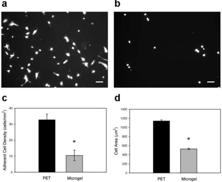
Figure 5. In vitro human primary macrophage adhesion to biomaterial surfaces.
Adherent cells were scored for viability, and cell density and spread area were quantified. Compared to unmodified PET substrates (a), microgel coatings (b) reduce macrophage adhesion to biomaterial surfaces. (c) Microgel coatings elicit a 3-fold reduction in cell adhesion compared to unmodified PET surfaces, * p < 1.1×10⁻⁴. (d) Adherent macrophages also exhibit more cell extensions and 2-fold larger surface areas on unmodified PET controls than on microgel-coated surfaces, * p < 9.5×10⁻⁷. Data is presented as the average value ± standard error of the mean using n = 5 samples per treatment group. Scale bar is 100 µm.

