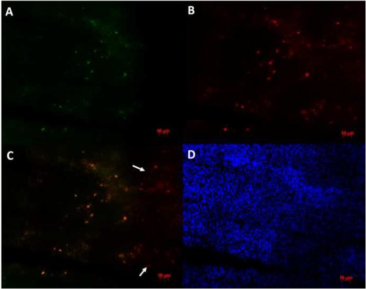
Figure 4.
Immunofluorescent staining of macrophages in the marrow of a femur injected with 10% BC particles. (A) Mouse anti-GFP monoclonal antibody is the primary antibody and Alexa Fluor 488 conjugated goat anti-mouse IgG is the secondary antibody; (B) Rat anti-MOMA monoclonal antibody is the primary antibody and Alexa Fluor 594 conjugated goat anti-rat IgG is the secondary antibody; (C) Image A overlaped with image B using Adobe Photoshop. Note the co-localization of the two antibodies. Arrows indicate the spots with only MOMA-2 immunostaining signal. (D) DAPI was used for nuclear staining. All three images were taken from the same field of vision. Scale bar is 10 microns.

