Abstract
The ability to quantify and characterize antigen-specific CD8+ T cells irrespective of functional readouts using fluorochrome-conjugated tetrameric peptide-MHC class I (pMHCI) complexes in conjunction with flow cytometry has transformed our understanding of cellular immune responses over the past decade. In the case of prevalent CD8+ T cell populations that engage cognate pMHCI tetramers with high avidities, direct ex vivo identification and subsequent data interpretation is relatively straightforward. However, the accurate identification of low frequency antigen-specific CD8+ T cell populations can be complicated, especially in situations where TCR-mediated tetramer binding occurs at low avidities. Here, we highlight a few simple techniques that can be employed to improve the visual resolution, and hence the accurate quantification, of tetramer-binding CD8+ T cell populations by flow cytometry. These methodological modifications enhance signal intensity, especially in the case of specific CD8+ T cell populations that bind cognate antigen with low avidity, minimize background noise and enable improved discrimination of true pMHCI tetramer binding events from nonspecific uptake.
Introduction
Adaptive immunity is mediated by a complex network of cellular and molecular interactions that sense and respond to antigenic stimuli derived from dangerous entities. In order to deconvolute this system, it is important that the necessary tools are available to enable the accurate and reproducible measurement of antigen-specific cell populations directly ex vivo; however, it is equally important that these tools are used with an understanding of their limitations. One of the most significant advances in immunotechnology over the past few years has been the development of soluble recombinant peptide-major histocompatibility complex class I (pMHCI) multimers (1,2). These reagents bind stably to cognate T cell receptors (TCRs) expressed on the surface of antigen-specific CD8+ T cells, despite the low affinity and rapid kinetics of monomeric TCR/pMHCI binding, and are internalized at physiological binding temperatures (3,4); thus, in fluorochrome-conjugated form, pMHCI multimers allow the visualization of cognate CD8+ T cells by flow cytometry. In general, pathogen-specific CD8+ T cell populations, which tend to express TCRs that bind cognate pMHCI with high affinity (5) and comprise a substantial proportion of the memory T cell pool, are easily identified with relatively basic flow cytometers and associated software. However, it is sometimes difficult to distinguish true antigen-specific CD8+ T cells from background binding events, especially when the target population is present at low frequency and binds cognate pMHCI with low avidity. Resolution of these issues is critical for the analysis and interpretation of pMHCI multimer-based data. In this brief review, which is necessarily selective in scope, we highlight a few techniques that might help to improve the ex vivo detection of true pMHCI multimer binding events.
What is real?
A priori, the simplest way to confirm both the presence and magnitude of a specific CD8+ T cell population detected by pMHCI multimer staining is to demonstrate and measure the functional consequences of cognate antigen engagement at the single cell level; this can be achieved relatively easily with a variety of readouts using flow cytometric platforms (Figure 1). However, there are a number of issues that complicate the use of functional verification as a "gold standard" test.
Figure 1. Physical identification of antigen-specific CD8+ T cells with pMHCI tetramers is independent of downstream functional consequences.
Peripheral blood mononuclear cells (PBMC) were incubated with pre-titered allophycocyanin (APC)-conjugated cytomegalovirus (CMV) pp65495–503/human leukocyte antigen (HLA) A*0201 tetramer at 37 °C for 20 minutes and then stimulated for 6 hours with the corresponding cognate peptide at 2 µg/ml in the presence of costimulatory monoclonal antibodies (mAbs; αCD28 and αCD49d, each at 1 µg/ml), brefeldin A (10 µg/ml), monensin (0.7 µg/ml) and pre-titered Alexa 680-conjugated αCD107a to detect degranulation as described previously (42). After stimulation, the cells were washed, and then stained sequentially for surface and intracellular markers prior to acquisition as described previously with minor modifications (28). A minimum of 200,000 live cells, identified within the "dump negative" population as depicted for a different dataset in Figure 2, were acquired on a modified LSRII flow cytometer (BD BioSciences) and analyzed with FlowJo software (TreeStar Inc.). The top row shows CD8+ T cells expressing cognate TCRs specific for the HLA A*0201-restricted CMV pp65495–503 epitope that were identified with the corresponding pMHCI tetramer (5.68% of CD8+ T cells); degranulation (CD107a mobilization) and cytokine production (IFNγ and TNFα) were then assessed in parallel (tetramer+ events are shown in red). Functional CD8+ T cells that do not stain with the CMV pp65495–503/ HLA A*0201 tetramer are observed; this could reflect either non-specific background activation in vivo or antigen-specific activation of CD8+ T cells ex vivo that fail to bind cognate tetramer (13–15). Discrimination between these possibilities is difficult due to the fact that the appropriate negative control cannot be conducted because pMHCI tetramers are not biologically inert. Thus, cognate CD8+ T cell activation in the absence of subsequent peptide stimulation can be induced by: (i) multimeric TCR engagement by pMHCI tetramers in isolation (26); and, (ii) peptide representation by endogenous MHCI molecules (43,44). The bottom row shows the proportion of non-functional tetramer+ cells by sequential gating (from left to right) on CD8+ T cells that lack TNFα and IFNγ production and CD107a mobilization in response to antigenic stimulation as described above; in this experiment, 1.30% of CD8+ T cells were tetramer+ and functionally inert within the limitations of the measured parameters. Atypically, a substantial proportion of cognate CD8+ T cells in this experiment failed to produce either TNFα or IFNγ, yet mobilized CD107a; this is consistent with the differential triggering sensitivities of these functions and probably reflects suboptimal stimulation due to pMHCI tetramer-induced down-regulation of cell surface TCRs that might be stimulated more effectively by membrane-bound antigen.
Antigen-experienced CD8+ T cells are primed to execute a programmed array of effector functions when triggered by cognate pMHCI molecules; nevertheless, not all cells and not all functions are equivalent. Thus, it is possible that a truly cognate CD8+ T cell might not be functional in a given ex vivo bioassay (Figure 1). Indeed, many studies have now shown that tetramer-binding CD8+ T cells can exhibit various degrees of dysfunction, for example due to the effects of persistent antigen exposure [(6) and citations therein]. Furthermore, such discrepancies between function and physical presence may vary with circumstance; thus, distinct changes in functional capabilities can accompany both generic T cell differentiation (7,8) and the unique ontogeny of individual antigen-specific T cells (9), thereby giving rise to a vast heterogeneity of phenotypic characteristics and functional profiles within the memory T cell pool. Similarly, certain functions are triggered at different thresholds and thus can be differentially elicited in CD8+ T cells with different antigen sensitivities (10–12).
A further complicating issue arises from the observation that CD8+ T cells bearing cognate TCRs can exhibit functional activation in response to agonist ligands, yet fail to bind the corresponding pMHCI multimer (13–15). Recent data indicate that these discrepancies can arise as a consequence of the differential TCR/pMHCI affinity thresholds and kinetic rules that govern pMHCI multimer binding and functional activation (15). Thus, pMHCI multimer binding is dependent on both the expression of cognate TCRs that bind monomeric pMHCI above a certain affinity and a corresponding compound cellular avidity for antigen that is determined by multiple additional factors (16). In contrast, pMHCI antigens with monomeric affinities for TCR that lie below the threshold required for binding of the corresponding multimer can act as agonist ligands in functional assays (15).
Thus, even with the assumption that an optimal antigenic stimulus is delivered with current protocols, functional measurements, either individually or in combination, are insufficient in isolation to determine the integrity of pMHCI multimer binding. How else, then, can we provide a "reality check" to discriminate signal from noise in pMHCI multimer-based assays? The purpose of what follows is to provide some potential solutions to these issues.
Reagent preparation, assay conditions and interfacing with the flow cytometric platform
Many factors can influence the reliability and reproducibility of pMHCI multimer-based assays. A complete discussion of these factors is beyond the scope of this article; however, we have recently reviewed many of these issues separately (16) and optimization of some related parameters is also considered in a companion article by Keeney and colleagues in this issue of Cytometry. For the purposes of this review, we will assume that: (i) all recombinant pMHCI reagents are produced to a high degree of purity and fully tetramerized, albeit with an integral degree of heterogeneity (17), by conjugation to high quality, bright fluorochrome-labeled streptavidin preparations; (ii) all stains are performed at physiological binding temperature (37 °C) for a maximum of 20 minutes as described previously (3); (iii) all reagents are optimally titrated to ensure maximal discrimination between signal and noise; (iv) all flow cytometry-related variables are optimally configured; and, (v) sufficient numbers of events are collected to allow accurate data interpretation (18). It should be noted that the techniques discussed below are applicable to pMHCI multimers of different valencies; the commonly used tetrameric scaffold is considered here for simplicity.
Noise reduction
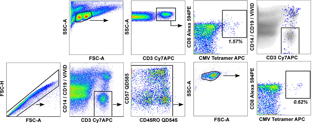
As the frequency of antigen-specific CD8+ T cells decreases, so the true pMHCI tetramer signal becomes increasingly obscured within the inherent background noise that assumes greater relative proportionality within the system; this can lead to erroneous measurements of quantity and quality. Thus, it is essential to minimize noise through the elimination of aberrant binding events from the analysis. This can be achieved through the incorporation of a "dump" channel, in which specific stains are used to identify confounding events that are subsequently gated out en masse (Figure 2). A viability marker should always be included in the dump channel because dead/dying cells with impaired membrane integrity are a major source of non-specific binding events; the fixable amine reactive dyes are particularly useful for this purpose as they are compatible with fixation/permeabilization procedures (19). Similarly, it is generally useful to exclude monocytes and B-cells with αCD14 and αCD19 mAbs respectively (20,21). Multiple additional markers can also be included within a single dump channel as circumstances dictate; for example, the addition of αCD33 and αCD34 might eliminate further nonspecific binding events in bone marrow mononuclear cell samples. Overall, the use of a dump channel increases measurement sensitivity by reducing compensation-induced spreading error and eliminating irrelevant cells that artifactually masquerade as true signal with traditional gating strategies (Figure 2).
Figure 2. Gating strategies to eliminate aberrant binding events.
Bone marrow mononuclear cells were washed with phosphate-buffered saline (PBS) and stained with the amine reactive viability dye ViViD (Invitrogen/Molecular Probes) as described previously (19). After a further wash, cells were stained at 37 °C for 20 minutes with pretitered APC-conjugated CMV pp65495–503/HLA A*0201 tetramer mutated in the α3 domain of the heavy chain (D227K/T228A) to abrogate CD8 binding (26); such CD8-null tetramers enable the selective identification of high avidity cognate CD8+ T cells (28,37,38). Cells were subsequently stained for surface markers prior to immediate acquisition as described in Figure 1; αCD14 and αCD19 were conjugated to Pacific Blue to fluoresce in the same channel as ViViD. The top row shows a "traditional" gating strategy used with six-parameter flow cytometers, in which lymphocyte populations are identified in a forward scatter-area (FSC-A) versus side scatter-area (SSC-A) plot; tetramer+ cells are then displayed in a bivariate plot with CD8 after gating to identify T cells on the basis of CD3 expression. The bottom row shows a polychromatic gating strategy to eliminate aberrant binding events. Singlet cells are first selected in a FSC-A versus FSC-height plot and then CD3+ T cells are identified against the "dump" channel to exclude cells that could bind tetramer and mAbs non-specifically (dead/dying cells with compromised membranes, monocytes and B-cells). Fluorochrome aggregates are then excluded from the analysis before gating small lymphocytes on the basis of standard light scatter properties. Finally, tetramer+ cells are displayed versus CD8 as shown in the top row. Both rows show the same dataset. The "true" tetramer+ CD8+ T cell population is cleanly identified with the polychromatic gating strategy (0.62%); the poorly resolved tetramer+ CD8+ population (1.57%) identified with the traditional gating strategy contains a high proportion of non-specific "background" binding events that are eliminated by inclusion of the "dump" channel (right panel, top row; tetramer+ events are shown in blue overlaid on the total population in a bivariate CD3 versus CD14/CD19/ViViD plot). It should be noted that in some cases it appears that activated CD8+ T cells can associate with CD14 within the small lymphocyte gate and that, consequently, some antigen-specific events might be excluded from the analysis inadvertently by the inclusion of αCD14 in the dump channel (Betts & Price, unpublished observation).
Signal/noise discrimination
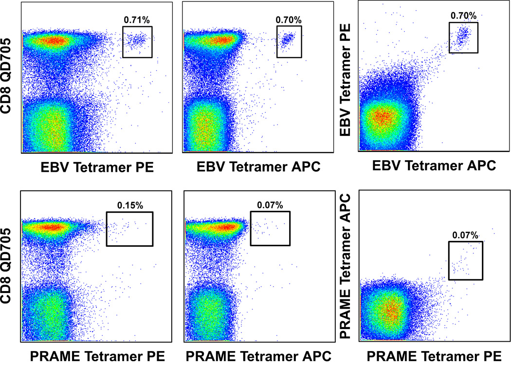
Even once aberrant binding events have been eliminated, a degree of background staining frequently persists within the CD8+ T cell population that can be difficult to resolve. A visual clue to the extent of the problem can be attained from simple inspection of the corresponding CD3+CD8− cell population; thus, if a similar degree of pMHCI tetramer staining is observed, then it is less likely that the signal emanating from the CD3+CD8+ cell population is real. Indeed, this information should always be disclosed in the presentation of pMHCI tetramer-based data. However, such observations do not necessarily exclude the presence of cognate CD8+ T cells; rather, they provide an indication of the general level of noise from which any true binding events must be extracted. Assuming that such non-specific staining is, in part, random, one empirical approach to this task is to use the same pMHCI tetrameric reagent labeled in different colors (Figure 3). The principle here is that cognate CD8+ T cells will bind specifically regardless of the label, whereas the random element of the staining pattern will not associate equally with each version of the reagent. Thus, although there is inevitably a concomitant reduction in the intensity of specific labeling with each individual pMHCI tetramer, a degree of confidence is added by the observation of dual fluorochrome uptake. This strategy can be especially useful in the case of infrequent CD8+ T cells that are not easily amenable to functional verification and exhibit poor separation as a consequence of low antigen binding avidities (22,23).
Figure 3. Discrimination of true tetramer binding events by double fluorochrome verification.
PBMC were stained with BiViD or ViViD, pre-titered Epstein-Barr virus (EBV) BMLF1259–267/HLA A*0201 (top row) or preferentially expressed antigen on melanomas (PRAME)100–108/HLA A*0201 (bottom row) tetramers and mAbs specific for cell surface markers as described in the legend to Figure 2. Antigen-specific CD8+ T cells identified with phycoerythrin (PE)-conjugated or APC-conjugated pHLA A*0201 tetramers are shown in the left and middle panels respectively. As shown, background staining can be more pronounced with PE-conjugated pMHCI tetramers due, in part, to greater spreading error and the tendency for heterogeneity within such preparations (17,20). Random background originating from individual tetramers representing a given antigen specificity can be discerned by staining with a mixture of differentially conjugated tetramers together (right panel). Cognate CD8+ T cells will capture pMHCI tetramers from solution regardless of the associated fluorochrome; singly stained cells likely represent aberrant binding events. In these experiments, pre-mixing of tetramer solutions prior to staining minimizes the bias that can arise from sequential additions due to rapid on-rates for binding (26). Nevertheless, it remains possible that some cognate CD8+ T cells will stain with only one of the corresponding pMHCI tetramers simply by chance, although this is unlikely given that many fluorochromes must be taken up for an individual cell to be visible by flow cytometry. Furthermore, doubly stained cognate CD8+ T cell populations exhibit reduced individual fluorescence intensities due to competition between tetramers for a finite number of surface TCRs. Thus, this technique is best considered as confirmatory for situations in which low frequency antigen-specific CD8+ T cells are difficult to discriminate reliably; this is especially pertinent when traditional gating trees are used (Figure 2).
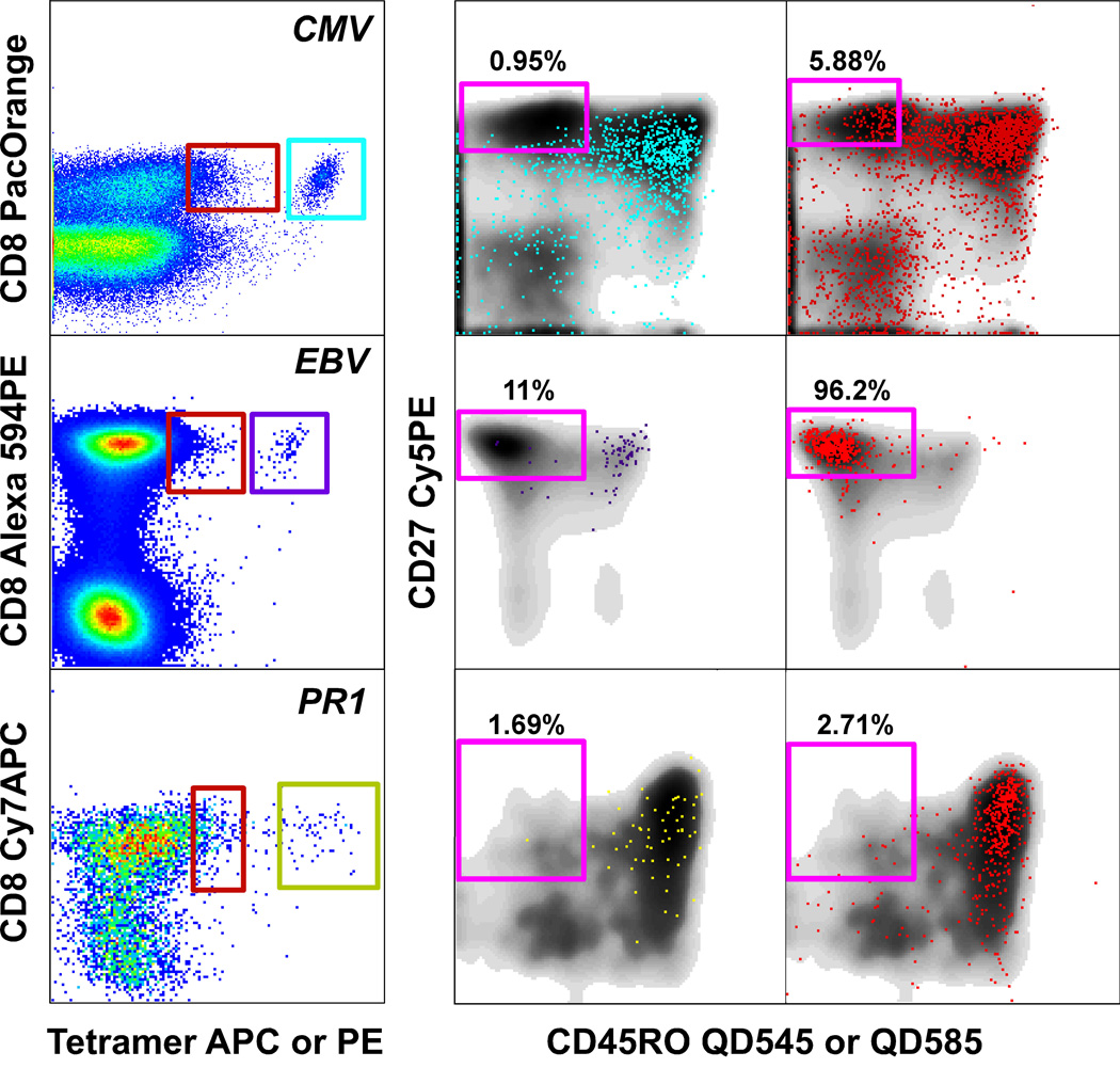
In addition, however, there is often an intrinsic element to background staining that can vary idiosyncratically with different pMHCI tetramers in different individuals. To some extent at least, this is a CD8 coreceptor-mediated effect (24). Consequently, tetramers produced from pMHCI mutants with impaired CD8 binding properties exhibit diminished background staining and can thereby lead to enhanced signal resolution (25–27). In general, this has proven to be an effective strategy for pathogen-specific CD8+ T cells that typically express high affinity TCRs and bind antigen with high avidity (5,28). However, CD8 stabilizes and enhances TCR/pMHCI interactions at the cell surface through effects on both the off-rate and the on-rate (15,24,29), despite the fact that these trimolecular interactions are structurally distinct and non-cooperative in three dimensions (30–32). Therefore, modification of CD8 coreceptor binding also alters the avidity range of antigen-specific CD8+ T cells that can bind pMHCI tetramers; given that avidity for antigen is at least partially dependent on the corresponding TCR/pMHCI monomeric affinity, intact or even enhanced CD8 binding properties (see below) are likely to be required for optimal pMHCI tetramer-based detection of tumor-specific and autoreactive CD8+ T cell populations (5,15). How, then, can we distinguish background staining from signal without compromising the detection of low avidity CD8+ T cells? Perhaps the most useful technique here is to include phenotypic markers in the flow cytometric panel. The "shoulder" that protrudes from the non-cognate CD8+ T cell population and represents background staining comprises a mixture of both memory and naïve cells; i.e. the phenotypic distribution follows that of the CD8+ T cell population as a whole. In contrast, true cognate CD8+ T cells that bind pMHCI tetramer generally exhibit an almost exclusive memory phenotype (Figure 4). It should be noted that there are some exceptions to this general dichotomy; for example, CD8+ T cell populations specific for Melan-A/MART-1 can contain substantial numbers of naïve cells (33). Nevertheless, "memory gating" can help to distinguish true binding events from background staining in most experimental settings.
Figure 4. Discrimination of true tetramer binding events on the basis of phenotypic characteristics.
PBMC were stained with ViViD, pre-titered pHLA A*0201 tetramers as indicated and mAbs specific for cell surface markers as described in the legend to Figure 2; the αCD8 mAbs used in these distinct experiments were conjugated to Pacific Orange (CMV pp65495–503), Alexa 594-PE (EBV BMLF1259–267) and Cy7-APC (PR1; proteinase-3, residues 169–177). Expression of CD27 and CD45RO was examined in the bright tetramer+ CD8+ T cell population and in the "shoulder" that comprises tetramerdim/neg CD8+ T cells. The vast majority of the bright tetramer+ CD8+ T cells express markers consistent with a memory phenotype (various colors; middle panels), whereas the tetramerdim/neg CD8+ T cells follow a phenotypic distribution similar to that of the overall CD8+ T cell population in the sample (red; right panels). Thus, the presence of substantial numbers of naïve CD8+ T cells helps to resolve non-specific background staining; this can be useful when staining patterns lack clarity, for example with low avidity cognate CD8+ T cell populations that tend to separate poorly. The incorporation of additional memory markers can also be helpful, especially with antigen-specific CD8+ T cell populations that tend to exhibit atypical phenotypic characteristics (45).
Signal amplification
The intensity of labeling with pMHCI tetramers reflects, at least in part, cellular avidity for antigen (34,35). This parameter is determined both by TCR/pMHCI affinity (15) and by factors beyond the direct interface with antigen, such as membrane flexibility, molecular density and cell surface topography [reviewed in (16)]. Although the clonotypic structure of a given antigen-specific CD8+ T cell population is fixed at the time of analysis, pMHCI tetramer binding to that population can be altered via manipulations that affect either cellular behaviour or the structure of the reagent; these technical modifications, which primarily impact the detection of low avidity CD8+ T cells, are not necessarily mutually exclusive and can exhibit useful compound effects in certain situations.
Cellular modification
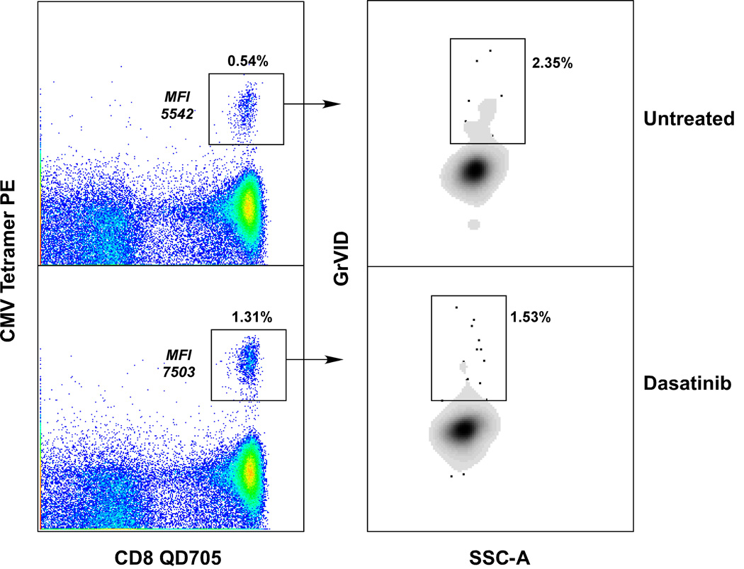
The extent of ligand binding is dependent on the concentration of both partners. For this reason, the maximum concentration of pMHCI tetramer that allows clear visualization with minimal background issues should be selected in reagent titrations for optimal experimental usage; this will facilitate the capture of cognate pMHCI tetramers by lower avidity CD8+ T cells (see below). It is also possible to alter the surface densities of TCR and CD8 by pre-treating cells with the protein tyrosine kinase inhibitor dasatinib (36). This manipulation can substantially enhance the intensity of pMHCI tetramer staining and preferentially benefits the detection of low avidity CD8+ T cells (Figure 5). These phenomena are not unexpected given the effects of CD8 on membrane-constrained TCR/pMHCI interactions and the impact of TCR density on the rate at which the second and third pMHCI components of the tetramer bind to form a stable complex with the cell [reviewed (16)]. However, it is surprising that dasatinib can exert these beneficial effects on tetramer binding with incubation times shorter than those required to observe any upregulation of TCR or CD8 on the cell surface; preliminary experiments suggest that the underlying mechanism relates to the prevention of "empty" TCR internalization during pMHCI binding, but other explanations remain possible (Lissina et al., submitted). Regardless, substantial signal enhancement with minimal background effects can be observed with this manipulation, especially in the context of low avidity CD8+ T cell populations. Furthermore, it appears from preliminary observations that dasatinib can reduce the degree of cell death induced by pMHCI tetramers during the course of the staining procedure (Figure 5; data not shown). Such effects may be conducive to the viable sorting of CD8+ T cells labeled with cognate pMHCI tetramers, especially as the effects of dasatinib are reversible (36).
Figure 5. Dasatinib pre-treatment enhances pMHCI tetramer staining.
PBMC were stained with ViViD, washed and then incubated with PBS alone or with PBS containing 100 nM dasatinib for 30 minutes at 37 °C. Subsequently, cells were stained at 4 °C for 20 minutes with pre-titered PE-conjugated CMV pp65495–503/HLA A*0201 tetramer; dasatinib was maintained in the medium at a concentration of 100nM throughout the staining procedure in the relevant experimental tubes. After a futher wash, cells were stained with mAbs specific for cell surface markers and then with GriViD, an amine reactive viability dye that is spectrally distinct from ViViD. Cells were acquired and analyzed as described in the legends to Figure 1 and Figure 2. The left panels show that tetramer staining is substantially enhanced in the presence of dasatinib; specifically, both greater signal intensity and improved resolution of intermediate binding events are observed. The right panels show the percentage of tetramer+ cells that die during the staining procedure. It is established that pMHCI multimer-induced signaling can trigger cell death (17,26,46,47), and it appears that dasatinib can mitigate these effects. In the experiment shown, few cells die during the assay; however, the extent of procedural cell death varies widely in different circumstances and can exert a substantial effect on the results. Similar data were obtained with dasatinib experiments conducted at 37 °C using pMHCI tetramers representing several different specificities (Lissina et al., submitted).
Reagent modification
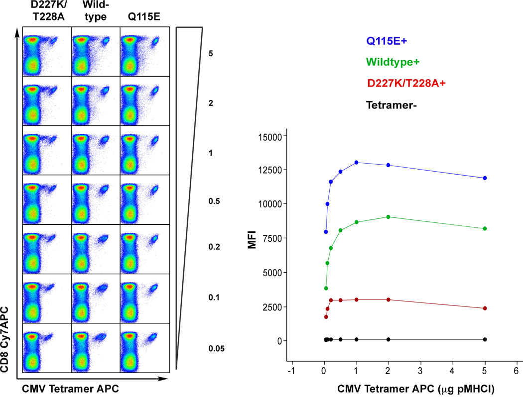
As discussed above, the CD8 coreceptor profoundly affects TCR/pMHCI binding at the cell surface by slowing the dissociation rate and enhancing the association rate (15,24,29). These effects influence the range of pMHCI ligands that can be bound in tetrameric form by a given CD8+ T cell. Thus, in the absence of CD8 binding, the monomeric TCR/pMHCI affinity threshold required to observe tetramer binding is substantially increased (15), thereby enabling the selective detection of high avidity antigen-specific CD8+ T cell populations with tetrameric forms of α3 domain-mutated (D227K/T228A) pMHCI molecules that retain TCR binding integrity but fail to engage CD8 (28,37,38). Recently, we have constructed pMHCI molecules that bind CD8 with enhanced affinity (24,39,40). The Q115E α2 domain mutation increases CD8 binding affinity from KD ≈ 120 µM to KD ≈ 85 µM (24) and extends the range of pMHCI ligands that can be bound in tetrameric form by a given CD8+ T cell; these effects enable the detection of low avidity antigen-specific CD8+ T cells by "coreceptor-enhanced" pMHCI tetramers (41). As discussed above, enhanced CD8 binding can be associated with concomitant increases in background staining; these can be minimized with appropriate reagent titration and resolved by the inclusion of phenotypic markers as shown in Figure 4 (41). However, while background staining is to some extent a CD8-mediated phenomenon, there are clearly additional factors that affect its penetrance in practice; thus, these effects appear to vary between individuals according to the nature of the antigen (41) and are not always a problematic accompaniment to the use of coreceptor-enhanced tetramers (Figure 6). In general, then, coreceptor-enhanced pMHCI tetramers can be usefully employed for signal amplification purposes and seem destined to find widespread applications in fields such as tumor immunology and autoimmunity where low avidity CD8+ T cell populations might prevail.
Figure 6. Coreceptor-enhanced pMHCI tetramers improve signal strength.
PBMC were stained with ViViD, pre-titered CMV pp65495–503/HLA A*0201 tetramers as indicated and mAbs specific for cell surface markers as described in the legend to Figure 2. The D227K/T228A α3 domain mutation abrogates CD8 binding (26), thereby enabling the selective identification of high avidity cognate CD8+ T cell populations (28,37,38); the Q115E α2 domain mutation increases CD8 binding from KD ≈ 120 µM to KD ≈ 85 mM (24,40) and enables the identification of low avidity cognate CD8+ T cells that lie below the threshold of detection with wildtype tetramers under identical conditions (41). In these experiments, tetrameric preparations were standardized for concentration and volume, such that conditions differed solely with respect to the nature of the pHLA A*0201/CD8 interaction; furthermore, all tetramers were prepared afresh from pMHCI monomers frozen at −80 °C to minimize confounding effects related to differential protein stability (48). The titrations shown range from 5 to 0.05 µg pMHCI with respect to the monomeric component in minimal staining volume; median fluorescence intensities (MFIs) of the tetramer+ populations are displayed in the right panel. Several effects are apparent. First, signal intensity improves as the affinity of the pMHCI/CD8 interaction increases; this is more apparent at lower pMHCI tetramer concentrations. Second, an "avidity separation" effect is visible within the cognate CD8+ T cell population; thus, at more limiting concentrations of wildtype pMHCI tetramer, two distinct populations emerge that presumably represent CD8+ T cells with differential avidities for antigen as described previously (28). This segregation is more apparent at higher concentrations with the D227K/T228A pMHCI tetramer as intrinsic avidities for antigen are exposed more clearly in the absence of CD8-mediated compensatory effects (28). In contrast, the low avidity CD8+ T cells are enveloped within the overall cognate population in the presence of enhanced CD8 binding; this is consistent with the observation that the Q115E mutation preferentially enhances pMHCI tetramer binding, and hence signal amplification, in the case of lower avidity CD8+ T cells (41).
Concluding remarks
As immunotechnology continues to advance with the development of multiplex applications for combined ex vivo measurements (21), it is increasingly important that signal and noise are accurately discriminated with basic reagents such as pMHCI tetramers in order to obtain reliable data. In this brief review, we have highlighted a few techniques that might contribute to the accurate detection of true antigen-specific CD8+ T cell populations with pMHCI tetramers directly ex vivo. These modifications to the standard assay approach are neither mutually exclusive nor definitive, but provide some options that can be tested in different scenarios to improve resolution.
Acknowledgements
This work was supported by the Intramural Research Program of the National Institutes of Health, Vaccine Research Center, National Institute of Allergy and Infectious Diseases. DAP is a Medical Research Council (UK) Senior Clinical Fellow.
Footnotes
This article is based, in part, on a talk given by David Price at the 3rd MASIR meeting, La Plagne, France.
References
- 1.Garboczi DN, Hung DT, Wiley DC. HLA-A2-peptide complexes: refolding and crystallization of molecules expressed in Escherichia coli and complexed with single antigenic peptides. Proc Natl Acad Sci U S A. 1992;89(8):3429–3433. doi: 10.1073/pnas.89.8.3429. [DOI] [PMC free article] [PubMed] [Google Scholar]
- 2.Altman JD, Moss PA, Goulder PJ, Barouch DH, McHeyzer-Williams MG, Bell JI, McMichael AJ, Davis MM. Phenotypic analysis of antigen-specific T lymphocytes. Science. 1996;274(5284):94–96. doi: 10.1126/science.274.5284.94. [DOI] [PubMed] [Google Scholar]
- 3.Whelan JA, Dunbar PR, Price DA, Purbhoo MA, Lechner F, Ogg GS, Griffiths G, Phillips RE, Cerundolo V, Sewell AK. Specificity of CTL interactions with peptide-MHC class I tetrameric complexes is temperature dependent. J Immunol. 1999;163(8):4342–4348. [PubMed] [Google Scholar]
- 4.Laugel B, Boulter JM, Lissin N, Vuidepot A, Li Y, Gostick E, Crotty LE, Douek DC, Hemelaar J, Price DA, et al. Design of soluble recombinant T cell receptors for antigen targeting and T cell inhibition. J Biol Chem. 2005;280(3):1882–1892. doi: 10.1074/jbc.M409427200. [DOI] [PubMed] [Google Scholar]
- 5.Cole DK, Pumphrey NJ, Boulter JM, Sami M, Bell JI, Gostick E, Price DA, Gao GF, Sewell AK, Jakobsen BK. Human TCR-binding affinity is governed by MHC class restriction. J Immunol. 2007;178(9):5727–5734. doi: 10.4049/jimmunol.178.9.5727. [DOI] [PubMed] [Google Scholar]
- 6.Rehr M, Cahenzli J, Haas A, Price DA, Gostick E, Huber M, Karrer U, Oxenius A. Emergence of polyfunctional CD8+ T cells after prolonged suppression of HIV replication by antiretroviral therapy. J Virol. 2008;82(7):3391–3404. doi: 10.1128/JVI.02383-07. [DOI] [PMC free article] [PubMed] [Google Scholar]
- 7.Wolint P, Betts MR, Koup RA, Oxenius A. Immediate cytotoxicity but not degranulation distinguishes effector and memory subsets of CD8+ T cells. J Exp Med. 2004;199(7):925–936. doi: 10.1084/jem.20031799. [DOI] [PMC free article] [PubMed] [Google Scholar]
- 8.Takata H, Takiguchi M. Three memory subsets of human CD8+ T cells differently expressing three cytolytic effector molecules. J Immunol. 2006;177(7):4330–4340. doi: 10.4049/jimmunol.177.7.4330. [DOI] [PubMed] [Google Scholar]
- 9.Seder RA, Darrah PA, Roederer M. T-cell quality in memory and protection: implications for vaccine design. Nat Rev Immunol. 2008;8(4):247–258. doi: 10.1038/nri2274. [DOI] [PubMed] [Google Scholar]
- 10.Valitutti S, Muller S, Dessing M, Lanzavecchia A. Different responses are elicited in cytotoxic T lymphocytes by different levels of T cell receptor occupancy. J Exp Med. 1996;183(4):1917–1921. doi: 10.1084/jem.183.4.1917. [DOI] [PMC free article] [PubMed] [Google Scholar]
- 11.Price DA, Sewell lAK, Dong T, Tan R, Goulder PJ, Rowland-Jones SL, Phillips RE. Antigen-specific release of beta-chemokines by anti-HIV-1 cytotoxic T lymphocytes. Curr Biol. 1998;8(6):355–358. doi: 10.1016/s0960-9822(98)70138-1. [DOI] [PubMed] [Google Scholar]
- 12.Betts MR, Price DA, Brenchley JM, Lore K, Guenaga FJ, Smed-Sorensen A, Ambrozak DR, Migueles SA, Connors M, Roederer M, et al. The functional profile of primary human antiviral CD8+ T cell effector activity is dictated by cognate peptide concentration. J Immunol. 2004;172(10):6407–6417. doi: 10.4049/jimmunol.172.10.6407. [DOI] [PubMed] [Google Scholar]
- 13.Rubio-Godoy V, Dutoit V, Rimoldi D, Lienard D, Lejeune F, Speiser D, Guillaume P, Cerottini JC, Romero P, Valmori D. Discrepancy between ELISPOT IFN-gamma secretion and binding of A2/peptide multimers to TCR reveals interclonal dissociation of CTL effector function from TCR-peptide/MHC complexes half-life. Proc Natl Acad Sci U S A. 2001;98(18):10302–10307. doi: 10.1073/pnas.181348898. [DOI] [PMC free article] [PubMed] [Google Scholar]
- 14.Lyons GE, Roszkowski JJ, Man S, Yee C, Kast WM, Nishimura MI. T-cell receptor tetramer binding or the lack there of does not necessitate antigen reactivity in T-cell receptor transduced T cells. Cancer Immunol Immunother. 2006;55(9):1142–1150. doi: 10.1007/s00262-005-0103-9. [DOI] [PMC free article] [PubMed] [Google Scholar]
- 15.Laugel B, van den Berg HA, Gostick E, Cole DK, Wooldridge L, Boulter J, Milicic A, Price DA, Sewell AK. Different T cell receptor affinity thresholds and CD8 coreceptor dependence govern cytotoxic T lymphocyte activation and tetramer binding properties. J Biol Chem. 2007;282(33):23799–23810. doi: 10.1074/jbc.M700976200. [DOI] [PubMed] [Google Scholar]
- 16.Wooldridge L, Lissina A, Cole DK, van den Berg HA, Price DA, Sewell AK. Tricks with tetramers: how to get the most from multimeric peptide-MHC. Immunology. 2008 doi: 10.1111/j.1365-2567.2008.02848.x. in press. [DOI] [PMC free article] [PubMed] [Google Scholar]
- 17.Guillaume P, Legler DF, Boucheron N, Doucey MA, Cerottini JC, Luescher IF. Soluble major histocompatibility complex-peptide octamers with impaired CD8 binding selectively induce Fas-dependent apoptosis. J Biol Chem. 2003;278(7):4500–4509. doi: 10.1074/jbc.M208863200. [DOI] [PubMed] [Google Scholar]
- 18.Roederer M. How many events is enough? Are you positive? Cytometry A. 2008;73A(5):384–385. doi: 10.1002/cyto.a.20549. [DOI] [PubMed] [Google Scholar]
- 19.Perfetto SP, Chattopadhyay PK, Lamoreaux L, Nguyen R, Ambrozak D, Koup RA, Roederer M. Amine reactive dyes: an effective tool to discriminate live and dead cells in polychromatic flow cytometry. J Immunol Methods. 2006;313(1–2):199–208. doi: 10.1016/j.jim.2006.04.007. [DOI] [PubMed] [Google Scholar]
- 20.Perfetto SP, Chattopadhyay PK, Roederer M. Seventeen-colour flow cytometry: unravelling the immune system. Nat Rev Immunol. 2004;4(8):648–655. doi: 10.1038/nri1416. [DOI] [PubMed] [Google Scholar]
- 21.Chattopadhyay PK, Price DA, Harper TF, Betts MR, Yu J, Gostick E, Perfetto SP, Goepfert P, Koup RA, De Rosa SC, et al. Quantum dot semiconductor nanocrystals for immunophenotyping by polychromatic flow cytometry. Nat Med. 2006;12(8):972–977. doi: 10.1038/nm1371. [DOI] [PubMed] [Google Scholar]
- 22.Lee PP, Yee C, Savage PA, Fong L, Brockstedt D, Weber JS, Johnson D, Swetter S, Thompson J, Greenberg PD, et al. Characterization of circulating T cells specific for tumor-associated antigens in melanoma patients. Nat Med. 1999;5(6):677–685. doi: 10.1038/9525. [DOI] [PubMed] [Google Scholar]
- 23.Zippelius A, Batard P, Rubio-Godoy V, Bioley G, Lienard D, Lejeune F, Rimoldi D, Guillaume P, Meidenbauer N, Mackensen A, et al. Effector function of human tumor-specific CD8 T cells in melanoma lesions: a state of local functional tolerance. Cancer Res. 2004;64(8):2865–2873. doi: 10.1158/0008-5472.can-03-3066. [DOI] [PubMed] [Google Scholar]
- 24.Wooldridge L, van den Berg HA, Glick M, Gostick E, Laugel B, Hutchinson SL, Milicic A, Brenchley JM, Douek DC, Price DA, et al. Interaction between the CD8 coreceptor and major histocompatibility complex class I stabilizes T cell receptor-antigen complexes at the cell surface. J Biol Chem. 2005;280(30):27491–27501. doi: 10.1074/jbc.M500555200. [DOI] [PMC free article] [PubMed] [Google Scholar]
- 25.Bodinier M, Peyrat MA, Tournay C, Davodeau F, Romagne F, Bonneville M, Lang F. Efficient detection and immunomagnetic sorting of specific T cells using multimers of MHC class I and peptide with reduced CD8 binding. Nat Med. 2000;6(6):707–710. doi: 10.1038/76292. [DOI] [PubMed] [Google Scholar]
- 26.Purbhoo MA, Boulter JM, Price DA, Vuidepot AL, Hourigan CS, Dunbar PR, Olson K, Dawson SJ, Phillips RE, Jakobsen BK, et al. The human CD8 coreceptor effects cytotoxic T cell activation and antigen sensitivity primarily by mediating complete phosphorylation of the T cell receptor zeta chain. J Biol Chem. 2001;276(35):32786–32792. doi: 10.1074/jbc.M102498200. [DOI] [PubMed] [Google Scholar]
- 27.Glick M, Price DA, Vuidepot AL, Andersen TB, Hutchinson SL, Laugel B, Sewell AK, Boulter JM, Dunbar PR, Cerundolo V, et al. Novel CD8+ T cell antagonists based on beta 2-microglobulin. J Biol Chem. 2002;277(23):20840–20846. doi: 10.1074/jbc.M201819200. [DOI] [PubMed] [Google Scholar]
- 28.Price DA, Brenchley JM, Ruff LE, Betts MR, Hill BJ, Roederer M, Koup RA, Migueles SA, Gostick E, Wooldridge L, et al. Avidity for antigen shapes clonal dominance in CD8+ T cell populations specific for persistent DNA viruses. J Exp Med. 2005;202(10):1349–1361. doi: 10.1084/jem.20051357. [DOI] [PMC free article] [PubMed] [Google Scholar]
- 29.Gakamsky DM, Luescher IF, Pramanik A, Kopito RB, Lemonnier F, Vogel H, Rigler R, Pecht I. CD8 kinetically promotes ligand binding to the T-cell antigen receptor. Biophys J. 2005;89(3):2121–2133. doi: 10.1529/biophysj.105.061671. [DOI] [PMC free article] [PubMed] [Google Scholar]
- 30.Gao GF, Tormo J, Gerth UC, Wyer JR, McMichael AJ, Stuart DI, Bell JI, Jones EY, Jakobsen BK. Crystal structure of the complex between human CD8alpha(alpha) and HLA-A2. Nature. 1997;387(6633):630–634. doi: 10.1038/42523. [DOI] [PubMed] [Google Scholar]
- 31.Wyer JR, Willcox BE, Gao GF, Gerth UC, Davis SJ, Bell JI, van der Merwe PA, Jakobsen BK. T cell receptor and coreceptor CD8 alphaalpha bind peptide-MHC independently and with distinct kinetics. Immunity. 1999;10(2):219–225. doi: 10.1016/s1074-7613(00)80022-9. [DOI] [PubMed] [Google Scholar]
- 32.Cole DK, Dunn SM, Sami M, Boulter JM, Jakobsen BK, Sewell AK. T cell receptor engagement of peptide-major histocompatibility complex class I does not modify CD8 binding. Mol Immunol. 2008;45(9):2700–2709. doi: 10.1016/j.molimm.2007.12.009. [DOI] [PubMed] [Google Scholar]
- 33.Pittet MJ, Valmori D, Dunbar PR, Speiser DE, Lienard D, Lejeune F, Fleischhauer K, Cerundoloq V, Cerottini JC, Romero P. High frequencies of naive Melan-A/MART-1-specific CD8(+) T cells in a large proportion of human histocompatibility leukocyte antigen (HLA)-A2 individuals. J Exp Med. 1999;190(5):705–715. doi: 10.1084/jem.190.5.705. [DOI] [PMC free article] [PubMed] [Google Scholar]
- 34.Busch DH, Pamer EG. T cell affinity maturation by selective expansion during infection. J Exp Med. 1999;189(4):701–710. doi: 10.1084/jem.189.4.701. [DOI] [PMC free article] [PubMed] [Google Scholar]
- 35.Yee C, Savage PA, Lee PP, Davis MM, Greenberg PD. Isolation of high avidity melanoma-reactive CTL from heterogeneous populations using peptide-MHC tetramers. J Immunol. 1999;162(4):2227–2234. [PubMed] [Google Scholar]
- 36.Weichsel R, Dix C, Wooldridge L, Clement M, Fenton-May A, Sewell AK, Zezula J, Greiner E, Gostick E, Price DA, et al. Profound inhibition of antigen-specific T-cell effector functions by dasatinib. Clin Cancer Res. 2008;14(8):2484–2491. doi: 10.1158/1078-0432.CCR-07-4393. [DOI] [PubMed] [Google Scholar]
- 37.Choi EM, Chen JL, Wooldridge L, Salio M, Lissina A, Lissin N, Hermans IF, Silk JD, Mirza F, Palmowski MJ, et al. High avidity antigen-specific CTL identified by CD8-independent tetramer staining. J Immunol. 2003;171(10):5116–5123. doi: 10.4049/jimmunol.171.10.5116. [DOI] [PubMed] [Google Scholar]
- 38.Pittet MJ, Rubio-Godoy V, Bioley G, Guillaume P, Batard P, Speiser D, Luescher I, Cerottini JC, Romero P, Zippelius A. alpha3 domain mutants of peptide/MHC class I multimers allow the selective isolation of high avidity tumor-reactive CD8 T cells. J Immunol. 2003;171(4):1844–1849. doi: 10.4049/jimmunol.171.4.1844. [DOI] [PubMed] [Google Scholar]
- 39.Gostick E, Cole DK, Hutchinson SL, Wooldridge L, Tafuro S, Laugel B, Lissina A, Oxenius A, Boulter JM, Price DA, et al. Functional and biophysical characterization of an HLA-A*6801-restricted HIV-specific T cell receptor. Eur J Immunol. 2007;37(2):479–486. doi: 10.1002/eji.200636243. [DOI] [PMC free article] [PubMed] [Google Scholar]
- 40.Wooldridge L, Lissina A, Vernazza J, Gostick E, Laugel B, Hutchinson SL, Mirza F, Dunbar PR, Boulter JM, Glick M, et al. Enhanced immunogenicity of CTL antigens through mutation of the CD8 binding MHC class I invariant region. Eur J Immunol. 2007;37(5):1323–1333. doi: 10.1002/eji.200636765. [DOI] [PMC free article] [PubMed] [Google Scholar]
- 41.Melenhorst JJ, Scheinberg P, Chattopadhyay PK, Lissina A, Gostick E, Cole DK, Wooldridge L, van den Berg HA, Bornstein E, Hensel NF, et al. Detection of low avidity CD8+ T cell populations with coreceptor-enhanced peptide-major histocompatibility complex class I tetramers. J Immunol Methods. 2008 doi: 10.1016/j.jim.2008.07.008. in press. [DOI] [PMC free article] [PubMed] [Google Scholar]
- 42.Betts MR, Brenchley JM, Price DA, De Rosa SC, Douek DC, Roederer M, Koup RA. Sensitive and viable identification of antigen-specific CD8+ T cells by a flow cytometric assay for degranulation. J Immunol Methods. 2003;281:65–78. doi: 10.1016/s0022-1759(03)00265-5. [DOI] [PubMed] [Google Scholar]
- 43.Ge Q, Stone JD, Thompson MT, Cochran JR, Rushe M, Eisen HN, Chen J, Stern LJ. Soluble peptide-MHC monomers cause activation of CD8+ T cells through transfer of the peptide to T cell MHC molecules. Proc Natl Acad Sci U S A. 2002;99(21):13729–13734. doi: 10.1073/pnas.212515299. [DOI] [PMC free article] [PubMed] [Google Scholar]
- 44.Schott E, Bertho N, Ge Q, Maurice MM, Ploegh HL. Class I negative CD8 T cells reveal the confounding role of peptide-transfer onto CD8 T cells stimulated with soluble H2-Kb molecules. Proc Natl Acad Sci U S A. 2002;99(21):13735–13740. doi: 10.1073/pnas.212515399. [DOI] [PMC free article] [PubMed] [Google Scholar]
- 45.Precopio ML, Betts MR, Parrino J, Price DA, Gostick E, Ambrozak DR, Asher TE, Douek DC, Harari A, Pantaleo G, et al. Immunization with vaccinia virus induces polyfunctional and phenotypically distinctive CD8(+) T cell responses. J Exp Med. 2007;204(6):1405–1416. doi: 10.1084/jem.20062363. [DOI] [PMC free article] [PubMed] [Google Scholar]
- 46.Xu XN, Purbhoo MA, Chen N, Mongkolsapaya J, Cox JH, Meier UC, Tafuro S, Dunbar PR, Sewell AK, Hourigan CS, et al. A novel approach to antigen-specific deletion of CTL with minimal cellular activation using alpha3 domain mutants of MHC class I/peptide complex. Immunity. 2001;14(5):591–602. doi: 10.1016/s1074-7613(01)00133-9. [DOI] [PubMed] [Google Scholar]
- 47.Cebecauer M, Guillaume P, Hozak P, Mark S, Everett H, Schneider P, Luescher IF. Soluble MHC-peptide complexes induce rapid death of CD8+ CTL. J Immunol. 2005;174(11):6809–6819. doi: 10.4049/jimmunol.174.11.6809. [DOI] [PubMed] [Google Scholar]
- 48.Hutchinson SL, Wooldridge L, Tafuro S, Laugel B, Glick M, Boulter JM, Jakobsen BK, Price DA, Sewell AK. The CD8 T cell coreceptor exhibits disproportionate biological activity at extremely low binding affinities. J Biol Chem. 2003;278(27):24285–24293. doi: 10.1074/jbc.M300633200. [DOI] [PubMed] [Google Scholar]


