Background
Female athletes are currently reported to be 4 to 6 times more likely to sustain a sports related non-contact ACL injury than male athletes in comparable high-risk sports.1–4 Altered or decreased neuromuscular control during the execution of sports movements, which manifests itself in resultant lower limb joint mechanics (motions and loads), may increase the risk of ACL injury in female athletes.5–10 The established links between lower limb mechanics and non-contact anterior cruciate ligament (ACL) injury risk led to the development of neuromuscular training interventions designed to prevent ACL injury by targeting deficits identified in specific populations.7, 10–15 Injury prevention protocols have resulted in positive preventative and biomechanical changes in female athletic populations at high risk for knee injury.12, 14, 16, 17 Pilot work also indicates that female athletes categorized as high risk for ACL injury, based on previous coupled biomechanical and epidemiologic studies7, may be more responsive to neuromuscular training.11 Yet, following neuromuscular training, the high-risk categorized females may not reduce risk predictors to levels similar to those of low-risk categorized athletes.11 In addition, Agel et al.18 performed a thirteen year (1989–2002) retrospective epidemiological study to determine the trends in ACL injury rates of National Collegiate Athletic Association soccer and basketball athletes. These authors reported significant decrease in ACL injuries in male soccer players, whereas female soccer athletes showed no change over the same time period. Both female basketball and soccer players showed no change in the rates of non-contact ACL injuries over the study period and the magnitude of difference in rates (3.6 X) between their male counterparts remained unchanged.18
There is evidence that neuromuscular training not only reduces the levels of potential biomechanical risk factors for ACL injury, but also decreases knee and ACL injury incidence in female athletes.12 However, reevaluation of ACL injury rates in female athletes indicates that this important health issue has yet to be resolved.18 The purpose of the current review of the literature is to provide evidence to outline a novel theory used to define the mechanisms related to increased risk of ACL injury in female athletes. In addition, this discussion will include theoretical constructs for the description of the mechanisms that lead to increased risk. Finally, a clinical application section will outline neuromuscular training techniques designed to target deficits that underlie the proposed mechanism of increased risk of knee injury in female athletes.
Biomechanics related to Increased Risk of ACL injury in Female Athletes
Altered or decreased neuromuscular control during the execution of sports movements, which result in excessive resultant lower limb joint motions and loads, may increase risk of ACL injury in female athletes.19 Hewett et al. (2005) prospectively demonstrated that measures of lower extremity valgus, including knee abduction motion and torque, during jump-landing tasks, predicted ACL injury risk in young female athletes with high sensitivity and specificity.7 Females also exhibit increased lower extremity valgus alignment and load compared to males during landing and pivoting movements.5, 9, 20–27 Similar lower extremity valgus alignments are often demonstrated by females at the time of injury.28–30 While the understanding of the biomechanics associated with ACL injuries that predict increased injury risk is important , it may be more relevant to define the mechanisms which actually induce the high-risk biomechanics. If this is determined, more effective and efficient neuromuscular intervention could be made available to high risk female populations.
The Relationship of Growth and Maturation to Development of High Risk Mechanisms
Contrary to the findings of sex differences in ACL injury risk in the adolescent female athlete, there is no evidence that a sex difference in ACL injury rates is present in pre-pubescent athletes.31–34 While knee injuries do occur in the pre-adolescent athlete, with up to 63% of the sports related injuries in children aged 6–12 years reported as joint sprains, and with the majority of these sprains occurring at the knee,34 specific sprains such as injuries to the anterior cruciate ligament are more rare, and sex differences do not appear to be present in children prior to their growth spurt.31–33 However, following their growth spurt, female athletes have higher rates of sprains than males, and this trend continues into maturity.35
During peak height velocity in pubertal athletes, the tibia and femur grow at relatively rapid rates in both sexes.36 Rapid growth of the two longest levers in the human body initiate height increases and, in turn, an increased height of the center of mass, making muscular control of trunk more difficult. In addition, increased body mass, concomitant with growth of joint levers, may initiate greater joint forces that are more difficult to balance and dampen during high velocity maneuvers.22, 37, 38 Thus, it can be hypothesized that following the onset of puberty and the initiation of peak height velocity, increased tibia and femur lever length, with increased body mass and height of the center of mass, in the absence of increases in strength and recruitment of the musculature at the hip and trunk, lead to decreased “core stability” or control of trunk motion during dynamic tasks.39 As female athletes reach maturity, decreased “core stability” may underlie their tendency to demonstrate increased dynamic lower extremity valgus load (hip adduction and knee abduction) during dynamic tasks (Figure 1).22, 37, 38, 40–42
Figure 1.
Theory linking growth, neuromuscular adaptation, neuromuscular control, valgus and joint load to ACL injury risk.
We have developed a concept of trunk and lower extremity function that identifies the body's “core” as a critical modulator of lower extremity alignments and loads during dynamic tasks. The trunk and hip stabilizers may pre-activate to counterbalance trunk motion and regulate lower extremity postures.40–42 Reduced pre-activation of the trunk and hip stabilizers may allow increased lateral trunk positions the can incite knee abduction loads.43 Decreased “core stability” and muscular synergism of the trunk and hip stabilizers may affect performance in power activities and may increase the incidence of injury secondary to lack of control of the center of mass, especially in female athletes.44, 45 Zazulak et al. reported that factors related to core stability predicted risk of knee injuries in female athletes but not in male athletes.46 Thus, the current evidence indicates that compromised function of the trunk and hip stabilizers, as they relate to “core” neuromuscular control, may underlie the mechanisms of increased ACL injury risk in female athletes.7, 28, 29, 46
Neuromuscular Training Targeted to the Trunk (TNMT)
Appendix I presents a neuromuscular training protocol to be instituted with female athletes in order to target deficits in trunk and hip control.47 Five exercise phases are utilized to facilitate progressions designed to improve the athletes' ability to control the trunk and improve “core stability” during dynamic activities (Table 1). All exercises in each phase progressively increase the intensity of the exercise techniques. End stage progressions incorporate lateral trunk perturbations that force the athlete to decelerate and to control the trunk in the coronal plane in order to successfully execute the prescribed technique. Selected protocol sets and repetitions should only be “soft” guidelines that an attainable goal for the athlete (Table 1). Initial volume selection should be low to allow the athlete the opportunity to learn to perform the exercise with excellent technique and relative ease. Volume (or resistance, when applicable) should be increased until the athlete can perform all of the exercises at the prescribed volume and intensity with near-perfect technique. The individual supervising the athletes should be skilled in recognizing the proper technique for a given exercise, and should encourage the athlete to maintain proper technique. If the athlete fatigues to a point that she can no longer perform the exercise with near-perfect form, or she displays a sharp decline in proficiency, then she should be instructed to stop. The repetitions for each completed exercise should be noted, and the goal of the next training session should be to continue to improve technique and to increase volume (number of repetitions) or intensity (resistance). Once an athlete becomes proficient with all exercises within a progression phase, she can advance to the next successive phase. To improve exercise techniques, instructors should give continuous and immediate feedback to the young athlete, both during and after each exercise bout. This will make the athlete aware of proper form and technique, as well as undesirable and potentially dangerous positions. All of the exercises selected for the initial phase prior to progression are adapted from previous epidemiological or interventional investigations that have reported reductions in ACL injury risk or risk factors (Table 2).48–51 The protocol progressions were developed from previous biomechanical investigations that reported reductions in knee abduction load in female athletes following their training protocols.10, 11, 13, 14 The novelty to this training approach is that the current protocol will incorporate exercises that perturb the trunk to improve control of trunk and improve “core stability” and decrease the mechanisms that induce high knee abduction loading in female athletes.
Table 1.
Suggested repetitions and sets of the selected exercise progressions.
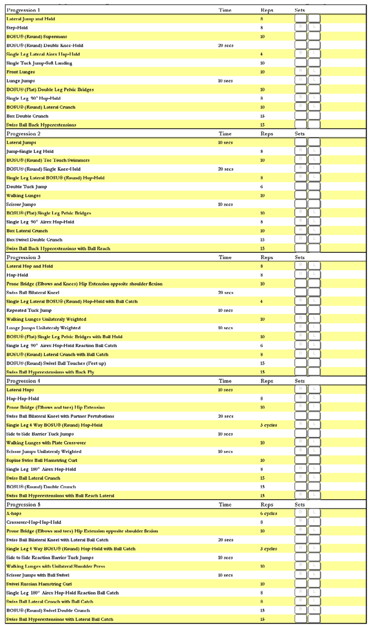 |
Table 2.
Exercise progressions and the published intervention it was derived and adapted from.
| Phase I | Exercise Adapted from Intervention |
|---|---|
| Lateral Jumping Progression | Hewett 1999, Mandelbaum 2004 |
| Single-Leg Anterior Progression | Hewett 1999 |
| Prone Trunk Stability Progression | Myer 2007 |
| Kneeling Trunk Stability Progression | Myer 2007 |
| Single Leg Lateral Progression | Myklebust 2003, Petersen 2006 |
| Tuck Jump Progression | Hewett 1999 |
| Lunge Progression | Mandelbaum 2004 |
| Lunge Jump Progression | Hewett 1999 |
| Hamstring Specific Progression | Mandelbaum 2004 |
| Single Leg Rotatory Progression | Myklebust 2003, Petersen 2006 |
| Lateral Trunk Progression | Myer 2007 |
| Trunk Flexion Progression | Myklebust 2003, Petersen 2006 |
| Trunk Extension Progression | Myer 2007 |
Effects of Trunk Neuromuscular Training on Hip Abduction Peak Torque
Pilot studies that utilized the proposed TNMT protocol indicate that increased standing hip abduction strength can be improved in female athletes (Table 3–Table 15).47 Hip abduction strength and recruitment may improve the ability of female athletes to increase control of lower extremity alignment and decrease knee abduction motion and loads resulting from increased trunk displacement during sports activities. Future investigations are needed to determine if improved hip strength following TNMT translates into reduced knee abduction load in high ACL injury risk female athletes. If this association is observed, then parallel investigations should be undertaken to determine if TNMT is effective in pubertal and pre-pubertal athletes to artificially induce “neuromuscular spurt” (defined as the natural adaptation of increased power, strength and coordination that occur with increasing chronological age and maturational stage in adolescent boys) especially related to relative hip strength and control which is often reduced as young female athletes mature.15, 52
Table 3.
Figures 2a–2z Exercise 1. Lateral Jumping Progression
|
Phase I – Lateral Jump and Hold The athlete prepares for this exercise by standing with her feet close together and her knees slightly bent. The athlete should jump laterally over a line keeping her knees bent and staying close to the line. When she lands on the opposite side, she should immediately descend into a deep hold. |
 |
|
Phase II – Lateral Jumps The athlete prepares for this exercise by standing with her feet close together and knees slightly bent on one side of the line. The athlete should jump sideways over the line keeping her knees bent and staying close to the line. When the athlete lands on the opposite side, she should immediately redirect back to the initial position. The athlete should repeat this sequence as quickly as she can while maintaining proper form. When teaching this exercise, encourage the athlete to achieve as many repetitions as possible in the allotted time by jumping close to the lines, shortening the ground contact time, and not using excessive height on the jumps. Do not allow the athlete to perform a double hop on the side of the line. Early in the training the athlete may focus on the line, as her technique improves encourage her to shift her visual focus away from the line to outside cues. |
 |
|
Phase III – Lateral Hop and Hold The athlete prepares for this exercise by standing on one foot and her knee slightly bent. The athlete should jump sideways over a line keeping her knee bent and staying close to the line. When she land on the opposite side, she should immediately descend into a single leg deep hold. |
 |
|
Phase IV – Lateral Hops The athlete prepares for this exercise by standing on one leg with her knee slightly bent on one side of the line. The athlete should jump sideways over the line keeping her knee bent and staying close to the line. When the athlete lands on the opposite side, she should immediately redirect back to the initial position. When teaching this exercise encourage the athlete to achieve as many repetitions as possible in the allotted time by jumping close to the lines, shortening the ground contact time, and not using excessive height on the jumps. Do not allow the athlete to perform a double hop on the side of the line. Early in the training the athlete may focus on the line, as her technique improves encourage her to shift her visual focus away from the line to outside cues. |
 |
|
Phase V – X Hops The athlete begins facing a quadrant pattern standing on a single limb with her support knee slightly bent. She will hop diagonally, landing in the opposite quadrant, maintaining forward stance, and holding the deep knee flexion landing for three seconds. The athlete then hops laterally into the side quadrant again holding the landing. Next, the athlete will hop diagonally backwards holding the landing. Finally, she hops laterally into the initial quadrant holding the landing. The athlete should repeat this figure 8 pattern for the required number of sets. Encourage the athlete to maintain balance during each landing, keeping her eyes up and her focus away from her feet. |
 |
Table 15.
Figures 14a–14s, Exercise 13. Trunk Extension Progression
|
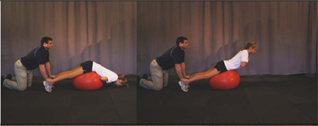
Phase I – Swiss Ball Back Hyperextensions The athlete begins in a prone position on the Swiss ball with her hips centered on top of the Swiss ball and a partner anchoring her feet to the floor. The movement is initiated by extending her hips and lower back to bring the athlete into a position of slight hyperextension. The position should be maintained for a short pause and then returned to the flexed position. |
 |
|
Phase II – Swiss Ball Back Hyperextensions with Ball Reach The athlete begins in a prone position on the Swiss ball with her hips centered on top of the Swiss ball and a partner anchoring her feet to the floor. The movement is initiated by extending hips and lower back to bring the athlete into a position of slight hyperextension. While performing this motion the athlete will also extend and return a medicine ball from the chest to full shoulder and elbow extension and back to the chest. |
 |
|
Phase III – Swiss Ball Hyperextensions with Back Fly The athlete begins in a prone position on the Swiss ball with her hips centered on top of the Swiss ball and a partner anchoring her feet to the floor. The movement is initiated by extending hips and lower back to bring the athlete into a position of slight hyperextension. The position should be maintained while the athlete brings dumbbells out to the side similar to a back fly exercise. |
 |
|
Phase IV – Swiss Ball Hyperextensions with Ball Reach Lateral The athlete begins in a prone position on the Swiss ball with her hips centered on top of the Swiss ball and a partner anchoring her feet to the floor. The movement is initiated by extending hips and lower back to bring the athlete into a position of slight hyperextension. The position should be maintained while the athlete brings a medicine ball above her head and slightly to the side. |
 |
|
Phase V – Swiss Ball Hyperextensions with Lateral Ball Catch The athlete begins in a prone position on the Swiss ball with her hips centered on top of the Swiss ball and a partner anchoring her feet to the floor. The movement is initiated by extending hips and lower back to bring the athlete into a position of slight hyperextension. The position should be maintained while the athlete brings a medicine ball above her head and slightly to the side. A ball should be tossed back and forth with a partner to increase the difficulty of this exercise. |
 |
Summary and Conclusions
Dynamic neuromuscular analysis oriented training appears to reduce ACL injuries in adolescent and mature female athletes.17, 48, 50, 51 Targeted neuromuscular training, at or near the onset of puberty, may simultaneously improve lower extremity strength and power, reduce dangerous biomechanics related to ACL injury risk and improve single leg balance.22, 53 Neuromuscular training could be advocated in pre- and early pubertal children to help prevent the development of high risk knee joint biomechanics that develop during this stage of maturation.54 A preemptive approach that institutes early interventional training may also reduce the peak rate of ACL injuries that occurs near age sixteen in young girls55. Due to the near 100% risk of osteoarthritis in the ACL injured population,56 with or without surgical reconstruction, prevention is currently the only effective intervention for these life-altering injuries. Additional efforts towards the development of more specific injury prevention protocols targeted towards the mechanism demonstrated in high risk athletes with the determination of the timing of when these interventions should most effectively be utilized is imperative. Specifically, neuromuscular training that focuses on trunk control instituted just prior to pubertal development may provide the most effective interventional approach alleviate high risk biomechanics in female athletes.
Table 4.
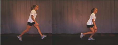
Figure 3a–3aa, Exercise 2. Single-Leg Anterior Progression
|
Phase I – Step-Hold The athlete starts by taking a quick step forward and continues by balancing in a deep hold position on the leg she stepped onto. |
 |
|
Phase II – Jump-Single Leg Hold The athlete will begin this exercise in the athletic position. The athlete proceeds to jump forward, landing and balancing on one leg in a deep hold position. |
 |
|
Phase III – Hop-Hold Starting in a balanced position on one foot, the athlete hops forward, landing and balancing on one leg in a deep hold position. |
 |
|
Phase IV – Hop-Hop-Hold The athlete hops forward twice quickly, landing and balancing on one leg in a deep hold position. |
 |
|
Phase V – Crossover-Hop-Hop-Hold The athlete hops forward while alternating legs three times quickly, landing and balancing on one leg in a deep hold position. |
 |
Table 5.
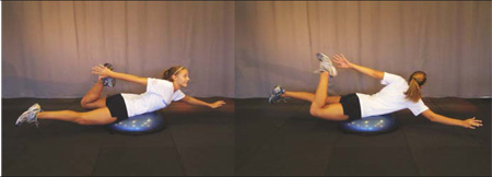
Figure 4a– 4i, Exercise 3. Prone Trunk Stability Progression
|
Phase I – BOSU® (Round) Toe Touch Swimmers The athlete begins in a prone position with her abdomen centered on the round side of the BOSU® and her arms overhead and legs extended. The athlete reaches back with one arm to touch opposite foot and returns to the outstretched superman position. |
 |
|
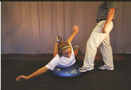
Phase II – BOSU® (Round) Swimmers with Partner Perturbations The athlete begins in prone position with abdomen centered on the round side of the BOSU® and with her arms overhead and legs extended. The movement is initiated by elevating the opposite arm and leg and held for three seconds. A partner will offer random perturbations by stepping on different sides of the BOSU® during the exercise. |
 |
|
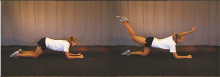
Phase III – Prone Bridge (Elbows and Knees) Hip Extension Opposed Shoulder Flexion The athlete begins in prone position with her elbows flexed and balanced on an Airex pad and knees on the ground. The movement is initiated by elevating the opposite arm and leg and held for a single count and finished by returning to the original position. |
 |
|
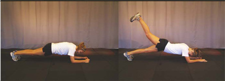
Phase IV – Prone Bridge (Elbows and Toes) Hip Extension The athlete begins in prone position with elbows flexed and balanced on an Airex pad and toes on the ground. The movement is initiated by elevating the each leg individually and held for a single count and finished by returning to the original position. |
 |
|
Phase V – Prone Bridge (Elbows and Toes) Hip Extension Opposite Shoulder Flexion The athlete begins in prone position with elbows flexed and balanced on an Airex pad and toes on the ground. The movement is initiated by elevating the opposite arm and leg and held for a single count and finished by returning to the original position. |
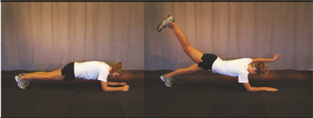 |
Table 6.
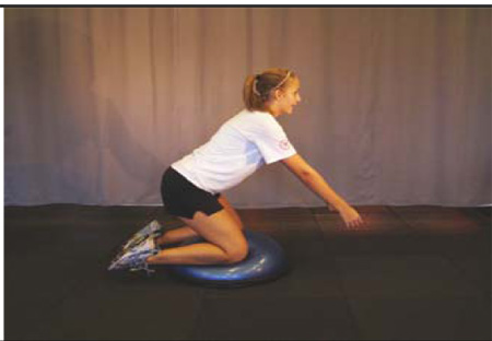
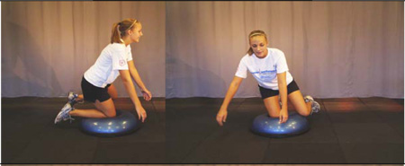
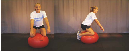
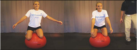
Figure 5a–5m, Exercise 4. Kneeling Trunk Stability Progression
|
Phase I – BOSU® (Round) Double Knee-Hold The athlete begins this exercise by balancing in a kneeling position with her knees on each side of the round side of the BOSU®. The athlete will maintain this balanced position with the hips slightly flexed for the duration of the exercise. |
 |
|
Phase II – BOSU® (Round) Single Knee-Hold The athlete begins this exercise by balancing in a kneeling position with one knee directly in the middle of the round side of the BOSU® and the other knee extended out to the side. The athlete will maintain this balanced position with the hip slightly flexed for the duration of the exercise. |
 |
|
Phase III – Swiss Ball Bilateral Kneel The athlete kneels and balances on Swiss ball with feet off the ground. A spotter should be available at all times in front of the athlete. |
 |
|
Phase IV – Swiss Ball Bilateral kneel with Partner Perturbations The athlete kneels and balances on Swiss ball with her feet off of the ground. Once the athlete is stabilized a partner can perturb the ball by kicking in unanticipated directions. A spotter should be available at all times in front of the athlete. |
 |
|
Phase V – Swiss Ball Bilateral Kneel with Lateral Ball Catch The athlete kneels and balances on Swiss ball with feet off the ground. A ball should be tossed back and forth with a partner to increase the difficulty of this exercise. |
 |
Table 7.
Figure 6a–6bb, Exercise 5. Single Leg Lateral Progression
|
Phase I – Single Leg Lateral AIREX Hop-Hold Athlete starts on one side of the Airex pad and hops laterally onto the Airex. The athlete should maintain balance and hold the knee in a flexed position. The athlete then hops off the other side of the Airex onto the ground, maintains balance and then repeats the exercise in the other direction. |
 |
|
Phase II – Single Leg Lateral BOSU® (Round) Hop-Hold Athlete starts on one side of the BOSU® and hops laterally onto the BOSU®. The athlete should maintain balance and hold the knee in a flexed position. The athlete then hops off the other side of the BOSU® onto the ground, maintains balance and then repeats the exercise in the other direction. |
 |
|
Phase III – Single Leg Lateral BOSU® (Round) Hop-Hold with Ball Catch The athlete starts on one side of the BOSU® and hops laterally onto the BOSU®. The athlete should maintain balance and hold the knee in a flexed position. The athlete then hops off the other side of the BOSU® onto the ground, maintains balance and then repeats the exercise in the other direction. The athlete is further challenged by having to catch and return a ball upon each landing. |
 |
|
Phase IV – Single Leg 4 Way BOSU® (Round) Hop-Hold The athlete starts in a single leg athletic position immediately behind the BOSU®. The athlete hops forward onto the round side of the BOSU® and lands in a balanced position. After achieving a balanced single leg stance on the BOSU®, the athlete proceeds to hop off the BOSU® laterally and assumes this same stance on the floor immediately next to the BOSU®. The athlete will then continue to hop on and off the BOSU®, achieving a balanced athletic position, in each of the four directions: forward, backwards, lateral and medial. |
 |
|
Phase V – Single Leg 4 Way BOSU® (Round) Hop-Hold with Ball Catch The athlete starts in a single leg athletic position immediately behind the BOSU®. The athlete hops forward onto the round side of the BOSU® and lands in a balanced position. After achieving a balanced single leg stance on the BOSU®, the athlete proceeds to hop off the BOSU® laterally and assumes this same stance on the floor immediately next to the BOSU®. The athlete will then continue to hop on and off the BOSU®, achieving a balanced athletic position, in each of the four directions: forward, backwards, lateral and medial. A ball should be tossed back and forth with a partner upon landing to increase the difficulty of this exercise. |
 |
Table 8.
Figure 7a–7x, Exercise 6. Tuck Jump Progression
| Phase I - Single Tuck Jump Soft Landing The athlete starts in the athletic position with her feet shoulder width apart. The athlete initiates a vertical jump with a slight crouch downward while she extends her arms behind her. The athlete then swings her arms forward as she simultaneously jumps straight up and pulls her knees up as high as possible. At the highest point of the jump the athlete should be positioned in the air with her thighs parallel to the ground. On landing, the athlete should land softly, using a toe to mid-foot rocker landing. The athlete should not continue this jump if she cannot control the high landing force or keep her knees aligned during landing. If the athlete is unable to raise the knees to the proper height, it may be valuable to instruct her to “grasp the knees and they bring the thighs to horizontal.” |
 |
|
Phase II – Double Tuck Jump Similar to the single tuck jump described above but with an additional jump performed immediately after the first jump. The athlete should focus on maintaining good form and minimizing time on the ground between jumps. |
 |
|
Phase III – Repeated Tuck Jumsp The athlete starts in the athletic position with her feet shoulder width apart. The athlete initiates a vertical jump with a slight crouch downward while she extends her arms behind her. The athlete then swings her arms forward as she simultaneously jump straight up and pull her knees up as high as possible. At the highest point of the jump the athlete should be positioned in the air with her thighs parallel to the ground. When landing the athlete should immediately begin the next tuck jump. |
 |
|
Phase IV – Side to Side Tuck Jumps The athlete starts in the athletic position with her feet shoulder width apart. The athlete initiates a vertical jump over a barrier with a slight crouch downward while she extends her arms behind her. The athlete then swings her arms forward as she simultaneously jumps straight up and pulls her knees up as high as possible. At the highest point of the jump the athlete should be positioned in the air with her thighs parallel to the ground. When landing, the athlete should immediately begin the next tuck jump back to the other side of the barrier. |
 |
|
Phase V – Side to Side Reaction Barrier Tuck Jumps The athlete starts in the athletic position with her feet shoulder width apart. The athlete initiates a vertical jump over a barrier with a slight crouch downward while she extends her arms behind her. The athlete then swings her arms forward as she simultaneously jump straight up and pull her knees up as high as possible. At the highest point of the jump the athlete should be positioned in the air with her thighs parallel to the ground. When landing the athlete should immediately begin the next tuck jump. When prompted, the athlete should jump to the other side of the barrier without breaking rhythm. |
 |
Table 9.
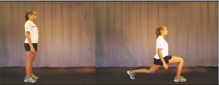
Figure 8a–8q, Exercise 7. Lunge Progression
|
Phase I – Front Lunges The athlete begins by stepping forward from a standing position. The step should be exaggerated in length to the point that her front leg is positioned with the knee flexed to 90° and the lower leg completely vertical. The back leg should be as straight as possible and the torso upright. Emphasis should be placed on getting the hips as low as possible while maintaining the previously described body position. The exercise is completed by driving off the front leg and returning to the original position. |
 |
|
Phase II – Walking Lunges The athlete performs a lunge and instead of returning to the start position she step through with the back limb and proceed forward with a lunge on the opposite limb. Encourage the athlete to lunge her front limb far enough out so that her knee does not advance beyond her ankle during the exercise. An alternative coaching method is to instruct the athlete to attempt to maintain a constant low center of gravity and roll through the lunges. This increases the intensity of the exercise and attempts to mimic motions frequently occurring in sports. |
 |
|
Phase III – Walking Lunges Unilaterally Weighted The athlete performs a lunge and instead of returning to the start position she steps through with the back limb and proceeds forward with a lunge on the opposite limb while holding a dumbbell in one hand. Encourage the athlete to lunge her front limb far enough out so that her knee does not advance beyond her ankle during the exercise. This exercise is then repeated with the dumbbell in the opposite hand. |
 |
|
Phase IV – Walking Lunges with Plate Crossover The athlete performs a lunge and instead of returning to the start position she steps through with the back limb and proceeds forward with a lunge on the opposite limb while reaching with a weight plate to the open side of the body. Encourage the athlete to lunge her front limb far enough out so that her knee does not advance beyond her ankle during the exercise. |
 |
|
Phase V – Walking Lunges with Unilateral Shoulder Press The athlete performs a lunge and instead of returning to the start position she steps through with the back limb and proceeds forward with a lunge on the opposite limb while pressing a dumbbell above her head. The weight should move up and down with the same tempo and direction as the lunge. Encourage the athlete to lunge her front limb far enough out so that her knee does not advance beyond her ankle during the exercise. |
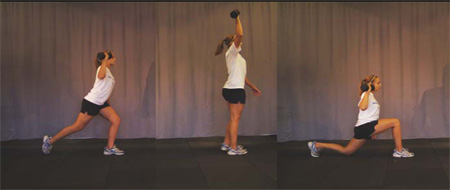 |
Table 10.
Figure 9a – 9s, Exercise 8. Lunge Jump Progression
|
Phase I – Lunge Jumps The athlete starts in an extended stride position with the hips pushed forward, and the front knee positioned directly above the ankle and flexed to 90°. The back leg is fully extended at the hip and knee providing minimal support for the stance. The athlete should jump vertically off of the front support leg maintaining the starting position during flight and landing. The jump is repeated as quickly as possible while still achieving maximum vertical height. To coach this jump, encourage the athlete to keep the back leg straight and use it only for balance support. Vertical power is obtained by the front leg. Stance support percentages are approximately 80% for the front leg and 20% for the back. |
 |
|
Phase II – Scissor Jumps The athlete starts in an extended stride position with the hips pushed forward, and the front knee positioned directly above the ankle and flexed to 90°. The back leg is fully extended at the hip and knee providing minimal support for the stance. The athlete should jump vertically off of the front support leg and switch the position of the legs while in flight. The jump is repeated as quickly as possible while still achieving maximum vertical height. The athlete will be jumping off alternate legs on each jump during this exercise. |
 |
|
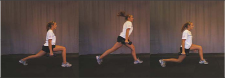
Phase III – Lunge Jumps Unilaterally Weighted The athlete starts in an extended stride position with her hips pushed forward, and the front knee positioned directly above the ankle and flexed to 90°. The back leg is fully extended at the hip and knee providing minimal support for the stance. The athlete should jump vertically off of the front support leg maintaining the starting position during flight and landing. The jump is repeated as quickly as possible while still achieving maximum vertical height. To unilaterally weight this exercise a dumbbell should be held in one hand. This exercise is then repeated with the dumbbell in the opposite hand. |
 |
|
Phase IV – Scissor Jumps Unilaterally Weighted The athlete starts in an extended stride position with the hips pushed forward, and the front knee positioned directly above the ankle and flexed to 90°. The back leg is fully extended at the hip and knee providing minimal support for the stance. The athlete should jump vertically off of the front support leg and switch the position of the legs while in flight. The jump is repeated as quickly as possible, while still achieving maximum vertical height. To unilaterally weight this exercise, a dumbbell should be held in one hand. The athlete will be jumping off alternate legs on each jump during this exercise. This exercise is then repeated with the dumbbell in the opposite hand. |
 |
|
Phase V – Scissor Jumps with Ball Swivel The athlete starts in an extended stride position with the hips pushed forward, and the front knee positioned directly above the ankle and flexed to 90°. The back leg is fully extended at the hip and knee providing minimal support for the stance. The athlete should jump vertically off of the front support leg and switch the position of the legs while in flight. The jump is repeated as quickly as possible while still achieving maximum vertical height. To unilaterally weight this exercise, a medicine ball should be swiveled to the open side of the body during each jump. The athlete will be jumping off alternate legs. |
 |
Table 11.
Figure 10a – 10n, Exercise 9. Hamstring Specific Progression
|
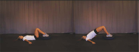
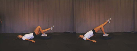
Phase I – BOSU® (Flat) Pelvic Bridge The athlete lays supine with her hip and knees flexed and her feet planted on the flat side of the BOSU®. The athlete then extends her hips and elevates her trunk off the ground to execute a pelvic bridge. This position should be held for 3 seconds prior to repeating the next repetition. |
 |
|
Phase II – BOSU® (Flat) Single Leg Pelvic Bridge The athlete lays supine with her hip and knees flexed and a single foot planted on the flat side of the BOSU® and the contralateral leg fully extended. The athlete then extends her hips and elevates her trunk off the ground to execute a pelvic bridge. This position should be held for 3 seconds prior to repeating the next repetition. |
 |
|
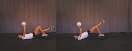
Phase III – BOSU® (Flat) Single Leg Pelvic Bridge The athlete lays supine with her hip and knees flexed and a single foot planted on the flat side of the BOSU® and the contralateral leg fully extended holding a ball directly above her in her hands. The athlete then extends her hips and elevates her trunk off the ground to execute a pelvic bridge. This position should be held for 3 seconds prior to repeating the next repetition. A weight plate is positioned on the hips to add resistance. |
 |
|
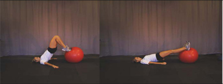
Phase IV – Supine Swiss Ball Hamstring Curl The athlete lays supine with her hip and knees flexed with both heels planted on top of a Swiss ball. The athlete then extends her hips and elevates her trunk off the ground while pulling her heels in to her buttocks. |
 |
|
Phase V – Russian Hamstring Curl with Lateral Touch The athlete begins in a kneeling position with a partner providing foot support and torso support (with band assistance). The athlete extends at the knee to lower their torso towards the ground. Once touching the BOSU® with their chest the athlete swivels their trunk and returns to the original position. The coach should provide enough assistance so that the exercise can be performed without flexing at the hip. |
 |
Table 12.
Figure 11a–11u, Exercise 10. Single Leg Rotatory Progression
|
Phase I – Single Leg 90° Hop-Hold The starting position for this jump is with the athlete in a semi-crouched position on the single limb being trained. The jump should focus on attaining maximum height while maintaining good form upon landing. During the flight phase, the athlete should rotate 90°. The landing occurs on the same leg and should be performed with deep knee flexion (to 90°). The landing should be held for a minimum of three seconds to be counted as a successful landing. Coach this jump with care to protect the athlete from injury. Start the athlete with a sub maximal effort so she can experience the difficulty of the jump. Continue to increase the intensity of the jump as the athlete improves her ability to stick and hold the final landing. Have the athlete keep her focus away from her feet, to help prevent too much forward lean. |
 |
|
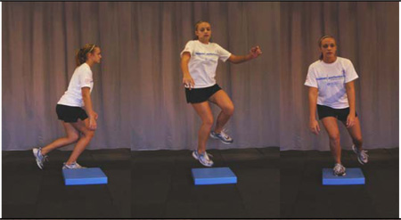
Phase II – Single Leg 90° AIREX Hop-Hold The starting position for this jump is with the athlete in a semi-crouched position on the single limb being trained. The jump should focus on attaining maximum height while maintaining good form upon landing. During the flight phase the athlete should rotate 90°. The landing occurs on the same leg and should be performed with deep knee flexion (to 90°). The landing should be held for a minimum of three seconds on an AIREX pad to be counted as a successful landing. Coach this jump with care to protect the athlete from injury. |
 |
|
Phase III – Single Leg 90° Hop-Hold Reaction Ball Catch The starting position for this jump is with the athlete in a semi-crouched position on the single limb being trained. The jump should focus on attaining maximum height while maintaining good form upon landing. During the flight phase the athlete should rotate 90°. The landing occurs on the same leg and should be performed with deep knee flexion (to 90°). The landing should be held for a minimum of three seconds on an AIREX pad to be counted as a successful landing. Upon landing a ball will be passed back and forth with the athlete to increase the difficulty of a successful landing. |
 |
|
Phase IV – Single Leg 180° AIREX Hop-Hold The starting position for this jump is with the athlete in a semi-crouched position on the single limb being trained. The jump should focus on attaining maximum height while maintaining good form upon landing. During the flight phase the athlete should rotate 180°. The landing occurs on the same leg and should be performed with deep knee flexion (to 90°). The landing should be held for a minimum of three seconds on an AIREX pad to be counted as a successful landing. |
 |
|
Phase V – Single Leg 180° AIREX Hop-Hold Reaction Ball Catch The starting position for this jump is with the athlete in a semi-crouched position on the single limb being trained. The jump should focus on attaining maximum height while maintaining good form upon landing. During the flight phase the athlete should rotate 180°. The landing occurs on the same leg and should be performed with deep knee flexion (to 90°). The landing should be held for a minimum of three seconds on an AIREX pad to be counted as a successful landing. Upon landing a ball will be passed back and forth with the athlete to increase the difficulty of a successful landing. |
 |
Table 13.
Figure 12a–12q, Exercise 11. Lateral Trunk Progression
|
Phase I – BOSU® (Round) Lateral Crunch The athlete starts lying on her side with her hip located in the center of the round side of the BOSU®. The athlete’s feet and legs must be anchored during this exercise by the trainer or a stationary object. The athlete will proceed to bend laterally at the waist back and forth for the prescribed repetitions. |
 |
|
Phase II – Box Lateral Crunch Athlete starts in a supine position on a plyo box with arms placed on the back of the head. The athlete flexes her trunk simultaneous with hip flexion. As the trunk and hip are maximally flexed, the athlete rotates at the trunk touching each elbow to the opposite knee. |
 |
|
Phase III – BOSU® (Round) Lateral Crunch with Ball Catch Athlete starts lying on side with hip located top of the round side of a BOSU®. The athlete’s feet and legs must be anchored during this exercise by the trainer or a stationary object. The athlete will proceed to bend laterally at the waist back and forth for the prescribed repetitions. A ball should be tossed back and forth with a partner to increase the difficulty of this exercise. |
 |
|
Phase IV – Swiss Ball Lateral Crunch Athlete starts lying on side with hip located top of a Swiss ball. The athlete’s feet and legs must be anchored during this exercise by the trainer or a stationary object. The athlete will proceed to bend laterally at the waist back and forth for the prescribed repetitions. |
 |
|
Phase V – Swiss Ball Lateral Crunch with Ball Catch Athlete starts lying on side with hip located top of a Swiss ball. The athlete’s feet and legs must be anchored during this exercise by the trainer or a stationary object. The athlete will proceed to bend laterally at the waist back and forth for the prescribed repetitions. A ball should be tossed back and forth with a partner to increase the difficulty of this exercise. |
 |
Table 14.
Figure 13a–13u, Exercise 12. Trunk Flexion Progression
|
Phase I – Box Double Crunch The athlete starts out supine on a plyometric box or similar object and flexes her trunk simultaneous with hip flexion. |
 |
|
Phase II – Box Swivel Double Crunch Athlete starts in a supine position on a plyo box with arms placed across chest. The athlete flexes her trunk simultaneous with hip flexion. As the trunk and hip are maximally flexed, the athlete rotates at the trunk touching each elbow to the opposite knee. |
 |
|
Phase III – BOSU® (Round) Swivel Ball Touches (Feet up) Athlete starts sitting on the round side of a BOSU® holding a medicine ball. The athlete will proceed to swivel at the trunk to touch the medicine ball to the floor for each repetition. |
 |
|
Phase IV – BOSU® (Round) Double Crunch Athlete starts sitting on the round side of a BOSU®. The athlete flexes her trunk simultaneous with hip flexion. |
 |
|
Phase V – BOSU® (Round) Swivel Double Crunch Athlete starts sitting on the round side of a BOSU®. The athlete flexes her trunk simultaneous with hip flexion. As the trunk and hip are maximally flexed, the athlete rotates at the trunk touching each elbow to the opposite knee. |
 |
ACKNOWLEDGEMENTS
The authors would like to acknowledge funding support from National Institutes of Health/NIAMS Grant R01-AR049735.
Footnotes
Publisher's Disclaimer: This is a PDF file of an unedited manuscript that has been accepted for publication. As a service to our customers we are providing this early version of the manuscript. The manuscript will undergo copyediting, typesetting, and review of the resulting proof before it is published in its final citable form. Please note that during the production process errors may be discovered which could affect the content, and all legal disclaimers that apply to the journal pertain.
References
- 1.Arendt E, Dick R. Knee injury patterns among men and women in collegiate basketball and soccer. NCAA data and review of literature. Am J Sports Med. 1995;23(6):694–701. doi: 10.1177/036354659502300611. [DOI] [PubMed] [Google Scholar]
- 2.Malone TR, Hardaker WT, Garrett WE, Feagin JA, Bassett FH. Relationship of gender to anterior cruciate ligament injuries in intercollegiate basketball players. Journal of Southern Orthopaedic Association. 1993;2(1):36–39. [Google Scholar]
- 3.Myklebust G, Maehlum S, Holm I, Bahr R. A prospective cohort study of anterior cruciate ligament injuries in elite Norwegian team handball. Scand J Med Sci Sports. 1998;8(3):149–153. doi: 10.1111/j.1600-0838.1998.tb00185.x. [DOI] [PubMed] [Google Scholar]
- 4.Mihata LC, Beutler AI, Boden BP. Comparing the incidence of anterior cruciate ligament injury in collegiate lacrosse, soccer, and basketball players: implications for anterior cruciate ligament mechanism and prevention. Am J Sports Med. 2006 Jun;34(6):899–904. doi: 10.1177/0363546505285582. [DOI] [PubMed] [Google Scholar]
- 5.Ford KR, Myer GD, Hewett TE. Valgus knee motion during landing in high school female and male basketball players. Med Sci Sports Exerc. 2003 Oct;35(10):1745–1750. doi: 10.1249/01.MSS.0000089346.85744.D9. [DOI] [PubMed] [Google Scholar]
- 6.Ford KR, Myer GD, Toms HE, Hewett TE. Gender differences in the kinematics of unanticipated cutting in young athletes. Medicine & Science in Sports. 2005 Jan;37(1):124–129. [PubMed] [Google Scholar]
- 7.Hewett TE, Myer GD, Ford KR, et al. Biomechanical Measures of Neuromuscular Control and Valgus Loading of the Knee Predict Anterior Cruciate Ligament Injury Risk in Female Athletes: A Prospective Study. Am J Sports Med. 2005 Feb 8;33(4):492–501. doi: 10.1177/0363546504269591. [DOI] [PubMed] [Google Scholar]
- 8.McLean SG, Lipfert SW, van den Bogert AJ. Effect of gender and defensive opponent on the biomechanics of sidestep cutting. Med Sci Sports Exerc. 2004 Jun;36(6):1008–1016. doi: 10.1249/01.mss.0000128180.51443.83. [DOI] [PubMed] [Google Scholar]
- 9.Chappell JD, Yu B, Kirkendall DT, Garrett WE. A comparison of knee kinetics between male and female recreational athletes in stop-jump tasks. Am J Sports Med. 2002 Mar–Apr;30(2):261–267. doi: 10.1177/03635465020300021901. [DOI] [PubMed] [Google Scholar]
- 10.Myer GD, Ford KR, McLean SG, Hewett TE. The Effects of Plyometric Versus Dynamic Stabilization and Balance Training on Lower Extremity Biomechanics. Am J Sports Med. 2006;34(3):490–498. doi: 10.1177/0363546505281241. [DOI] [PubMed] [Google Scholar]
- 11.Myer GD, Ford KR, Brent JL, Hewett TE. Differential neuromuscular training effects on ACL injury risk factors in "high-risk" versus "low-risk" athletes. BMC Musculoskelet Disord. 2007;8(39):1–7. doi: 10.1186/1471-2474-8-39. [DOI] [PMC free article] [PubMed] [Google Scholar]
- 12.Hewett TE, Ford KR, Myer GD. Anterior Cruciate Ligament Injuries in Female Athletes: Part 2, A Meta-analysis of Neuromuscular Interventions Aimed at Injury Prevention. Am J Sports Med. 2006 Dec 28;34(3):490–498. doi: 10.1177/0363546505282619. [DOI] [PubMed] [Google Scholar]
- 13.Myer GD, Ford KR, Brent JL, Hewett TE. The Effects of Plyometric versus Dynamic Balance Training on Power, Balance and Landing Force in Female Athletes. J Strength Cond Res. 2006;20(2):345–353. doi: 10.1519/R-17955.1. [DOI] [PubMed] [Google Scholar]
- 14.Myer GD, Ford KR, Palumbo JP, Hewett TE. Neuromuscular training improves performance and lower-extremity biomechanics in female athletes. J Strength Cond Res. 2005 Feb;19(1):51–60. doi: 10.1519/13643.1. [DOI] [PubMed] [Google Scholar]
- 15.Myer GD, Ford KR, Hewett TE. Rationale and Clinical Techniques for Anterior Cruciate Ligament Injury Prevention Among Female Athletes. J Athl Train. 2004 Dec;39(4):352–364. [PMC free article] [PubMed] [Google Scholar]
- 16.Myer GD, Ford KR, Brent JL, Hewett TE. The Effects of Plyometric versus Dynamic Balance Training on Landing Force and Center of Pressure Stabilization in Female Athletes. Br J Sports Med. 2005;39(6):397. [Google Scholar]
- 17.Hewett TE, Lindenfeld TN, Riccobene JV, Noyes FR. The effect of neuromuscular training on the incidence of knee injury in female athletes. A prospective study. Am J Sports Med. 1999 Nov–Dec;27(6):699–706. doi: 10.1177/03635465990270060301. [DOI] [PubMed] [Google Scholar]
- 18.Agel J, Arendt EA, Bershadsky B. Anterior cruciate ligament injury in national collegiate athletic association basketball and soccer: a 13-year review. Am J Sports Med. 2005 Apr;33(4):524–530. doi: 10.1177/0363546504269937. [DOI] [PubMed] [Google Scholar]
- 19.Hewett TE, Myer GD, Ford KR, et al. Biomechanical measures of neuromuscular control and valgus loading of the knee predict anterior cruciate ligament injury risk in female athletes. Am J Sports Med. 2005;33(4) doi: 10.1177/0363546504269591. [DOI] [PubMed] [Google Scholar]
- 20.Ford KR, Myer GD, Smith RL, Vianello RM, Seiwert SL, Hewett TE. A comparison of dynamic coronal plane excursion between matched male and female athletes when performing single leg landings. Clinical Biomechanics. 2006;21(1):33–40. doi: 10.1016/j.clinbiomech.2005.08.010. [DOI] [PubMed] [Google Scholar]
- 21.Malinzak RA, Colby SM, Kirkendall DT, Yu B, Garrett WE. A comparison of knee joint motion patterns between men and women in selected athletic tasks. Clin Biomech. 2001 Jun;16(5):438–445. doi: 10.1016/s0268-0033(01)00019-5. [DOI] [PubMed] [Google Scholar]
- 22.Hewett TE, Myer GD, Ford KR. Decrease in neuromuscular control about the knee with maturation in female athletes. J Bone Joint Surg Am. 2004;86-A(8):1601–1608. doi: 10.2106/00004623-200408000-00001. [DOI] [PubMed] [Google Scholar]
- 23.McLean SG, Huang X, Su A, van den Bogert AJ. Sagittal plane biomechanics cannot injure the ACL during sidestep cutting. Clin Biomech. 2004;19:828–838. doi: 10.1016/j.clinbiomech.2004.06.006. [DOI] [PubMed] [Google Scholar]
- 24.Kernozek TW, Torry MR, H VH, Cowley H, Tanner S. Gender differences in frontal and sagittal plane biomechanics during drop landings. Med Sci Sports Exerc. 2005 Jun;37(6):1003–1012. discussion 1013. [PubMed] [Google Scholar]
- 25.Zeller BL, McCrory JL, Kibler WB, Uhl TL. Differences in Kinematics and Electromyographic Activity Between Men and Women during the Single-Legged Squat. Am J Sport Med. 2003;31(3):449–456. doi: 10.1177/03635465030310032101. [DOI] [PubMed] [Google Scholar]
- 26.Pappas E, Hagins M, Sheikhzadeh A, Nordin M, Rose D. Biomechanical differences between unilateral and bilateral landings from a jump: gender differences. Clin J Sport Med. 2007 Jul;17(4):263–268. doi: 10.1097/JSM.0b013e31811f415b. [DOI] [PubMed] [Google Scholar]
- 27.Hewett TE, Ford KR, Myer GD, Wanstrath K, Scheper M. Gender Differences in Hip Adduction Motion and Torque During a Single Leg Agility Maneuver. J Orthop Res. 2006;24(3):416–421. doi: 10.1002/jor.20056. [DOI] [PubMed] [Google Scholar]
- 28.Olsen OE, Myklebust G, Engebretsen L, Bahr R. Injury mechanisms for anterior cruciate ligament injuries in team handball: a systematic video analysis. Am J Sports Med. 2004 Jun;32(4):1002–1012. doi: 10.1177/0363546503261724. [DOI] [PubMed] [Google Scholar]
- 29.Krosshaug T, Nakamae A, Boden BP, et al. Mechanisms of anterior cruciate ligament injury in basketball: video analysis of 39 cases. Am J Sports Med. 2007 Mar;35(3):359–367. doi: 10.1177/0363546506293899. [DOI] [PubMed] [Google Scholar]
- 30.Boden BP, Dean GS, Feagin JA, Garrett WE. Mechanisms of anterior cruciate ligament injury. Orthopedics. 2000;23(6):573–578. doi: 10.3928/0147-7447-20000601-15. [DOI] [PubMed] [Google Scholar]
- 31.Andrish JT. Anterior cruciate ligament injuries in the skeletally immature patient. Am J Orthop. 2001;30(2):103–110. [PubMed] [Google Scholar]
- 32.Buehler-Yund C. A longitudinal study of injury rates and risk factors in 5 to 12 year old soccer players. Cincinnati: Environmental health, University of Cincinnati; 1999. [Google Scholar]
- 33.Clanton TO, DeLee JC, Sanders B, Neidre A. Knee ligament injuries in children. J Bone Joint Surg Am. 1979;61(8):1195–1201. [PubMed] [Google Scholar]
- 34.Gallagher SS, Finison K, Guyer B, Goodenough S. The incidence of injuries among 87,000 Massachusetts children and adolescents: results of the 1980–81 Statewide Childhood Injury Prevention Program Surveillance System. Am J Public Health. 1984;74(12):1340–1347. doi: 10.2105/ajph.74.12.1340. [DOI] [PMC free article] [PubMed] [Google Scholar]
- 35.Tursz A, Crost M. Sports-related injuries in children. A study of their characteristics, frequency, and severity, with comparison to other types of accidental injuries. Am J Sports Med. 1986;14(4):294–299. doi: 10.1177/036354658601400409. [DOI] [PubMed] [Google Scholar]
- 36.Tanner JM, Davies PS. Clinical longitudinal standards for height and height velocity for North American children. J Pediatr. 1985;107(3):317–329. doi: 10.1016/s0022-3476(85)80501-1. [DOI] [PubMed] [Google Scholar]
- 37.Hewett TE, Myer GD, Ford KR, J.L. S. Preparticipation Physical Exam Using a Box Drop Vertical Jump Test in Young Athletes: The Effects of Puberty and Sex. Clin J Sport Med. 2006;16(4):298–304. doi: 10.1097/00042752-200607000-00003. [DOI] [PubMed] [Google Scholar]
- 38.Hewett TE, Biro FM, McLean SG, Van den Bogert AJ. Cincinnati Children's Hospital: National Institutes of Health; Identifying Female Athletes at High Risk for ACL Injury. 2003
- 39.Ford KR, Myer GD, Hewett TE. Increased Trunk Motion in Female Athletes Compared to Males during Single Leg Landing. Med Sci Sports Exerc. 2007;39(5):S70. [Google Scholar]
- 40.Wilson JD, Dougherty CP, Ireland ML, Davis IM. Core Stability and Its Relationship to Lower Extremity Function and Injury. J Am Acad Orthop Surg. 2005;13:316–325. doi: 10.5435/00124635-200509000-00005. [DOI] [PubMed] [Google Scholar]
- 41.Hodges PW, Richardson CA. Contraction of the abdominal muscles associated with movement of the lower limb. Phys Ther. 1997 Feb;77(2):132–142. doi: 10.1093/ptj/77.2.132. discussion 142–134. [DOI] [PubMed] [Google Scholar]
- 42.Hodges PW, Richardson CA. Feedforward contraction of transversus abdominis is not influenced by the direction of arm movement. Exp Brain Res. 1997 Apr;114(2):362–370. doi: 10.1007/pl00005644. [DOI] [PubMed] [Google Scholar]
- 43.Winter DA. Biomechanics and Motor Control of Human Movement. 3rd ed. New York: John Wiley & Sons, Inc.; 2005. [Google Scholar]
- 44.Ireland ML. The female ACL: why is it more prone to injury? Orthop Clin North Am. 2002 Oct;33(4):637–651. doi: 10.1016/s0030-5898(02)00028-7. [DOI] [PubMed] [Google Scholar]
- 45.Zatsiorsky VM. Science and practice of strength training. Champaign, IL: Human Kinetics; 1995. [Google Scholar]
- 46.Zazulak BT, Hewett TE, Reeves NP, Goldberg B, Cholewicki J. The effects of core proprioception on knee injury: a prospective biomechanical-epidemiological study. Am J Sports Med. 2007 Mar;35(3):368–373. doi: 10.1177/0363546506297909. [DOI] [PubMed] [Google Scholar]
- 47.Myer GD, Brent JL, Ford KR, Hewett TE. A pilot study to determine the effect of trunk and hip focused neuromuscular training on hip and knee isokinetic strength. British Journal of Sports Medicine. 2008 doi: 10.1136/bjsm.2007.046086. In Press. [DOI] [PMC free article] [PubMed] [Google Scholar]
- 48.Mandelbaum BR, Silvers HJ, Watanabe D, et al. Effectiveness of a neuromuscular and proprioceptive training program in preventing the incidence of ACL injuries in female athletes: two-year follow up. Am J Sport Med. 2005;33(6) doi: 10.1177/0363546504272261. [DOI] [PubMed] [Google Scholar]
- 49.Hewett TE, Stroupe AL, Nance TA, Noyes FR. Plyometric training in female athletes. Decreased impact forces and increased hamstring torques. Am J Sports Med. 1996;24(6):765–773. doi: 10.1177/036354659602400611. [DOI] [PubMed] [Google Scholar]
- 50.Petersen W, Braun C, Bock W, et al. A controlled prospective case control study of a prevention training program in female team handball players: the German experience. Arch Orthop Trauma Surg. 2006 Feb 10; doi: 10.1007/s00402-005-0793-7. [DOI] [PubMed] [Google Scholar]
- 51.Myklebust G, Engebretsen L, Braekken IH, Skjolberg A, Olsen OE, Bahr R. Prevention of anterior cruciate ligament injuries in female team handball players: a prospective intervention study over three seasons. Clin J Sport Med. 2003 Mar;13(2):71–78. doi: 10.1097/00042752-200303000-00002. [DOI] [PubMed] [Google Scholar]
- 52.Brent JL, Myer GD, Ford KR, Hewett TE. A Longitudinal Examination of Hip Abduction Strength in Adolescent Males and Females. Medicine and Science in Sports and Exercise. 2008;39(5) [Google Scholar]
- 53.Myer GD, Ford KR, Divine JG, Hewett TE. Specialized dynamic neuromuscular training can be utilized to induce neuromuscular spurt in female athletes. Med Sci Sports Exerc. 2004;36(5):343–344. [Google Scholar]
- 54.Ford KR, Myer GD, Divine JG, Hewett TE. Landing differences in high school female soccer players grouped by age. Med Sci Sports Exerc. 2004;36(5):S293. [Google Scholar]
- 55.Shea KG, Pfeiffer R, Wang JH, Curtin M, Apel PJ. Anterior cruciate ligament injury in pediatric and adolescent soccer players: an analysis of insurance data. J Pediatr Orthop. 2004 Nov–Dec;24(6):623–628. doi: 10.1097/00004694-200411000-00005. [DOI] [PubMed] [Google Scholar]
- 56.Myklebust G, Bahr R. Return to play guidelines after anterior cruciate ligament surgery. Br J Sports Med. 2005 Mar;39(3):127–131. doi: 10.1136/bjsm.2004.010900. [DOI] [PMC free article] [PubMed] [Google Scholar]


