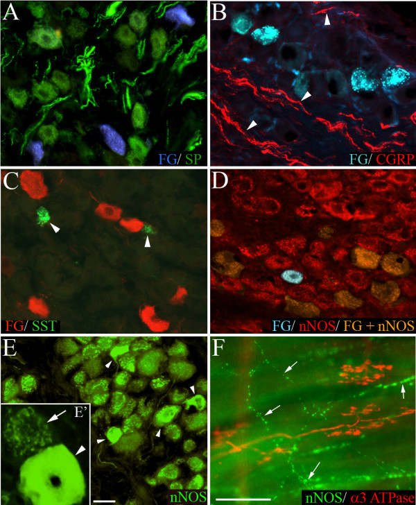
Figure 7.

Representative photomicrographs showing nodose neurons retrogradely labeled from the trachea with fluorogold (FG) overlaid with immunoreactivity for (A) substance P (SP), (B) calcitonin gene-related peptide (CGRP), (C) somatostatin (SST), and (D) neuronal nitric oxide synthase (nNOS). The arrow heads in panels (B) and (C) point out CGRP-labeled nerve fibers and SST-labeled neurons, respectively. Panel (E) shows low and higher (E') magnification of nNOS immunoreactive cells in the nodose ganglia without FG overlaid. Note the clustered labeling associated with most neurons (arrow, E') and smaller population of intensely fluorescent cells (arrow heads, E and E'). In tracheal wholemounts (F), cough receptors identified with α3 Na+/K+ ATPase were not immunoreactive for nNOS, whereas fine varicose fibers (arrows) were nNOS positive (representative of 4 similar experiments). Traced neurons appear red in panel (C) as the tissue underwent secondary immunoprocessing for FG. Scale bar in panel E represents 50 μm in panels (A-E) and 20 μm in panel (E'). Scale bar in panel (F) represents 50 μm.
