



Abstract
The expression patterns of PPARβ/δ have been described, but the majority of these data are based on mRNA data. To date, there are no reports that have quantitatively examined the expression of PPARβ/δ protein in mouse tissues. In the present study, a highly specific PPARβ/δ antibody was developed, characterized and used to examine tissue expression patterns of PPARβ/δ. As compared to commercially available anti-PPARβ/δ antibodies, one of six polyclonal anti-PPARβ/δ antibodies developed was significantly more effective for immunoprecipitation of in vitro translated PPARβ/δ. This antibody was used for quantitative western blot analysis using radioactive detection methods. Expression of PPARβ/δ was highest in colon, small intestine, liver and keratinocytes as compared to other tissues including heart, spleen, skeletal muscle, lung, brain and thymus. Interestingly, PPARβ/δ expression was localized in the nucleus and RXRα can be co-immunoprecipitated with nuclear PPARβ/δ. Results from these studies demonstrate that PPARβ/δ expression is highest in intestinal epithelium, liver and keratinocytes, consistent with significant biological roles in these tissues.
Keywords: peroxisome proliferator-activated receptor-β/δ, antibody, expression, biochemistry
Introduction
Peroxisome proliferator-activated receptor-β/δ (PPARβ/δ) is a ligand activated transcription factor with important biological functions. Ligand activation of PPARβ/δ improves glucose tolerance [1], increases skeletal muscle fatty acid catabolism [2] and mediates terminal differentiation of a number of cell types [3; 4]. Thus, PPARβ/δ is a potential target for the treatment of diseases including dyslipidemias, diabetes and cancer. One level of regulation of PPARβ/δ is the presence of ligands that can activate the receptor through classic nuclear hormone receptor mechanisms [3]. However, another level of PPARβ/δ-dependent function is the relative expression.
To date, there is limited quantitative data demonstrating the relative expression of PPARβ/δ protein in tissues. The majority of studies that have examined relative expression patterns of PPARβ/δ have focused on mRNA expression, which may or may not accurately reflect actual protein levels. There are at least six studies that have examined tissue expression patterns of PPARβ/δ in rodents [5; 6; 7; 8; 9; 10]. Escher et al focused primarily on mRNA expression of PPARs in rat but did perform one confirmatory western blot for PPARβ/δ demonstrating higher expression of PPARβ/δ in rat liver after feeding [7]. More comprehensive analysis of PPARβ/δ expression was performed using tissue microarray-based immunohistochemistry and characterized cellular localization of PPARβ/δ [8]. However, PPARβ/δ protein expression has not been examined using highly quantitative methods. For this reason, the focus of the present study was to develop a highly specific PPARβ/δ antibody, and to use the antibody to examine PPARβ/δ protein expression in mice using the most quantitative method for protein levels in mammalian tissues, radioactive detection of protein blots.
Materials and methods
Anti-PPAR β/δ antibody characterization
Anti-serum from six rabbits (designated 8095–8100) immunized with a PPARβ/δ-specific peptide (amino acids 1–29; sequence NP-035275) was obtained from Affinity Bioreagents. Anti-serum was used to screen for PPARβ/δ immunoreactivity using western blot analysis. COS-1 cells were transfected with a mouse PPARβ/δ expression vector, kindly provided by Drs. Walter Wahli and Pallavi Devchand, and cell lysate used for a positive control for these experiments. COS-1 cell lysate was separated using SDS-PAGE, transferred to a PVDF membrane and blocked using 5% non-fat milk in Tris-buffered saline with 0.1% Tween-20. After blocking, membranes were washed then incubated with anti-serum from one of the six different rabbits. Following incubation in the primary antibody, membranes were washed three times, incubated with biotinylated secondary antibody and then washed again before incubation with 125I-streptavidin. Membranes were exposed to phosphorimager plates and the level of radioactivity quantified with a Packard phosphorimager.
Antisera were then screened for PPARβ/δ-specific cross-reactivity using western blot and immunoprecipitation techniques. Three antisera having good reactivity and specificity for mouse PPARβ/δ were selected (8095, 8099 and 8100) and affinity purified. These three positive antibodies were further analyzed using western blot and immunoprecipitation techniques using in vitro translated protein, as well as in primary mouse keratinocyte samples. In vitro translations were carried out using TNT T7 coupled reticulocyte lysate system (Promega, Madison, WI) using 35S-methionine. To initially characterize the relative ability of the three affinity purified antibodies to immunoprecipitate, in vitro-translated PPARβ/δ was precleared with 25–60 µL of protein A/G-agarose beads (Santa Cruz Biotechnologies, Santa Cruz, CA) for 45 minutes at 4 °C followed by immunoprecipition with either 1, 2, or 3 µg of affinity-purified anti-PPARβ/δ antibodies prebound with protein A/G-agarose beads for 1 hour at 4 °C. Immunoprecipitations were then washed 4X in 0.5X RIPA buffer and resolved by SDS-PAGE. To characterize the ability of the affinity purified antibodies to immunoprecipitate from complex cellular extracts, lysate from COS-1 cells transfected with a mouse PPARβ/δ expression vector, in vitro-translated PPARβ/δ or soluble keratinocyte protein was precleared with 25–60 µL of protein A/G-agarose beads for 45 minutes at 4 °C. After preclearing, protein samples were immunoprecipitated with 3 µg of affinity-purified 8099 anti-PPARβ/δ antibody prebound with protein A/G-agarose beads for 1 hour at 4 °C. Immunoprecipitated protein was resolved by SDS-PAGE and subjected to western blot analysis as described above. To compare the relative efficacy of immunoprecipitation, affinity purified 8099 anti-PPARβ/δ antibody was compared with commercially available anti-PPARβ/δ antibodies (Affinity Bioreagents PA1–823; Santa Cruz sc-1987; Santa Cruz sc-7197). In vitro-translated 35S-methionine-labeled PPARβ/δ was precleared with 25–60 µL of protein A/G-agarose beads for 45 minutes at 4 °C followed by immunoprecipition with three µg of affinity purified 8099 anti-PPARβ/δ antibody or the commercial antibodies prebound with protein A/G-agarose beads for 1 hour at 4 °C. Immunoprecipitations were then washed 4X in 0.5X RIPA buffer, resolved using SDS-PAGE and exposed to a phosphorimager plate as described above to quantify relative immunoprecipitated PPARβ/δ.
Expression levels of PPAR β/δ in mouse tissues
Male C57BL6 mice, 8–10 weeks of age were euthanized by over-exposure to carbon dioxide. Tissues were dissected and flash frozen in liquid nitrogen. Tissues were homogenized in RIPA buffer with protease inhibitors and then centrifuged at 14,000 rpm for 30 minutes in 4 °C. For examination of PPARβ/δ expression in keratinocytes, cells were obtained and protein isolated as described previously [11]. Protein concentration was determined using a BCA kit (Pierce, Rockford, IL). The supernatant from this centrifugation was used to assess relative tissue distribution of PPARβ/δ using western blotting. Fifty micrograms of protein extract was separated using SDS-PAGE, transferred to a PVDF membrane and blocked using 5% non-fat milk in Tris-buffered saline with 0.1% Tween-20 (TBST). After blocking, membranes were washed three times with TBST and then incubated with either an affinity purified 8099 anti-PPARβ/δ antibody or an anti-LDH antibody in TBST. Following incubation in the primary antibody, membranes were washed three times with TBST, incubated with biotinylated secondary antibody diluted in TBST and then washed again before incubation with 125I-streptavidin. Membranes were exposed to phosphorimager plates and the level of radioactivity quantified with a Packard phosphorimager. Hybridization signals for PPARβ/δ were quantified and normalized to the amount of protein loaded. Rather than normalizing to a loading control such as LDH, hybridization signals for PPARβ/δ were normalized to the amount of protein to allow for comparison between tissues since the level of LDH can vary from tissue to tissue. This is feasible since the same amount of protein was loaded per lane per tissue sample, and the loading control demonstrated relatively equal loading.
Examination of nuclear and cytosolic PPARβ/δ
To determine whether PPARβ/δ was present in either the nucleus or cytosol, differential centrifugation of tissue protein samples was performed followed by western blot analysis. Briefly, tissue from male mice, 8–10 weeks of age, were homogenized in buffer containing 250 mM sucrose, 10 mM Tris HCl, 1 mM EDTA and centrifuged at 1,000 X g for 10 minutes at 4 °C. The supernatant from this centrifugation was then centrifuged at 100,000 X g for 60 minutes and this supernatant was used for analysis of cytosolic PPARβ/δ. For the nuclear fraction, the pellet from the first centrifugation at 1,000 X g was resuspended in the buffer described above, overlaid on a 1 M sucrose gradient, and centrifuged for 5 minutes at 1,600 X g at 4 °C. The pellet from this centrifugation was resuspended in buffer containing 150 mM sucrose, 10 mM Tris HCl, 1 mM EDTA and 1% NP-40 and centrifuged at 21,000 X g for 10 minutes at 4 °C. The supernatant from this centrifugation was used for analysis of nuclear PPARβ/δ. Western blot analysis was performed as described above, except that an anti-lamin antibody was used to confirm the relative purity of the nuclear preparations and an anti-LDH antibody was used to confirm the relative purity of the cytosolic preparations.
To determine whether the nuclear PPARβ/δ detected was potentially heterodimerized with retinoid X receptor-α (RXRα), nuclear extracts from liver were subjected to immunoprecipitation. Nuclear extracts were precleared with 25–60 µL of protein A/G-agarose beads for 45 minutes at 4 °C followed by immunoprecipitation with 3 µg of affinity-purified 8099 anti-PPARβ antibody or IgG control prebound with protein A/G-agarose beads for 1 hour at 4 °C. Immunoprecipitated protein was resolved by SDS-PAGE and subjected to western blot analysis for detection of either PPARβ/δ or RXRα (Santa Cruz Biotechnologies, Santa Cruz, CA) as described above.
Results
All six of the anti-serum obtained from Affinity Bioreagents were immunoreactive with the COS-1 cell lysate with high PPARβ/δ expression (Fig.1A). Of the six anti-sera examined, rabbit sera 8095, 8099 and 8100 were selected for further characterization after affinity purification. All three affinity purified antibodies immunoprecipitated in vitro translated PPARβ/δ, with the percentage of immunoprecipitated protein ranging from 12–32% (Fig. 1B), which is quite efficient relative to other antibodies. The affinity purified 8099 anti-PPARβ/δ antibody was most efficient at immunoprecipitating PPARβ/δ from soluble keratinocyte protein (Fig. 1C). This antibody was also markedly better for immunoprecipitation of in vitro translated PPARβ/δ as compared to three other commercially available anti-PPARβ/δ antibodies (Fig. 1D). The affinity purified 8099 anti-PPARβ/δ antibody was used for the remaining quantitative analysis of PPARβ/δ expression in mouse tissues.
Fig. 1.

Characterization of specific anti-PPARβ/δ antibody. (A) Western blot analysis using the six different anti-sera. Lysate from COS-1 cells transfected with a mouse PPARβ/δ expression vector was used as a positive control. (B) Immunoprecipitation of in vitro translated PPARβ/δ using three different affinity purified anti-PPARβ/δ antibodies. % IP = percentage of immunoprecipitation based on the 10% input. (C) Western blot analysis of immunoprecipitated PPARβ/δ. Lysate from COS-1 cells transfected with a mouse PPARβ/δ expression vector was used for the first lane (COS-1), in vitro translated PPARβ/δ was used for the second lane (in vitro) and soluble protein from mouse primary keratinocytes was used for the third lane (keratinocyte) for each of the three different affinity purified antibodies (8095, 8099 and 8100). Immunoprecipitated protein was detected by western blot analysis as described in Materials and Methods. (D) Comparison of immunoprecipitation of several commercially available anti- PPARβ/δ antibodies. Affinity purified 8099 anti-PPARβ/δ antibody was compared with Affinity Bioreagents anti-PPARβ/δ antibody (ABR PA1–828), Santa Cruz anti-PPARβ/δ antibody (sc-7197) or Santa Cruz anti-PPARβ/δ antibody (sc-1987) for their relative ability to immunoprecipitate using 35S-labeled in vitro translated PPARβ/δ.
The relative expression of PPARβ/δ was highest in colon, small intestine, liver and keratinocytes (Fig. 2, Table 1). Expression of PPARβ/δ was also detected in brain, skin, kidney, lung and testes. Relatively weak expression of PPARβ/δ was detected in spleen and thymus. In quadricep muscle, no immunoreactive PPARβ/δ protein was detected at the appropriate molecular weight, but a weak immunoreactive protein of a slightly higher molecular weight was detected (Fig. 2). Similarly, in heart, no immunoreactive PPARβ/δ protein was detected at the appropriate molecular weight, but an immunoreactive protein of a slightly higher molecular weight was detected (Fig. 2). To determine whether PPARβ/δ was present in either the nucleus or cytosol, western blot analysis of protein samples subjected to differential centrifugation was performed. Results from this analysis clearly demonstrate that PPARβ/δ was detected in the nuclear fraction of protein in colon, small intestine, liver, skin, brain, heart, testis and kidney (Fig. 3A). Very weak or no expression of PPARβ/δ was observed in cytosolic fractions. In contrast to the results obtained from the tissue extracts, relatively weak expression of PPARβ/δ protein was detected in heart and this protein was the correct molecular weight (Fig. 3A). The reason for this difference is uncertain but may reflect differences in the protein preparations and/or increased sensitivity of PPARβ/δ detection in the nuclear protein preparations that may be more enriched for PPARβ/δ. The results obtained from all of this analysis were highly consistent with the relative expression of lamin, a marker of nuclear proteins, and LDH, a marker of cytosolic proteins. Interestingly, western blot analysis of immunoprecipitated nuclear PPARβ/δ using the affinity purified 8099 anti-PPARβ/δ antibody demonstrates the immunoprecipitation pulled down both PPARβ/δ and its heterdimerization partner, RXRα in liver (Fig. 3B) and intestine (data not shown). Results from analysis of protein samples from PPARβ/δ-null mice also demonstrate the specificity of the affinity purified 8099 anti-PPARβ/δ antibody as no PPARβ/δ expression was detected in nuclear fractions from the null mouse samples (Fig. 4).
Fig. 2.
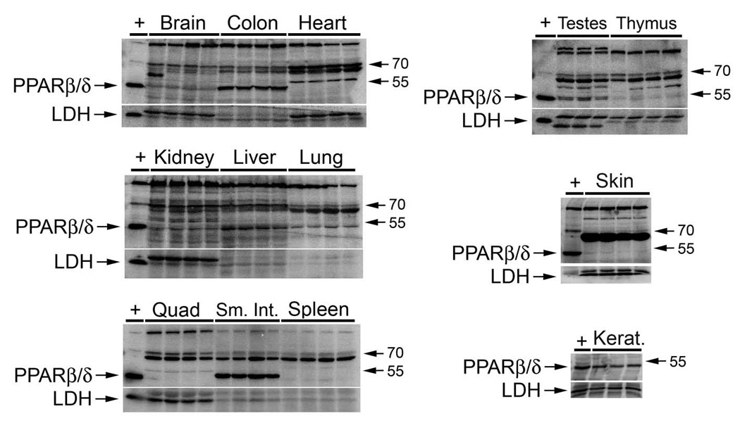
Quantitative expression of PPARβ/δ in male mouse tissues. Crude protein samples were prepared and analyzed by quantitative western blot analysis as described in Materials and Methods. Hybridization signals for PPARβ/δ protein was normalized to LDH and the normalized values are presented as the mean ± S.E.M. below the tissue samples. + = lysate from COS-1 cells transfected with a mouse PPARβ/δ expression vector as a positive control.
Table 1.
Relative expression patterns of PPARβ/δ in male mouse tissues.
| Tissue | Relative PPARβ/δ expression* |
|---|---|
| Small intestine | 27584 ± 2121 |
| Keratinocyte | 20491 ± 2509 |
| Liver | 10910 ± 685 |
| Colon | 7496 ± 305 |
| Kidney | 6414 ± 153 |
| Skin | 6220 ± 897 |
| Brain | 3742 ± 180 |
| Heart | 3156 ± 102 |
| Lung | 3069 ± 173 |
| Testes | 2936 ± 485 |
| Quadricep muscle | 1756 ± 72 |
| Spleen | 1190 ± 130 |
| Thymus | 666 ± 197 |
Values are presented as the average number of digital light units per µg of protein ± S.E.M based on analysis of four independent samples.
Fig. 3.

Nuclear and cytosolic localization of PPARβ/δ in male mouse tissues. (A) Nuclear and cytosolic protein preparations were prepared and analyzed by quantitative western blot analysis as described in Materials and Methods. + = lysate from COS-1 cells transfected with a mouse PPARβ/δ expression vector as a positive control. Blots were probed with anti-lamin antibody to demonstrate enriched nuclear fractions and anti-LDH to demonstrate enriched cytosolic fractions. (B) Immunoprecipitation of PPARβ/δ was performed using the 8099 anti-PPARβ/δ antibody or IgG control and nuclear extract from liver and then separated by SDS-PAGE. Immunoblots were then probed with either the 8099 anti-PPARβ/δ antibody or RXRα. Digital light units (DLU) for each immunoprecipitation (PPARβ/δ or RXRα) were corrected for background and non-specific binding from the IgG control.
Fig. 4.

Lack of PPARβ/δ expression in PPARβ/δ-null mouse small intestine. Nuclear and cytosolic protein preparations from small intestine were prepared from wild-type and PPARβ/δ-null mice and analyzed by quantitative western blot analysis as described in Materials and Methods. + = lysate from COS-1 cells transfected with a mouse PPARβ/δ expression vector as a positive control. Blots were probed with anti-lamin antibody to demonstrate enriched nuclear fractions and anti-LDH to demonstrate enriched cytosolic fractions.
Discussion
Previous studies by others have described the expression patterns of PPARβ/δ mRNA in rat and mouse tissues with some variability noted. For example, one of the first studies that examined expression of PPARβ/δ mRNA by northern blot analysis of rat samples noted the highest expression in adrenal gland, heart and intestine, with modest expression in brain, kidney and spleen, and low expression in liver and testis [10]. In mice, expression of PPARβ/δ mRNA was highest in mouse liver, with modest expression being observed in kidney and brown adipose tissue, and low expression in brain, spleen, intestine, heart and spleen, as measured by an RNase protection assay [9]. In a more limited analysis, expression of PPARβ/δ mRNA was found to be highest in colon as compared to small intestine and liver in mice, using an RNase protection assay [12]. More comprehensive analysis of PPARβ/δ expression was accomplished using in situ hybridization and immunohistochemistry of rat tissues [5]. In this work, expression patterns of PPARβ/δ were described ranging from tissue to cellular distribution. These studies showed that expression of PPARβ/δ was highest in testicular Sertoli cells, followed by moderately high expression of PPARβ/δ in brain, intestine, kidney and spleen [5]. Expression of PPARβ/δ was modest in heart and liver and reportedly low in colon [5]. More recently, the same group described quantitative mRNA data from tissue from fasted and fed rats [7]. Some differences were observed between fed and fasted rat tissue, but the highest expression of PPARβ/δ mRNA was found in colon, intestine, kidney, heart, esophagus and liver; although expression of PPARβ/δ mRNA in kidney and liver was lower in tissues from fasted rats [7]. Expression of PPARβ/δ mRNA was less than 50% of the highest expression in adipose, brain, adrenal, lung, skeletal muscle, testis, thymus and spleen [7]. Combined, all of these studies have demonstrated some modest variability of PPARβ/δ expression, primarily at the mRNA level in different tissues, with some variation being reported between studies. Results from the present studies extend these findings by establishing the relative quantitative expression levels of PPARβ/δ protein in similar tissues. This is particularly true because the present studies used the most quantitative methodology for accurately determining protein levels on protein blots using biotinylated secondary antibody and 125I-streptavidin. Expression of PPARβ/δ protein is highest in small intestine, keratinocytes, liver, colon, skin and kidney based on quantitative western blotting (Table 1). Significantly high expression of PPARβ/δ protein was also observed in brain, heart, lung and testis (Table 1). Expression of PPARβ/δ in quadricep muscle, spleen and thymus was lower as compared to all of the other tissues examined (Table 1). Several studies support the results from the present studies at the mRNA level. For example, expression of mRNA encoding PPARβ/δ is relatively high in rat and mouse intestine in a number of studies [5; 7; 10; 12], but other findings suggest low expression of mRNA in the intestine [9] or colon [5]. The reason for this difference could be related to differences in mRNA translation or possible issues relating to degradation of mRNA. Data from these studies also illustrate that the 8099 anti-PPARβ/δ antibody can be used to examine expression of the receptor in specific cell types within a specific tissue. For example, while modest expression of PPARβ/δ was detected in skin, significantly higher expression of PPARβ/δ was detected in keratinocytes.
The relatively high expression of PPARβ/δ protein in colon, small intestine, keratinocytes and liver is of interest, because significant roles for PPARβ/δ in these tissues have recently been established [3; 4; 13; 14]. For example, PPARβ/δ can modulate terminal differentiation in the intestine and skin [3; 4; 14], which is consistent with the observed expression pattern. PPARβ/δ can also inhibit inflammation, which may be important in epithelial tissue and liver [3; 4; 13]. Additionally, PPARβ/δ is important in the liver for protecting against liver toxicity [13] and for regulating glucose homeostasis [1]. Collectively, the relatively high expression of PPARβ/δ in colon, small intestine, keratinocytes and liver supports the idea that this receptor has important constitutive roles in these tissues.
The observation that PPARβ/δ localized in the nuclear fraction of tissues examined is consistent with less quantitative analysis performed using immunohistochemistry where similar nuclear localization was observed in many tissues [8]. Given the nuclear localization, this provides further support for the idea that constitutive PPARβ/δ may be transcriptionally active. This idea is strongly supported by the observation from the present study that immunoprecipitated PPARβ/δ from nuclear extracts is also bound with its heterodimerization partner, RXRα, coupled with the finding by others that fatty acids, which may serve as endogenous ligands, are found tightly bound with PPARβ/δ [15]. However, nuclear PPARβ/δ could also be transcriptionally silent in the absence of ligands, via association with co-repressors [16]. Alternatively, nuclear PPARβ/δ could be both constitutively active on some target genes via association with co-activators and silent on others because of association with co-repressors. A similar mechanism has recently been described for PPARγ whereby aP2 expression occurs constitutively in adipocytes but glycerol kinase expression is only increased in response to ligand activation [17]. These ideas should be examined in greater detail.
It is also worth noting that there was significant cross-reactivity of the effective anti-PPARβ/δ antibody using cellular extracts from different tissues. This illustrates the need for including a positive control for all western blots using anti-PPARβ/δ antibodies. This cross-reactivity may cause problems for immunoprecipitation of PPARβ/δ, in particular when using heterogeneous cellular preparations. This also suggests that immunohistochemistry using anti-PPARβ/δ antibodies may not be suitable unless analysis of null mouse samples is included for control.
In closing, results from the present study clearly demonstrate that expression of PPARβ/δ is relatively high in colon, small intestine, keratinocytes and liver and that PPARβ/δ expression is localized in the nucleus of tissues examined in these studied. Further studies are needed to examine potential differences in PPARβ/δ expression in different cell types within tissues, to determine the potential constitutive role of PPARβ/δ expression in these tissues in addition to the effect of endogenous and exogenous ligand activation.
Acknowledgement
Supported in part by the National Institutes of Health grants CA97999 and CA124533 (J.M.P.).
Footnotes
Publisher's Disclaimer: This is a PDF file of an unedited manuscript that has been accepted for publication. As a service to our customers we are providing this early version of the manuscript. The manuscript will undergo copyediting, typesetting, and review of the resulting proof before it is published in its final citable form. Please note that during the production process errors may be discovered which could affect the content, and all legal disclaimers that apply to the journal pertain.
References
- 1.Lee CH, Olson P, Hevener A, Mehl I, Chong LW, Olefsky JM, Gonzalez FJ, Ham J, Kang H, Peters JM, Evans RM. PPARdelta regulates glucose metabolism and insulin sensitivity. Proc Natl Acad Sci U S A. 2006;103:3444–3449. doi: 10.1073/pnas.0511253103. [DOI] [PMC free article] [PubMed] [Google Scholar]
- 2.Grimaldi PA. Regulatory functions of PPARbeta in metabolism: implications for the treatment of metabolic syndrome. Biochim Biophys Acta. 2007;1771:983–990. doi: 10.1016/j.bbalip.2007.02.006. [DOI] [PubMed] [Google Scholar]
- 3.Burdick AD, Kim DJ, Peraza MA, Gonzalez FJ, Peters JM. The role of peroxisome proliferator-activated receptor-beta/delta in epithelial cell growth and differentiation. Cell Signal. 2006;18:9–20. doi: 10.1016/j.cellsig.2005.07.009. [DOI] [PubMed] [Google Scholar]
- 4.Peters JM, Hollingshead HE, Gonzalesc FJ. Role of peroxisome proliferator-activated receptor-β/δ (PPARβ/δ) in gastrointestinal tract function and disease. Clinical Science. 2008 doi: 10.1042/CS20080022. [DOI] [PMC free article] [PubMed] [Google Scholar]
- 5.Braissant O, Foufelle F, Scotto C, Dauca M, Wahli W. Differential expression of peroxisome proliferator-activated receptors (PPARs): tissue distribution of PPAR-alpha, -beta, and -gamma in the adult rat. Endocrinology. 1996;137:354–366. doi: 10.1210/endo.137.1.8536636. [DOI] [PubMed] [Google Scholar]
- 6.Braissant O, Wahli W. Differential expression of peroxisome proliferator-activated receptor- alpha, -beta, and -gamma during rat embryonic development. Endocrinology. 1998;139:2748–2754. doi: 10.1210/endo.139.6.6049. [DOI] [PubMed] [Google Scholar]
- 7.Escher P, Braissant O, Basu-Modak S, Michalik L, Wahli W, Desvergne B. Rat PPARs: quantitative analysis in adult rat tissues and regulation in fasting and refeeding. Endocrinology. 2001;142:4195–4202. doi: 10.1210/endo.142.10.8458. [DOI] [PubMed] [Google Scholar]
- 8.Higashiyama H, Billin AN, Okamoto Y, Kinoshita M, Asano S. Expression profiling of peroxisome proliferator-activated receptor-delta (PPAR-delta) in mouse tissues using tissue microarray. Histochem Cell Biol. 2007;127:485–494. doi: 10.1007/s00418-007-0279-5. [DOI] [PubMed] [Google Scholar]
- 9.Jones PS, Savory R, Barratt P, Bell AR, Gray TJ, Jenkins NA, Gilbert DJ, Copeland NG, Bell DR. Chromosomal localisation, inducibility, tissue-specific expression and strain differences in three murine peroxisome-proliferator-activated-receptor genes. Eur J Biochem. 1995;233:219–226. doi: 10.1111/j.1432-1033.1995.219_1.x. [DOI] [PubMed] [Google Scholar]
- 10.Kliewer SA, Forman BM, Blumberg B, Ong ES, Borgmeyer U, Mangelsdorf DJ, Umesono K, Evans RM. Differential expression and activation of a family of murine peroxisome proliferator-activated receptors. Proc Natl Acad Sci U S A. 1994;91:7355–7359. doi: 10.1073/pnas.91.15.7355. [DOI] [PMC free article] [PubMed] [Google Scholar]
- 11.Kim DJ, Murray IA, Burns AM, Gonzalez FJ, Perdew GH, Peters JM. Peroxisome proliferator-activated receptor-β/δ (PPAR-β/δ) inhibits epidermal cell proliferation by down-regulation of kinase activity. J Biol Chem. 2005;280:9519–9527. doi: 10.1074/jbc.M413808200. [DOI] [PubMed] [Google Scholar]
- 12.Mansen A, Guardiola-Diaz H, Rafter J, Branting C, Gustafsson JA. Expression of the peroxisome proliferator-activated receptor (PPAR) in the mouse colonic mucosa. Biochem Biophys Res Commun. 1996;222:844–851. doi: 10.1006/bbrc.1996.0832. [DOI] [PubMed] [Google Scholar]
- 13.Shan W, Nicol CJ, Ito S, Bility MT, Kennett MJ, Ward JM, Gonzalez FJ, Peters JM. Peroxisome proliferator-activated receptor-beta/delta protects against chemically induced liver toxicity in mice. Hepatology. 2008;47:225–235. doi: 10.1002/hep.21925. [DOI] [PubMed] [Google Scholar]
- 14.Schmuth M, Jiang YJ, Dubrac S, Elias PM, Feingold KR. Thematic Review Series: Skin Lipids. Peroxisome proliferator-activated receptors and liver X receptors in epidermal biology. J Lipid Res. 2008;49:499–509. doi: 10.1194/jlr.R800001-JLR200. [DOI] [PubMed] [Google Scholar]
- 15.Fyffe SA, Alphey MS, Buetow L, Smith TK, Ferguson MA, Sorensen MD, Bjorkling F, Hunter WN. Reevaluation of the PPAR-beta/delta ligand binding domain model reveals why it exhibits the activated form. Mol Cell. 2006;21:1–2. doi: 10.1016/j.molcel.2005.12.001. [DOI] [PubMed] [Google Scholar]
- 16.Shi Y, Hon M, Evans RM. The peroxisome proliferator-activated receptor delta, an integrator of transcriptional repression and nuclear receptor signaling. Proc Natl Acad Sci U S A. 2002;99:2613–2618. doi: 10.1073/pnas.052707099. [DOI] [PMC free article] [PubMed] [Google Scholar]
- 17.Guan HP, Ishizuka T, Chui PC, Lehrke M, Lazar MA. Corepressors selectively control the transcriptional activity of PPARgamma in adipocytes. Genes Dev. 2005;19:453–461. doi: 10.1101/gad.1263305. [DOI] [PMC free article] [PubMed] [Google Scholar]