Abstract
Objective
C2 laminar screw fixation is considered as an excellent alternative to Magerl's transfacetal approach or Harms construct for the atlantoaxial stabilization. However, to our knowledge, there is no report on the feasibility of the new approach to Korean population. We investigated morphometric parameters of the dorsal arch of the C2 to provide the quantitative data for the feasibility of laminar screw fixation.
Methods
One-hundred-and-two patients' cervical computed tomography had been reconstructed and investigated on the anatomical parameters related with C2 laminar screw placement. Sixty patients were male and forty-two patients were female. Measurements included the laminar thickness and slope, spino-laminar angle, and maximal screw length.
Results
Ages ranged from 20 to 81 and the mean age was 48.4. Mean laminar thickness was 5.7 mm (±1.0) (5.8 mm in male and 5.4 mm in female). Fifty-one patients (50%) had a laminar thickness smaller than 5.5 mm at least unilaterally, therefore the patients were considered as inappropriate candidates for the laminar screw fixation in the smaller side of the laminae. Mean value of maximal length of screw was 33.3 mm (34.3 mm in male and 31.9 mm in female). Mean spino-laminar angle was 43.2° and mean slope angle was 32.9°.
Conclusion
Half of patients had inappropriate laminar profiles to accommodate a 3.5 mm screw in at least one side of the axis. The three-dimensional computed tomography reconstruction is mandatory for the preoperative assessment for the feasibility of the C2 lamina.
Keywords: Axis, Laminar screw
INTRODUCTION
Eighteen to twenty-three percent of C2 pars/pedicle is not safe to accept 3.5-mm diametered screw because of high-riding vertebral artery7,10). In such cases, crossing laminar screw fixation can provide excellent mechanical strength comparable to transarticular or pedicle/pars screw fixation with minimal risk of vertebral artery injury3,8,9,11,15). In addition, the placement of a laminar screw is relatively straightforward because anatomic structures of interest are exposed without fluoroscopic guidance. For this reason, the approach became popular among spine surgeons in Korea.
However, the new technique is not always suitable for the patients as well as pars/pedicle screw fixation. According to a cadaveric study in a western country, at least one side of axial laminae would not be able to accommodate a 3.5-mm diametered screw with a 1 mm margin of bone around it at the thinnest portion in 37% of the specimens14). The C2 laminar size of Asian may differ from non-Asian's. Therefore, it is important to know the C2 profile in Korean population for surgeon to plan a surgical approach.
The three dimensional computed tomography reconstruction has been a reliable tool in the analysis for the feasibility of the C1-C2 complex for various surgical techniques8,9,12,18). We investigated anatomical parameters of the dorsal arch of the C2 using three-dimensional cervical computed tomography reconstruction to provide the quantitative data for the feasibility of laminar screw fixation in Korean population.
MATERIALS AND METHODS
Patient selection and 3-D reconstruction of the cervical computed tomographies
All scans were obtained for the purpose of diagnosing the presence or absence of cervical spine disease at a clinic or emergency room of our hospital from January to April, 2008. We reviewed all the medical records and radiologists' documents. CT scans of patients with systemic pathology known to effect bone morphology including rheumatoid disease, chronic renal failure, infection, trauma, and tumor were excluded. Finally, 102 patients aged from 20 to 81 were included in current study. 3-D cervical CT reconstruction was performed on a CT unit equipped with a 16-detector array (LightSpeed ultra-16, GE healthcare, TN, U.S.A) using the software (Advantage window 4.2, GE healthcare, TN, U.S.A).
Measurements of morphometric parameters
Linear measurements were taken using an electronic caliper accurate to 0.1 mm, and angular measurements were taken using a goniometer accurate to 0.1 degree. The CT unit has been evaluated for the accuracy every third month by Korean institute for accreditation of medical image (Seoul, Korea).
To compare with results from western people, measurements followed previously reported methodology and included the laminar thickness, slope angle, spino-laminar angle and maximal screw length14,17).
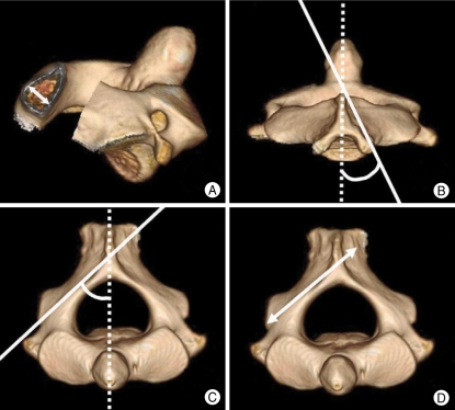
Laminar thickness measurement was made at the thinnest portion of the lamina bilaterally on axial view (Fig. 1A). The slope angle of the screw starting point was determined on dorsal view (Fig. 1B). The measurement of spino-laminar angle was made on axial view bilaterally (Fig. 1C). Spino-laminar angle implies the lateral angulation of the screw trajectory. Maximal screw length was determined from the screw entry point to the foramen transversarium on axial view (Fig. 1D). Two observers measured individually for the whole panel of 102 3-D CT images, and the average values were adopted. Analysis was performed for the application of a 3.5-mm diametered screw.
Fig. 1.
Measurement methods for the morphometric parameters of the axis vertebra. A : Oblique view showing maximal thickness of the lamina at the thinnest portion. B : Dorsal view showing the slope angle of the screw starting point. C : Axial view showing the lateral angulation of the screw trajectory. D : Maximal screw length.
Statistical analysis
Statistical analysis of parametric data was performed using Student's t test with p value < 0.05 considered significant.
RESULTS
One-hundred-and-two CT reconstructions of sixty male and forty-two female patients were investigated in the study. Measurement values including the means and standard deviations are shown in Table 1. The age ranged from 20 to 81 and the mean was 48.4. There was no statistically significant age-related difference between male and female patients (p = 0.565).
The mean laminar thickness was 5.7 mm. Seventy-nine of 204 laminas (38.7%) could not accommodate a 3.5-mm diametered screw, assuming the need for a 1 mm margin on each side in ventro-dorsal dimension. The numbers of patients with bilateral laminas thicker than 5.5 mm were 35 of male patients (58.3%) and 16 of female patients (38.1%). Fourteen of male patients (23.3%) and 9 of female patients (21.4%) had only one side lamina thicker than 5.5 mm. 11 of male patients (18.3%) and 17 of female patients (40.5%) could not be a candidate for 3.5-mm diametered laminar screw fixation bilaterally.
The laminar surface had an average slope of 32.9 degrees off the sagittal plane. The spino-laminar angle implying lateral angulation of the screw trajectory averaged 43.2 degrees (43.6 in males and 42.7 in females). The mean maximal screw length was 33.3 mm (34.3 mm in male and 31.9 mm in female) with wide range from 22.7 mm to 41.1 mm. None of the patients had maximal screw length shorter than 22 mm. The maximal screw length was significantly different between male and female patients (p < 0.01).
DISCUSSION
The major limitation of the transarticular screw technique that has been widely adopted for the atlantoaxial fixation is the potential risk of vertebral artery injury estimated up to 2.6-4.1%2,16).
The crossing laminar screw fixation can avoid the catastrophic complication15). The technique is possible because the C2 has greatest laminar dimension to accept 3.5-mm diametered screws in the cervical spine8,14,17). Then, atlantoaxial complex has unique anatomy and the axis has significant variations in the laminar profile1,14,17). In addition to anatomical variations, Asians have smaller profiles in morphometric parameters of the C218). However, the clinical applicability of laminar screw fixation technique in Korean adult population is not known. The purpose of the study was to provide a set of quantitative data on anatomical parameters of the C2 in Korean population.
In our measurements, only the maximal screw length of four parameters showed statistically significant gender-related differences with p value < 0.05. Regarding the laminar thickness, the most important parameter, male patients had greater thickness, however, the p value (0.067) did not clearly demonstrate a statistically significant difference between males and females. Then, it is not likely that the difference is clinically important because the difference is not sufficient to state that male patients are safe for the laminar screw fixation. There were 25 male patients (35%) with at least one of the laminae unsuitable for 3.5-mm diametered screw. This emphasizes the necessity of individual analysis using the 3-D CT reconstruction before placing screws.
In addition, our investigation demonstrated that at least one side of the laminae would not be able to accommodate a 3.5-mm diametered screw with 1 mm bony tolerance at the thinnest portion of the lamina in 50% of the patients. The result was higher than 37% in western population14).
To our knowledge, there is only one Asian study from Japan recently published study results on the feasibility for the laminar screw fixation8). According to the study, insertion of screws with diameter of 4 mm was possible in 50% of the males and only 24% of the females. Nakanishi and colleagues'8) performed the study with a different method and evaluated only one side of laminae. We once analyzed our data for the unilateral application of a 4-mm diametered screw although the direct comparison with their study was not proper. Sixty-two percent of males and 38% of females could accommodate 4 mm screw in at least one side of the laminae.
On the other hand, at least one side of the laminae would not be able to accommodate 4 mm screws in 47% of western patients included in Wang's14) investigation. Our data showed 72% of Korean patients had at least one side of the laminae unsuitable for 4-mm diametered screw. The comparisons have limitations of different methods and different sizes of study. Nevertheless, the results suggest that Korean patients should be carefully assessed for the C2 laminar screw fixation because of the narrow laminar profile.
The laminar slope of Korean population was bigger than that of western people. Assuming it would be ideal if the slope is orthogonal to the screw trajectory, careful drilling of entry site to reduce the slope angle is more required to Korean population. It is because less steepness of the screw starting point would result in inferiorly directed screw as placement of a screw is orthogonal to the entry surface on the bone. It is also ideal, if the laminar screw is placed in the thickest mid-level, equidistant from the superior and inferior margins, to achieve intraosseous placement17). Modification of the screw trajectory or identification of screw tip placed intraosseously through a cortical window would be required in some occasions5,13).
Finally, there was no patient with maximal screw length shorter than 22 mm. Therefore, insertion of the screw at the junction of the contralateral spino-laminar junction will permit placement of at least 22 mm screw in virtually all patients without entering the foramen transversarium.
CONCLUSION
Koreans have smaller profiles for the axis than western people. Half of patients had too thin lamina to accommodate 3.5-mm diametered screw, at least unilaterally on the axis. Careful preoperative assessment using the 3-D CT reconstruction is mandatory for the safe purchase of the laminar screw in Korean population.
Table 1.
Measurement values of morphometric parameters
Values are mean±standard deviation
References
- 1.Cassinelli EH, Lee M, Skalak Anthony, Ahn NU, Wright NM. Anatomic considerations for the placement of C2 laminar screws. Spine. 2006;31:2767–2771. doi: 10.1097/01.brs.0000245869.85276.f4. [DOI] [PubMed] [Google Scholar]
- 2.Gluf WM, Schmidt MH, Apfelbaum RI. Atlantoaxial transarticular screw fixation : a review of surgical indications, fusion rate, complication, and lessons learned in 191 adult patients. J Neurosurg Spine. 2005;2:155–163. doi: 10.3171/spi.2005.2.2.0155. [DOI] [PubMed] [Google Scholar]
- 3.Gorek J, Acaroglu E, Berven S, Yousef A, Puttlitz C. Constructs incorporating intralaminar C2 screws provide rigid stability for atlantoaxial fixation. Spine. 2005;30:1513–1518. doi: 10.1097/01.brs.0000167827.84020.49. [DOI] [PubMed] [Google Scholar]
- 4.Hong JT, Lee SW, Son BC, Park CK. Posterior C1-2 stabilization using translaminar screw fixation of the axis. J Korean Neurosurg Soc. 2006;40:387–390. [Google Scholar]
- 5.Jea A, Sheth RN, Vanni S, Green BA, Levi AD. Modification of Wright's technique for placement of bilateral crossing C2 translaminar screws : technical note. Spine J. 2007;8:656–660. doi: 10.1016/j.spinee.2007.06.008. [DOI] [PubMed] [Google Scholar]
- 6.Kim SY, Jang JS, Lee SH. Posterior atlantoaxial fixation with a combination of pedicle screws and a laminar screw in the axis for a unilateral high-riding vertebral artery. J Korean Neurosurg Soc. 2007;41:141–144. [Google Scholar]
- 7.Madawi A, Casey A, Solanki G, Tuite G, Veres R, Crockard H. Radiological and anatomical evaluation of the atlantoaxial transarticular screw fixation technique. J Neurosurg. 1997;86:961–968. doi: 10.3171/jns.1997.86.6.0961. [DOI] [PubMed] [Google Scholar]
- 8.Nakanishi K, Tanaka M, Sugimoto Y, Misawa H, Takigawa T, Fujiwara K, et al. Application of laminar screws to posterior fusion of cervical spine. Spine. 2008;33:620–623. doi: 10.1097/BRS.0b013e318166aa76. [DOI] [PubMed] [Google Scholar]
- 9.Nottmeier EW, Foy AB. Placement of C2 laminar screws using three-dimensional fluoroscopy-based image guidance. Eur Spine J. 2008;17:610–615. doi: 10.1007/s00586-007-0557-x. [DOI] [PMC free article] [PubMed] [Google Scholar]
- 10.Paramore C, Dickman C, Sonntag VKH. The anatomical suitability of the C1-2 complex for transarticular screw fixation. J Neurosurg. 1996;85:221–224. doi: 10.3171/jns.1996.85.2.0221. [DOI] [PubMed] [Google Scholar]
- 11.Reddy C, Ingalhalikar AV, Channon S, Lim TH, Torner J, Hitchon PW. In vitro biomechanical comparison of transpedicular versus translaminar C-2 screw fixation in C2-3 instrumentation. J Neurosurg Spine. 2007;7:414–418. doi: 10.3171/SPI-07/10/414. [DOI] [PubMed] [Google Scholar]
- 12.Resnick DK, Lapsiwala S, Trost GR. Anatomic suitability of the C1-C2 complex for pedicle screw fixation. Spine. 2002;27:1494–1498. doi: 10.1097/00007632-200207150-00003. [DOI] [PubMed] [Google Scholar]
- 13.Rhee WT, You SH, Jang YG, Lee SY. Modified trajectory of C2 laminar screw-double bicortical purchase of the inferiorly crossing screw. J Korean Neurosurg Soc. 2008;43:119–122. doi: 10.3340/jkns.2008.43.2.119. [DOI] [PMC free article] [PubMed] [Google Scholar]
- 14.Wang MY. C2 crossing laminar screws : cadaveric morphometric analysis. Neurosurgery. 2006;59(1 Suppl 1):ONS84–ONS88. doi: 10.1227/01.NEU.0000219900.24467.32. discussion ONS84-88. [DOI] [PubMed] [Google Scholar]
- 15.Wright NM. Posterior C2 fixation using bilateral, crossing C2 laminar screws : case series and technical note. J Spinal Disord Tech. 2004;17:158–162. doi: 10.1097/00024720-200404000-00014. [DOI] [PubMed] [Google Scholar]
- 16.Wright NM, Lauryssen C. Vertebral artery injury in C1-C2 transarticular screw fixation : results of a survey of the AANS/CNS section on disorders of the spine and peripheral nerves : American association of neurological surgeons/congress of neurological surgeons. J Neurosurg. 1998;88:634–640. doi: 10.3171/jns.1998.88.4.0634. [DOI] [PubMed] [Google Scholar]
- 17.Xu R, Burgar A, Ebraheim N, Yeasting R. The quantitative anatomy of the laminas of the spine. Spine. 1999;24:107–113. doi: 10.1097/00007632-199901150-00002. [DOI] [PubMed] [Google Scholar]
- 18.Yusof MI, Ming LK, Abdullah MS, Yusof AH. Computerized tomographic measurements of the cervical pedicles diameter in a Malaysian population and the feasibility for transpedicular fixation. Spine. 2006;31:E221–E224. doi: 10.1097/01.brs.0000210263.87578.65. [DOI] [PubMed] [Google Scholar]


