Abstract
Objective
To understand the anatomic characteristics of the aortic arch (AA) and its major branches to build a foundation toward performing endovascular surgery safely.
Methods
A total of 25 formalin fixed Korean adult cadavers were used. The authors investigated : anatomical variations of the AA and its major branches; curvature of the AA; distance from the mid-vertebrae line to the origin of the major branches; distances from the origin of the major branches of AA to the origin of its distal branches; and the angle of the three major branches, the brachiocephalic trunk (BCT), the left common carotid artery (LCCA) and the left subclavian artery (LSCA) arising from AA.
Results
The three major branches directly originated from AA in 21 (84%) of the cadavers. In two (8%) of remaining four cadavers, orifice of LCCA was slightly above the stem of BCT. In remaining two (8%) cadavers, the left vertebral artery (LVA) was directly originated from AA. Average angle of AA curvature to the coronal plane was 62.2 degrees. BCT originated 0.92 mm on the right of the mid-vertebrae line. LCCA and LSCA originated from 12.3 mm and 22.8 mm on the left of the mid-vertebrae line. Mean distance from the origin of the BCT to the origin of the RCCA was 32.5 mm. Mean distance from the origin of the LSCA to the origin of the LVA was 33.8 mm. Average angles at which the major branches arise from the AA were 65.3, 46.9 and 63.8 degrees.
Conclusion
This study may provides a basic anatomical information to catheterize AA and its branches for safely performing endovascular surgery.
Keywords: Aorta, Cadaver, Brachiocephalic trunk, Common carotid artery, Subclavian artery, Atherectomy
INTRODUCTION
In performing endovascular surgery, the most common technique is to puncture the femoral artery and advance a catheter toward the aortic arch through the abdominal aorta, as well as the major branches originating from the aortic arch (AA), brachiocephalic trunk (BCT), the left common carotid artery (LCCA) and the left subclavian artery (LSCA). However, despite the improvement of catheter quality and the rapid development of fluoroscopic imaging, this usual technique may be very difficult to perform in some cases due to the anatomical variations of the aortic arch and its major branches. Also, serious complications may develop due to these procedures19).
This study was initiated to seek the anatomical basis of endovascular surgery in order to obtain the optimal direction and configuration when inserting a catheter into the blood vessels without injuring neighboring structures in Korean population. Therefore, the purpose of this study was to understand the three-dimensional location and anatomical characteristics of the aortic arch and its major branches of blood vessels. These were evaluated by macroscopic examination and morphometric measurement of the exterior and interior morphology of the Korean adult cadaveric aortic arch and its major branches. This knowledge will assist surgeons in performing a safe and effective endovascular surgery through fluoroscopic imaging.
MATERIALS AND METHODS
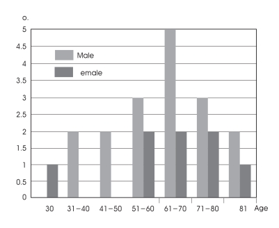
The study was performed on 25 Korean adult cadavers, 17 male and 8 female, fixated in formalin with a mean age of 63 years (range, 28-87 years) (Fig. 1).
Fig. 1.
Histogram showing the 25 adult cadaver's demography (male : 17, female : 8).
The thoracic cavity and abdomen of the cadavers were opened and the lung was removed to observe the aortic arch originating from the heart. The course from the right lower ilioinguinal ligament to the iliac artery, the descending aorta, the aortic arch, and the ascending aorta was examined. Subsequently, the aortic arch and its major branches were resected and measured. The BCT, LCCA, LSCA, LVA, right subclavian artery (RSCA), right common carotid artery (RCCA), and right vertebral artery (RVA) were exposed. The results of the examination are as follows.
Examination of the variation of the aortic arch and its major branches
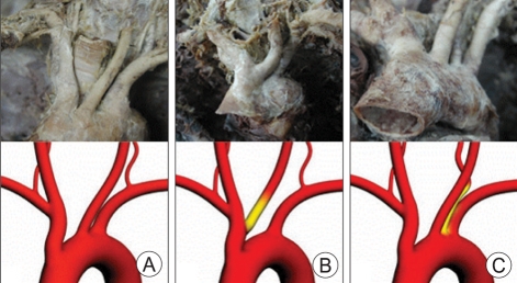
The course and variation of the BCT, LCCA, LSCA, and other arteries originating from the aortic arch were examined (Fig. 2).
Fig. 2.
Photographs and schematics showing the types of the major branches arising from the aortic arch. A : The 3 major branches are directly originated from the aortic arch. B : The brachiocephalic trunk and left subclavian artery are directly originated from the aortic arch. The orifice of the left common carotid artery is slightly distal to the origin of the brachiocephalic trunk. C : The 3 major branches and left vertebral artery are directly originated from the aortic arch. The left vertebral artery is situated between the left common carotid artery and left subclavian artery.
The external measurement of the aortic arch and its major branches
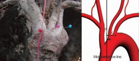
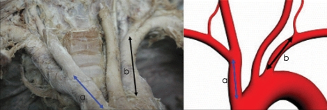
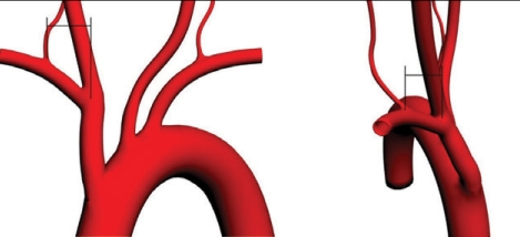
By observing the course from the descending aorta to the aortic arch, the angle of the aortic arch formed from the coronal plane was measured (Fig. 3). The distance from the mid-vertebrae line, from which the major branches of the aortic arch originated, was measured (Fig. 4). The distance from the origin of the BCT to the origin of the RCCA and the distance from the origin of the LSCA to the origin of the LVA were measured (Fig. 5). The vertical and anteroposterior distance from the origin of the RCCA to the origin of the RVA were measured (Fig. 6). The vertebrae level corresponding to the origin of the right and left vertebral artery was also examined.
Fig. 3.
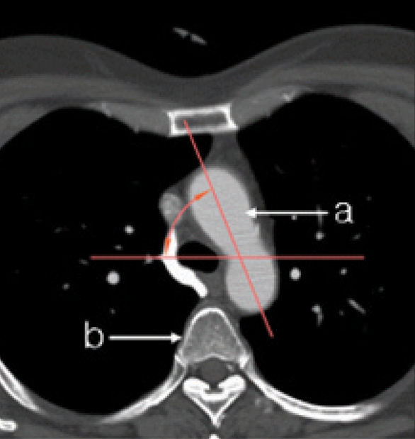
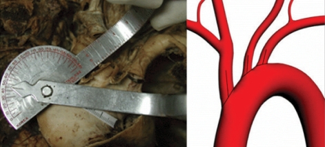
Photograph (left) and illustration (right) showing the measurement method for the degree (*) of the aortic arch curvature to the coronal plane. The average angle was 62.2° ± 14.82 (min=30°, max=90°). a : aortic arch, b : vertebral body.
Fig. 4.
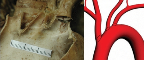
Photograph (left) and schematic (right) showing the measurement for the distance from the mid-vertebrae line to the origin of the major branches. Red, pink and blue pins indicate the orifice of the brachiocephalic trunk, left common carotid artery and left subclavian artery, respectively. a : distance between the brachiocephalic trunk and the mid-vertebrae line, b : distance between the left common carotid artery and the mid-vertebrae line, c : distance between the left subclavian artery and the mid-vertebrae line.
Fig. 5.
Photograph (left) and the schematic (right) illustrating the measurement for the distance from the origin of the brachiocephalic trunk to the origin of the right common carotid artery (a) and the distance from the origin of the left subclavian artery to the origin of the left vertebral artery (b).
Fig. 6.
Schematic illustrating the measurement of the distance from the origin of the right common carotid artery to the origin of the right vertebral artery. Left : anterior-posterior view. Right : lateral view.
Examination of the internal shape of the aortic arch and its major branches and measurement.
After the examination and external measurement of the blood vessels, the superior border of the curved portion of the aortic arch was resected by using a number 15 scalpel. The inside of the blood vessel was examined, while the inner diameter of the major branches of the aortic arch were measured (Fig. 7), along with the angle formed by each major branch from the aortic arch (Fig. 8).
Fig. 7.
Photograph (left) and schematic (right) showing the measurement of the inner diameters of the orifice of major branches.
Fig. 8.
Photograph (left) and schematic (right) showing the measurement for the angles at which the major branches arising from the aortic arch.
RESULTS
Anatomical variation of the aortic arch
The origin of the BCT, LCCA and LSCA from the AA was at the level of the 4th thoracic vertebra. Of the 25 cadavers, the three major branches, BCT, LCCA and LSCA, independently originated from the AA in 21 of the cadavers (84%). In two cadavers (8%), the BCT and LCCA originated together from the AA. In the remaining two cadavers, the major branches and left vertebral arteries independently branched directly from the aortic arch (Fig. 2).
The external shape of the aortic arch and its major branches and their measurement
The angle formed by the aortic arch and the coronal plane was an average of 62.2 (range, 30-90) degrees (Fig. 3) and the three major branches originated from the initial third of the anterior length of the aortic arch. The BCT originating from the branching site deviated by an average of 0.92 mm (range, from the right side at 19.2 mm to the left side at 11.8 mm) from the midvertebrae line. The LCCA and LSCA originating from the branching site deviated by an average of 12.3 mm to the left of the mid-vertebrae line (range, from the right side at 9.1 mm to the left side at 28.3 mm) and 22.8 mm (range, from the left side at 10.9 mm to the left side at 35.0 mm), respectively (Table 1, Fig. 4).
Table 1.
Distance from the mid-vertebrae line to the origin of the major branches
A : Distance from the mid-vertebrae line to the origin of the brachiocephalic trunk, B : Distance from the mid-vertebrae line to the origin of the left common carotid artery, C : Distance from the mid-vertebrae line to the origin of the left subclavian artery, SD : standard deviation, negative (-) : right side based on the mid-vertebral, line positive (+) : left side based on the mid-vertebral line
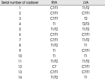
The distance from the origin of the BCT to the origin of the RCCA was an average of 32.5 mm (range, 24.2-46.0 mm) and the distance from the origin of the LSCA to the origin of the LVA was an average of 33.8 mm (range, 6.0-45.5 mm) (Table 2). The vertical and anteroposterior distance from the origin of the RCCA to the origin of the RVA were an average of 4.6 mm (range, 0.0-10.5 mm) and 16.7 mm (range, 10.9-23.3 mm), respectively (Table 3). The vertebrae position of the origin of the right and left vertebral artery could be assessed in 14 cadavers. In respect to the origin of the LVA, the cases positioned between the 7th cervical vertebra and the 1st thoracic vertebra were most prevalent, while there were also four cases positioned between the 1st and 2nd thoracic vertebra. The RVA was positioned between the 7th cervical vertebra and the 1st thoracic vertebra in five cases, which was most prevalent. There were four cases between the 1st and 2nd thoracic vertebra (Table 4).
Table 2.
Distance from the origin of the brachiocephalic trunk to the origin of the right common carotid artery and the distance from the origin of the left subclavian artery to the origin of the left vertebral artery
A : Distance from the origin of the brachiocephalic trunk to the origin of the right common carotid artery, B : Distance from the origin of the left subclavian artery to the origin of the left vertebral artery, SD : standard deviation
Table 3.
Distance from the origin of the right common carotid artery to the origin of the right vertebral artery
A : Distance from the origin of the right common carotid artery to the origin of the right vertebral artery on anterior-posterior view, B : Distance from the origin of the right common carotid artery to the origin of the right vertebral artery on lateral view, SD : standard deviation
Table 4.
Vertebrae level corresponding to the vertebral artery orifice
RVA : right vertebral artery, LVA : left vertebral artery, C : cervical, T : thoracic
Internal measurements of the aortic arch and its major branches
After resecting the external side of the AA, the internal shape was examined and found that the ridges formed by fibrous tissues were present between the blood vessel of the three major branches (Fig. 9). The inner diameter of the BCT, LCCA and LSCA were 18.3 mm (range, 11.4-31.5 mm), 9.5 mm (range, 6.9-14.1 mm), and 10.6 mm (range, 5.3-16.0 mm), respectively (Table 5). The angle formed by the BCT, LCCA, and LSCA with the AA was an average of 65.3 (range, 30-90), 46.9 (range, 5-97) and 63.8 (range, 2-102) degrees, respectively (Table 6).
Fig. 9.
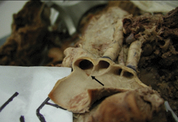
Photograph showing the inner surface of the aortic arch. The ridge of orifice (arrow) are visible between the major branches of the aortic arch.
Table 5.
Inner diameters of the orifice of brachiocephalic trunk, left common carotid artery and the left subclavian artery
SD : standard deviation, BCT : brachiocephalic trunk, LCCA : left common carotid artery, LSCA : left subclavian artery
Table 6.
Angles at which the major branches arise from the aortic arch
A : Angles at which the brachiocephalic trunk arise from the aortic arch. B : Angles at which the left common carotid artery arise from the aortic arch. C : Angles at which the left subclavian artery arise from the aortic arch, SD : standard deviation
DISCUSSION
The brain develops from the neuroectoderm during the third week of gestation and blood vessels develop from the mesoderm in the order of the anterior circulation system and the posterior circulation system16). The aortic arch is formed in the ventral and dorsal area as a pair, and six arches between the aorta are formed at the initial period of gestation and undergo a degeneration process. During the 4 mm embryonic period, the 1st and 2nd aortic arch degenerate as the 3rd and the 4th aortic arch appears. In the 12 mm embryonic period, the dorsal blood vessel in the 3rd aortic arch degenerates and the remaining segment of the 3rd aortic arch in the dorsal area forms the left carotid artery as well as the LSCA in the cervical vertebral segment16). Reaching the 40 mm embryonic period, among the dorsal blood vessels of the 3rd aortic arch, the residual part forms the BCT and the 4th aortic arch forms the normal adult type aortic arch, and thus the BCT, the LCCA, and the LSCA6).
It has been shown that in 30% of the total cases, there are some variations of the branches of the aortic arch and in its major blood vessels1,3,9,12,13). The findings show that the cases whose BCT and LCCA originated together are the most prevalent type of variation, and the remaining cases of variation showing either the left carotid artery originating from the aortic arch together with the BCT, or that the left carotid artery originated directly from the BCT13). Although it is rare, the aortic arch may be branched directly to the inner and the external aspects of the artery4).
In this study, all subjects were adult cadavers and the mean age was 63 years; however, the height and weight could not be measured. Among the 25 cadavers, 21 had the BCT, LCCA, and LSCA independently originating from the aortic arch. Thus, it was determined that 84% of the time, the aortic arch and its major branches develop normally. In two cadavers, the BCT and the LCCA originated simultaneously from the aortic arch. In two cadavers, the left vertebral artery was branched directly from the aortic arch. Comparing with other reports, Yamaki et al.20) examined the branch of the vertebral artery in 515 Japanese adult cadavers. In one cadaver, the right vertebral artery originated from the BCT. In 30 cadavers (5.8%), it directly originated from the aortic arch between the left vertebral artery and the left common carotid artery. Concerning angiographic findings, Best et al.3) reported a case in which the right vertebral artery originated directly from the aortic arch, while Matula et al.14) reported that among 190 cases, there were four cases (2.5%) in which the left vertebral artery originated directly from the aortic arch. Such results were comparable to our results. Therefore, during endovascular surgery, if the LCCA or the left vertebral artery cannot be visualized, then possible variations in which the LCCA originates from the BCT or that the left vertebral artery is originating directly from the aortic arch should be considered. It may be necessary to perform an aortogram in these cases.
CT angiogram and MR angiogram are non-invasive methods that reveal the structures of cerebrovascular diseases. In comparison with the catheter angiogram, the thoracic CT angiogram is useful for the diagnosis of calcified lesions. With respect to the aortic arch and its major branches, it clearly shows the BCT and the total carotid artery forming a rectangular angle on the imaging plane7,10). However, it cannot clearly draw the vertical segment of the subclavian artery or the vertebral artery5). Therefore, we concluded that CT and MR angiogram had limitations in understanding the three-dimensional structure for catheter manipulation. Thus, through cadavers, the aortic arch and its major branches were directly examined.
The results of this study show that the three major branches originating from the aortic arch were located in the anterior third of the aortic arch, and the angle formed by the aortic arch with the coronal plane was an average of 62.2 (range, 30-90) degrees. Therefore, during endovascular surgery, the three major branches appear to be located in the center of aortic arch on the true anteroposterior fluoroscopic images.
The BCT originates from the mid-vertebrae line with a right side deviation of an average of 0.92 mm and thus it is located almost in the mid-vertebrae area. The LCCA and the LSCA originating from the mid-vertebrae line deviated to the left by an average of 12.3 mm and an average of 22.8 mm, respectively. Therefore, on fluoroscopic images, it appears that the BCT is originating from the mid-vertebrae line. In addition, catheter manipulation may be performed considering that the distance from the origin of the BCT to the origin of the RCCA is an average of 32.5 mm, and the origin of the LSCA to left vertebral artery is an average of 33.8 mm.
The cerebral angiography of the posterior circulation system is performed generally through the left vertebral artery. Selection of the right vertebral artery for a catheter is much more difficult procedure. This study shows, however, that the vertical and anteroposterior distance from the origin of the RCCA to the right vertebral artery was an average of 4.6 mm and 16.7 mm, respectively. By anticipating that the origin of the right vertebral artery would be primarily between the 7th cervical vertebra and the 1st-2nd thoracic vertebra, it is easier to select the origin of the right vertebral artery. Based on such anticipation, it would assist in the examination the right vertebral artery during cerebral angiography if the anterior-posterior angle of the x-ray are rotated to the right side.
In endovascular surgery that requires the insertion of a guiding catheter within a major branch, the inner diameter of the blood vessel should be considered. The inner diameter of the major branches of the aortic arch varies depending on investigators8). In the results of this study, the inner diameter of the origin of the BCT, LCCA and LSCA were an average of 18.3 mm, 9.5 mm and 10.6 mm, respectively. Therefore, referring to such data would be helpful in selecting the appropriate size of catheter for each blood vesse2,15).
Zamir et al.21) reported the measurements of the angles from the aortic arch to the major branches and found that the BCT, LCCA, and LSCA had an average of 56.4, 58.4, and 66.5 degrees, respectively. However, the angle formed by each blood vessel did not correlate with age. According to this study, the angle formed by the brachiocephalic artery, LCCA and LSCA were an average of 65.3 degrees, 46.9 degrees, and 63.8 degrees, respectively. The angle formed by the brachiocephalic artery and the subclavian artery was similar to the results reported by Zamir et al.; however, the LCCA showed a more acute angle in our study.
Many kinds of cerebral angiographic catheters have been developed in diverse sizes and shapes15). The selection may be different according to the preference of surgeons; however, we have conventionally used the number 5 French, J type catheter in adults. Considering the flexion and angle of the blood vessels and the different types that may be selected, the Simons type may prove to be more applicable when the left common carotid artery has an acute angle18). Examining the inside of the blood vessels revealed that ridges formed by fibrous tissues were present between the three major branches. While manipulating a catheter within the aortic arch under fluoroscopic imaging, the pattern of the moving catheter could be observed. This was thought to be due to these fibrous tissue ridges and is also considered the point at which the major branches of the aortic arch originate. According to a recent report, after performing cerebral angiography on 2,899 cases, neurological complications were developed in 39 cases (1.3%) and among them, 25 cases (0.8%) were temporary and 14 cases (0.5%) had permanent impairment. The cases reporting patients 55 years and older, cardiovascular disease, and prolonged fluoroscopic imaging time showed the highest incidences of complications19). Qureshi et al.17) reported that after cerebral aneurysm embolization, embolism was induced in approximately 8% of the patients. In the insertion of the carotid artery stent, embolism was induced during the initial surgery period when a guide catheter was advanced to the common carotid artery.
Cases with a severely curved blood vessel, poor angulation of the cervical blood vessel branching from the aortic arch, or poor location of branching, difficulty in advancement due to the selection of a guide catheter were reported. This is because a selection vessel could contribute to an embolism from the atheroma of the aortic arch and its major branches to the intracranial artery11). Based on the above reports, the complications could be reduced by performing the procedures only after careful consideration of anatomical variations of the aortic arch in elderly patients with cardiovascular disease.
This study was initiated to address the question "what is the anatomical basis for the insertion of catheters into the major blood vessels within the aortic arch?" Although it is the result of initial studies, the three-dimensional structure and variation of the aortic arch and its major branches are now better understood. The result of this study is anticipated to provide important information for the general understanding and education of endovascular surgery.
CONCLUSION
We were able to find the anatomical basis for the insertion of catheters into the aortic arch and its major branches by studying adult cadavers in 25 Koreans. We were also able to obtain a further understanding of the three-dimensional aspects of the external and internal structures of the blood vessel, allowing endovascular surgery to be performed more safely. The use of the cadavers in this study reflected the anatomical aspects of endovascular surgery, but was limited in reflecting the vascular dynamics. Therefore, considering that in endovascular surgery the aortic arch and the major branches reflect the heart beat and the blood dynamic factors in the cervical area and the cerebral blood vessel, the use of brain magnetic resonance imaging or CT angiography should be considered in future studies.
References
- 1.Beigelman C, Mourey-Gerosa I, Gamsu G, Grenier P. New morphologic approach to the classification of anomalies of the aortic arch. Eur Radiol. 1995;5:435–442. [Google Scholar]
- 2.Berstein A, Lasjaunias P, Brugge KG. Surgical Neuroangiography. New York: Springer Co.; 2003. pp. 913–1181. [Google Scholar]
- 3.Best IM, Bumpers HL. Anomalous origins of the right vertebral, subclavian, and common carotid arteries in a patient with a four-vessel aortic arch. Ann Vasc Surg. 2002;16:231–234. doi: 10.1007/s10016-001-0041-8. [DOI] [PubMed] [Google Scholar]
- 4.Cakirer S, Karaarslan E, Kayabali M, Rozanes I. Separate origins of the left internal and external carotid arteries from the aortic arch : MR angiographic findings. AJNR Am J Neuroradiol. 2002;23:1600–1602. [PMC free article] [PubMed] [Google Scholar]
- 5.Chung JW, Park JH, Im JG, Chung MJ, Han MC, Ahn H. Spiral CT angiography of the thoracic aorta. Radiographics. 1996;16:811–824. doi: 10.1148/radiographics.16.4.8835973. [DOI] [PubMed] [Google Scholar]
- 6.Edwards JE. Anomalies of the derivatives of the aortic arch system. Med Clin North Am. 1948;32:925–949. doi: 10.1016/s0025-7125(16)35662-0. [DOI] [PubMed] [Google Scholar]
- 7.Gandhi D. Computed tomography and magnetic resonance angiography in cervicocranial vascular disease. J Neuroophthalmol. 2004;24:306–314. doi: 10.1097/00041327-200412000-00008. [DOI] [PubMed] [Google Scholar]
- 8.Gupta M, Sodhi L. Variations in branching pattern, shape, size and relative distances of arteries arising from arch of aorta. Nepal Med Coll J. 2005;7:13–17. [PubMed] [Google Scholar]
- 9.Haughton VM, Rosenbaum AE. Radiology of the skull and brain. St. Louis: Mosby Co.; 1974. pp. 1145–1163. [Google Scholar]
- 10.Jayaraman MV, Mayo-Smith WW. Multi-detector CT angiography of the intra-cranial circulation : normal anatomy and pathology with angiographic correlation. Clin Radiol. 2004;59:690–698. doi: 10.1016/j.crad.2003.12.004. [DOI] [PubMed] [Google Scholar]
- 11.Kim HJ, Suh DC. Atherosclerotic carotid stenosis : recent update of carotid stenting. Neurointervention. 2006;1:7–17. [Google Scholar]
- 12.Lambiase RE, Haas RA, Carney WI, Jr, Rogg J. Anomalous branching of the left common carotid artery with associated atherosclerotic changes : a case report. AJNR Am J Neuroradiol. 1991;12:187–189. [PMC free article] [PubMed] [Google Scholar]
- 13.Layton KF, Kallmes DF, Cloft HJ, Lindell EP, Cox VS. Bovine aortic arch variant in humans : clarification of a common misnomer. AJNR Am J Neuroradiol. 2006;27:1541–1542. [PMC free article] [PubMed] [Google Scholar]
- 14.Matula C, Trattnig S, Tschabitscher M, Day JD, Koos WT. The course of the prevertebral segment of the vertebral artery : anatomy and clinical significance. Surg Neurol. 1997;48:125–131. doi: 10.1016/s0090-3019(97)90105-1. [DOI] [PubMed] [Google Scholar]
- 15.Morris P. Practical Neuroangiography. Philadelphia: Lippincott Williams & Wilkins; 2007. pp. 36–85. [Google Scholar]
- 16.Padget DH. The development of the cranial arteries in the human embryo. Contrib Embryol. 1922;32:205–262. [Google Scholar]
- 17.Qureshi AI, Luft AR, Sharma M, Guterman LR, Hopkins LN. Prevention and treatment of thromboembolic and ischemic complications associated with endovascular procedures : Part II-Clinical aspects and recommendations. Neurosurgery. 2000;46:1360–1375. doi: 10.1097/00006123-200006000-00014. discussion 1375-1376. [DOI] [PubMed] [Google Scholar]
- 18.Simmons CR, Tsao EC, Thompson JR. Angiographic approach to the difficult aortic arch : a new technique for transfemoral cerebral angiography in the aged. Am J Roentgenol Radium Ther Nucl Med. 1973;119:605–612. doi: 10.2214/ajr.119.3.605. [DOI] [PubMed] [Google Scholar]
- 19.Willinsky RA, Taylor SM, TerBrugge K, Farb RI, Tomlinson G, Montanera W. Neurologic complications of cerebral angiography : prospective analysis of 2,899 procedures and review of the literature. Radiology. 2003;227:522–528. doi: 10.1148/radiol.2272012071. [DOI] [PubMed] [Google Scholar]
- 20.Yamaki K, Saga T, Hirata T, Sakaino M, Nohno M, Kobayashi S, et al. Anatomical study of the vertebral artery in Japanese adults. Anat Sci Int. 2006;81:100–106. doi: 10.1111/j.1447-073x.2006.00133.x. [DOI] [PubMed] [Google Scholar]
- 21.Zamir M, Sinclair P. Origin of the brachiocephalic trunk, left carotid, and left subclavian arteries from the arch of the human aorta. Invest Radiol. 1991;26:128–133. doi: 10.1097/00004424-199102000-00007. [DOI] [PubMed] [Google Scholar]


