

Abstract
Lysine methylation plays a central role in the “histone code” that regulates chromatin structure, impacts transcription, and responds to DNA damage. A single lysine can be mono-, di-, trimethylated, or unmethylated, with different functional consequences for each of the four forms. This review describes structural aspects of methylation of histone lysine residues by two enzyme families with entirely different structural scaffolding (the SET proteins and Dot1p), and the protein motifs that recognize (decode) these methyl-lysine signals. We also discuss the recently discovered protein lysine de-methylating enzymes (LSD1 and JmjC domains).
Keywords: protein lysine methylation and demethylation, SET domain proteins, S-adenosyl-L-methionine (AdoMet)
INTRODUCTION
AdoMet-dependent MTases are involved in biosynthesis, signal transduction, protein repair, chromatin regulation, and gene silencing. Methylation substrates range in size from arsenite through DNA and proteins, and the atomic targets can be carbon, oxygen, nitrogen, sulfur, or even halides. Histones can be modified in many ways that affect gene expression, including acetylation, phosphorylation, ubiquitination, methylation, and sumoylation [1-3]. Evidence accumulated over the past few years suggests that such modifications constitute a “histone code” that directs a variety of processes involving chromatin [4-13]. For example, one chromatin modification, the phosphorylation of histone H2A, links the recruitment of histone modifiers and ATP-dependent chromatin remodelling complexes to sites of DNA damage [14]. There are currently many known sites of lysine and arginine methylation on histones, and additional sites of modification are still being uncovered. Methylation at these sites, in combination with other modifications (or demodifications) at nearby residues, generates “modification cassettes” [15,16] that yield distinct patterns on chromatin for signaling downstream events [17]. The best-characterized sites of histone methylation are located on the N-terminal tails of histones (such as at Lys-4 and Lys-9 of histone H3 and Arg-3 of histone H4) that protrude from the nucleosome. In contrast, Lys-79 of histone H3 is located in the core of the histone, exposed on the nucleosome disk surface. The protein arginine methylation has been recently reviewed extensively in book chapters [18-20], and therefore in this review, we focus on progresses in the structural studies of histone lysine methylation enzmyes, methyl-lysine recognition domains and methyl-lysine demethylases.
SET Domain Proteins
With one exception (Dot1p, see below), histone lysine (K)methyltransferases (HKMTs) contain a SET domain of approximately 130 amino acids. The SET domain was originally identified in three Drosophila genes involved in epigenetic processes, the suppressor of position-effect variegation 3--9, Su(var)3--9; an enhancer of the eye color mutant zeste, En(zeste); and the homeotic gene regulator Trithorax[21]. Mammalian homologues of Drosophila Su(var) 3--9 were the first HKMTs identified, and they specifically methylate H3 at Lys-9 [22]. So far, SET-containing HKMTs that methylate Lys-4, -9, -27, or -36 of histone H3 and Lys-20 of histone H4 have been identified. The SET domain is found in a large number of eukaryotic proteins (see pfam database) as well as in a few bacterial proteins [23] and is not limited to HKMTs. HKMTs can be classified according to the presence or absence and the nature of sequences surrounding the SET domain that are conserved within families [24,25]. Representatives of the major families include SUV, SET1, SET2, EZ, and RIZ (for example, see [26]). The SET7/9 and SET8 proteins do not fit into these families. The SUV family includes the greatest number of HKMTs.
Structures of SET Domain Proteins
Currently known structures of SET proteins include the crystal structures of two SUV family proteins, Neurospora crassa DIM-5 [23,27] (Figure 1a) and Schizosaccharomyces pombe Clr4 [28] (Figure 1b); six human SET7/9 structures in various configurations [29-34](Figure 1c); two human SET8 (or Pr-SET7) structures [35,36] (Figure 1d); an NMR structure of a viral protein that contains only the SET domain (vSET) [37]; and a nonhistone Rubisco MTase [38,39]. These structures revealed that the SET domain forms a novel β-fold with a series of curved β-strands forming several small sheets, packed together with post-SET, pre-SET, or an additional domain (i-SET) inserted into SET domain.
Figure 1. SET domain Prtoein (histone) Lysine MTase structures.
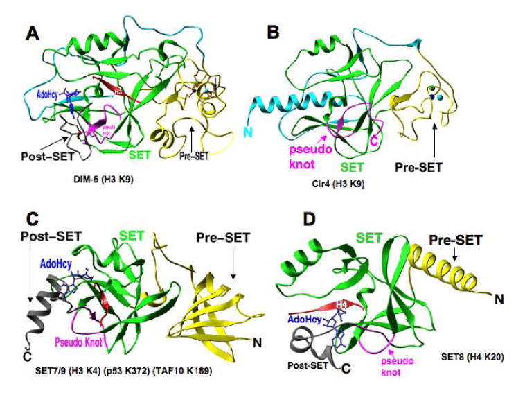
(a) Ribbon diagram of DIM-5 protein (one of the smallest members of the SUV family) contains four segments: a weakly conserved N-terminal region (cyan), a pre-SET domain containing nine invariant cysteines (yellow), the SET region containing four signature motifs (green and magenta), and the post-SET domain containing three invariant cysteines (grey). (b)S. pombeClr4. (c) Human SET7/9, and (d) Human SET8 (or PR-SET7).
The SET Domain Forms a Knot-Like Active Site
The SET domain folds into several small β-sheets that surround a knot-like structure by threading a C terminus through an opening of a short loop formed by a preceding stretch of the sequence (Figures 1a-d and Figure 2a). This remarkable knot-like structure brings together the two most-conserved SET signature motifs, RFINHxCxPN (III) and ELxFDY (IV) (Figure 2b), of the SET domain to form an active site in a location immediately next to the methyl-donor-binding pocket and peptide-binding cleft (Figure 1). Of the handful known protein structures that contain a knot, two are AdoMet-related proteins: Escherichia coli AdoMet synthetase [40] and the SPOUT family of RNA MTases [41-43] (for review on knotted protein structures, see Reference [44].
Figure 2. Domain structure of SET HKMT families.

(a) The knot-like structure. (b) Sequence alignment of SET proteins revealed four strongly conserved sequence motifs. We have termed them SET motif I (GxG), SET motif II (YxG), SET motif III (RFINHxCxPN), and SET motif IV (ELxFDY). The tertiary structure of SET proteins shows that these conserved residues are clustered together and involved in one of the three steps in the methylation reaction: methyl donor AdoMet binding (motif I: GxG; the first half of motif III: RFINH; and the last Tyr of motif IV, catalysis of methyl transfer (the Tyr of motif II), and formation of the hydrophobic target lysine-binding channel (the second half of motif III: CxPN, and motif IV: ELxFDY). (c) Illustration of pre-SET Zn3Cys9triangular zinc cluster. (d) Illustration of post-SET zinc center.
DIM-5: The Pre-SET Domain Forms a Triangular Zinc Cluster
The pre-SET domain of the SUV family HKMTs contains nine invariant cysteine residues that arrange into two segments of five and four cysteines separated by various numbers of amino acids (46 in DIM-5 and 28 in Clr4) (Figure 2b). The nine pre-SET cysteines coordinate three zinc ions to form an equilateral triangular cluster (Figure 2c). Each zinc ion is coordinated by two unique cysteines (six total) and the remaining three cysteine residues are each shared by two zinc atoms, thus serving as bridges to complete the tetrahedral coordination of the metal atoms. A similar metal-thiolate cluster can be found in metallothioneins that are involved in zinc metabolism, zinc transfer, and apoptosis [45]. Although the significance of this apparent similarity is unclear, it is intriguing to speculate that the zinc can be transferred from the pre-SET triangular cluster to the post-SET zinc center (see below), analogous to metallothioneins.
DIM-5: The Post-SET Zinc-Binding is essential for activity
The post-SET region contains three conserved cysteine residues that are essential for DIM-5 HKMT activity [23]. The structure of DIM-5 in a ternary complex with histone H3 Lys-9 peptide and AdoHcy [27] revealed that the three post-SET cysteines C306, C308, and C313, together with C244 of SET signature motif III, tetrahedrally coordinate a zinc ion near the active site (Figure 2d). Consequently, a narrow channel is formed to accommodate the target lysine side chain (see below).
The post-SET metal center observed in DIM-5 is universal among all SET proteins with the cysteine-rich post-SET including the SUV, SET1, and SET2 families. For almost all SET proteins, there appears to be an absolute correlation between the presence of the post-SET and a cysteine corresponding to C244 of DIM-5 from the knotted loop formed by the SET signature motif III (Figure 2b). As this metal center is absolutely required for enzymatic activity, it represents a good target to design inhibitors that disrupt metal coordination, perhaps analogous to the clinically successful examples of other metalloenzymes such as matrix metalloproteinases [46,47] and histone deacetylases (HDACs) [48].
Comparison of the structure of DIM-5 [27] with that of SET7/9 [32-34], and SET8 [35,36], two SET proteins that do not have a cysteine-rich post-SET domain, reveals a remarkable example of convergent evolution. In particular, like DIM-5, these two enzymes rely on residues C-terminal to the SET domain for the formation of a lysine channel, but do so by packing an α-helix, rather than a metal center, onto the active site (Figures 1c-d).
The Active-Site Channel
Five ternary structures, DIM-5 in complex with histone H3 Lys-9 peptide [27], SET7/9 in complex with a peptide containing either histone H3 Lys-4, the tumor suppressor p53 Lys-372, or the TBP-associated factor TAF10 Lys-189 [32-34], and SET8 in complex with histone H4 Lys-20 [35,36], revealed the target lysine is inserted into a narrow channel so that the target nitrogen lies in close proximity to the methyl-donor AdoMet at the opposite end of the channel (Figures 3b-c). The aromatic side chains form the channel wall and make van der Waals contacts to the methylene part of the target lysine side chain (Figure 3e). At the bottom of the channel, the terminal ε-amino group of the substrate lysine hydrogen bonds the hydroxyl of catalytic Tyr of SET signature motif II (Y178 in DIM-5 and Y245 in both SET7/9 and SET8) and is ~4 Å from the AdoHcy sulfur atom, where the transferable methyl group would be attached on AdoMet.
Figure 3. Active site activity of SET domain.
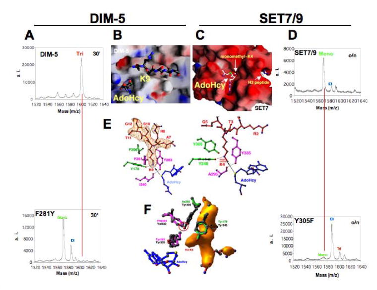
(a) Mass spectrometry analysis of methylation products for WT DIM-5 and its F281Y variant. (b) H3 peptide-binding site in DIM-5 with the target Lys-9 inserted into a channel (PDB 1PEG). (c) The AdoHcy-binding site in SET7/9, located at the opposite end of the target lysine-binding channel (PDB 1O9S) (right panel). (d) Mass spectrometry analysis of methylation products for WT SET7/9 and its Y305F variant. (e) The active sites in DIM-5 (PDB 1PEG) (left panel) and SET7/9 (PDB 1MT6) (right panel). The arrow indicates the movement of the methyl group transferred from the AdoMet methylsulfonium group to the target amino group. (f) Structural comparison of active sites in DIM-5 and SET7/9: either two tyrosines and one phenylalanine (DIM-5) or three tyrosines (SET7/9) surrounds the target lysine.
A Tyrosine/Phenylalanine Switch Controls Product Specificity
HKMTs differ both in their substrate specificity for the various acceptor lysines and in their product specificity for the number of methyl groups (one, two or three) they transfer. Human SET7/9 protein generates exclusively monomethyl Lys-4 of H3 [27,32]. DIM-5 of N. crassa, on the other hand, generates primarily trimethyl Lys-9, which marks chromatin regions for DNA methylation [49]. Considering that different methylation products might have different signaling properties [49-51], it is important to understand the structural basis for this product specificity [27].
DIM-5 and SET7/9 generate distinct products: DIM-5 forms trimethyl-lysine (Figure 3a)[27,49] and SET7/9 forms only monomethyl-lysine (Figure 3d)[27,32]. A likely structural explanation for their different product specificities is that residues in the lysine-binding channel of SET7/9 sterically exclude the target lysine side chain with methyl group(s). Comparison of the two active sites pinpointed the difference to a single amino acid that occupies a structurally similar position in both enzymes (F281 of DIM-5 and Y305 of SET7/9) (Figure 3f). Although the two residues are not aligned at the primary sequence level, the edge of the F281 phenyl ring in DIM-5 points to the same position as the Y305 hydroxyl in SET7/9, both in close proximity to the terminal ε-amino group of target lysine (Figure 3f). It was hypothesized that the Y305 hydroxyl in SET7/9 may be the source of steric hindrance limiting methylation [27]. Therefore, DIM-5 and SET7/9 mutants that swapped these residues were created. Remarkably, this swap almost completely inverts methylation product specificity [27]. Importantly, neither the lysine target specificity (Lys-9 versus Lys-4) nor the overall reaction rate for each enzyme was changed. Thus, DIM-5 was converted from a Lys-9 tri-MTase to a Lys-9 mono/di-MTase by F281Y mutation (Figure 3a, lower panel), while SET7/9 Y305F generated dimethylated instead of monomethylated Lys-4 (Figure 3d, lower panel) [27]. From a structural perspective, it appears the tyrosine hydroxyl can block substrate lysines with methyl group(s) attached from rotating into a position where they can be further methylated.
To further test the hypothesis that a single residue in the active site of SET-containing HKMTs, which aligns to F281 in DIM-5, is a major determinant of product specificity, F1205 was replaced with tyrosine (F1205Y) in human G9a, a predominant mammalian H3 Lys-9 HKMT that directs euchromatic mono- and dimethylation [52-54] but can generate trimethyl-H3K9 in some situations [55]. The mutation did not affect the catalytic activity of G9a; however, the reaction by F1205Y stalled at the monomethyl stage [56]. Thus, the F1205Y mutation changed the product specificity of G9a from a fast mono/di-MTase with a slow tri-MTase activity to a predominantly mono-MTase without affecting overall catalytic activity, analogous to the F281Y mutation for DIM-5. Conversely, mutation of Y334 to phenylalanine (Y334F) in human SET8 changed a mono-MTase to a predominantly di-MTase with virtually no effect on substrate binding or methylation [35].
Sequence alignment including HKMTs with known product specificity suggests that the tyrosine/phenlyalanine switch rule may be generalized (see Table 1 of reference [56]). Both Arabidopsis KYP and SUVH6 have a tyrosine at the switch position and are primarily mono-MTases [57]. Neurospora crassa SET2 includes F296 at the switch position and the ezyme can add up to three methyl groups on histone H3 Lys-36 [58]. Chlamydomonas Set1 contains Y1768 at the switch position which would predict to generate mono-methylated Lys-4 on histone H3, an epigenetic mark for repressed euchromatin in Chlamydomonas [59].
Table 1.
Known structures of methyl-lysine recognition domains
| Domain | Protein | Target methyllysine | Rreferences |
|---|---|---|---|
| Chromo | HP1 | H3K9me3 | [81] |
| Chromo | Polycomb | H3K27me3 | [17,82] |
| Double chromo | CHD | H3K4me3 | [83] |
| WD40 | WDR5 | H3K4me2 | [84] |
| Double Tudor | JMJD2A | H3K4me3 H4K20me2,3 | [87] |
| Double Tudor | 53BP1 | H4K20me2 | [89] |
| 3MBT | h-L3MBTL1 | H4K20me2 H3K4me1 | (PDB 1Z7O) |
| PHD | BPTF | H3K4me3 | [113] (PDB 2F6J) (PDB 2FUU) |
| PHD | ING2 | H3K4Me3 | [114] (PDB 2G6Q) |
At face value, Saccharomyces cerevisiae SET1 protein appears to conflict with our Phe/Tyr switch hypothesis. It has a tyrosine at the position comparable with Phe281 of DIM-5, and yet appears to be responsilnle for di- and tri-methylation of H3 Lys-4 in vivo [51,60]. It should be noted, however, that the published studies in 2002 did not reveal whether SET1 also produces mono-methyl-Lys-4, presumably because the mono-specific antibody was not yet available. However, more recent findings have indicated that an N-terminal RNA-recognition motif (RRM) is required for the histone H3 Lys-4 trimethylation activity of the yeast Set1 [61-63]. In its absence or when mutated, S. cerevisiae Set1 behaves mostly as a mono-MTase and di-MTase. Others suggested that histone H2B ubiquitylation controls processive methylation (transition from monomethylation to subsequent methylation states) but not monomethylation by Set1 [64]. It has recently been shown that the SET domain of a mammalian SET1 family protein, MLL, contains only activity on mono-methyl-Lys-4 of histone H3 [65].
Substrate specificity of SET7/9: (K/R)-(S/T/A)-K motif
Despite recent advances in identifying MTases, we still know little about what regulates their activities or determines their specificity. This is evident by recent reports that SET7/9 activity is not limited to histones, it is also able to methylate the tumour suppressor p53 [33] and the TBP-associated factor TAF10 [66]. The three ternary structures of SET7/9 in complex with mono-methylated-Lys-containing peptide - either from histone H3 [32], p53 [33], or TAF10 [34] - and the methyl donor byproduct AdoHcy revealed that SET7/9 recognizes a conserved K/R-S/T/A motif preceding the target lysine and has a propensity to bind aspartates and asparagines on the C-terminal side of the lysine target. Using the derived consensus sequence motif, several putative substrates for this prtoein lysine (K)MTase (PKMT) were identified [34] that will undoubtedly provide guidence for future studies aimed at identifying cellular functions for SET7/9-mediated mono-methylation.
Dot1p: Non-SET Domain HKMT
Histone H3 Lys-79 is methylated by Dot1p [60,67-69], a protein originally identified as a disruptor of telomeric silencing in S. cerevisiae [70]. Methylation of H3 Lys-79 in S. cerevisiae is important for the proper localization of the SIR (silent information regulator) complex [60,69] and DNA damage signaling [71,72]. In human, mistargeting of hDOT1L to Hoxa9 (a leukemia-relevant gene) plays an important role in MLL (mixed lineage leukemia)-AF10-mediated leukemogenesish [73]. A sequence analysis [74] suggested that Dot1p possesses AdoMet-binding motifs characteristic of class-I MTases [75], similar to those in protein arginine MTases that modify arginines on many proteins including histones H3 and H4 [20,26]. Class-I MTases such as Dot1p are distinct from and do not contain the SET domain. Thus, entirely different structural scaffolding and unrelated local active-site spatial arrangements can catalyze AdoMet-dependent methyl transfer to a protein lysine side chain.
Yeast Dot1p contains a core region conserved among human, Caenorhabditis elegans, Drosophila, and Anopheles gambiae Dot1p homologues (Figure 4a). The length of these Dot1 proteins varies from 582 amino acids in yeast to 2237 amino acids in Drosophila. The conserved Dot1p core is located at the C terminus in yeast but is at the N terminus in human, C. elegans, Drosophila, and Anopheles gambiae Dot1p homologues.
Figure 4. Dot1p family (non-SET HKMTs).
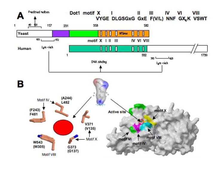
(a) Schematic representation of Dot1 homologues from yeast and human. (b) Dot1 core structure: (right panel) yeast Dot1p (residues 176--567) in complex with reaction by-product AdoHcy (PDB 1U2Z). Five hydrophobic residues of yeast Dot1p form the active-site pocket (the corresponding residue from human Dot1L is in parenthesis) (left panel). The opening of the pocket is approximately 4 × 5 Å, into which the target lysine could be inserted (middle panel). A surface representation of yeast Dot1p core showing the conserved motifs (X, VI, IV, and VIII) surrounding the active-site pocket, through which only the AdoHcy sulfur atom is visible (right panel). Conserved motifs (I, II, and III) involved in interacting with AdoHcy are buried and invisible from the surface.
The Dot1p conserved core contains an N-terminal helical domain and a seven-stranded catalytic domain that harbors the binding site for the methyl-donor and an active-site pocket sided with conserved hydrophobic residues (Figure 4b). Dot1p has several unique biochemical properties. Yeast Dot1p and its human homolog Dot1L methylate only nucleosomal substrates, but not free histone H3 protein [60,67-69]. A stretch of positively charged residues C-terminal to the human Dot1L core or N-terminal to the yeast Dot1p core (Figure 4a) were critical for nucleosome binding and therefore for enzymatic activity [76,77].
Methylation of Lys-79 of H3 in S. cerevisiae requires ubiquitination of Lys-123 of histone H2B in vivo [60,78]. H2B Lys-123 is located on the same nucleosome disk surface, ~30 Å away from the target Lys-79 of histone H3. Contrary to the in vivo data, recombinant yeast Dot1p was active on nucleosomes assembled in vitro from bacterially expressed, recombinant core histones [77], indicating that ubiquitination is not absolutely required for Dot1p activity. More recent data suggested that Dot1 binding to chromatin occurs normally in the absence of histone H2B Lys-123 ubiquitylation, suggesting that ubiquitylation does not regulate enzyme recruitment and monomethylation of H3 Lys-79 but does regulate the processive activity of Dot1p, i.e., the transition from monomethylation to subsequent methylation states [64]. Interestingly, a stretch of ~60 amino acids (residues 40-100) of yeast Dot1p have repeated hydrophobic residues every four to five positions and are predicted to form short helices by secondary structure prediction (Figure 4a). This stretch of Dot1p is similar in size to the ~50-amino-acid ubiquitin-binding domains which have a three-helix bundle structure [79]. Dot1p may interact, via this region, directly with H2B ubiquitinated nucleosome or indirectly through other ubiquitin-binding proteins. Such an interaction could be significant in vivo, recruiting Dot1p to specific high-order chromatin where ubiquitinated histone H2B might serve as a spacer between adjacent nucleosome disk surfaces, allowing Dot1p access to its target lysine [80].
Structures of the domains that read methyl-lysine code
The first structure of a methyl-lysine “decoder” is the chromodomain of HP1β in complex with a peptide containing methylated H3 lysine 9 [81]. Methyllysine recognition is achieved by a cage consists of three aromatic residues. The chromodomain of Polycomb, recognizes methylated H3 lysine 27 in a similar way [17,82]. The binding of trimethyl H3 lysine 4 by CHD1 protein involves double chromodomains, again using an aromatic cage (with two aromatic residues) [83]. A totally unrelated domain, WD40 of WDR protein, recognizes dimethylated lysine 4 of H3 [84]. In this case, the methyllysine is recognized in an entrirely different way: it lies on the surface of the protein and is primarily stabilized by a pair of nonconventional hydrogen bonds (C-H…O) between the two z-methyl groups of the dimethylated Lys-4 and the carboxylate oxygen of a glutamine residue in WDR5.
Chromodomain belongs to the “royal” superfamily of protein domains including tudor, MBT and PWWP domains that all contain a β–β–β–α310 core structure [85]. A recent study used a protein array technology to identify methyllysine binding domains from this family [86]. Among them, the double tudor domains of a histone lysine demethylase, JMJD2A (see below), was found to bind H3 trimethyl-Lys-4 and H4 trimethyl-Lys-20. A subsequent structure of this double tudor domains with an H3 peptide containing trimethyl-Lys-4 was determined [87] (see Figure 8d). Surprisingly, the two tudor domains form an interdigitated structure, so that the histone peptide was bound a hybrid tudor domain contributed by both tudor domains at the primary sequence level. Again, recognition of methyllysine involves an aromatic cage. The double tudor domain of 53BP1 was found to bind dimethylated H4 Lys-20 and H3 Lys-4 and Lys-9 in the protein array experiment. This domain has previously been found to bind H3 dimethyl-Lys-79 [88], Arg-Gly-rich peptide and DNA [89], so the biologically relevant binding activity of 53BP1 tudor is not clear. An NMR structure showed that the two tudor domains lie tandemly and NMR titration shows that both contribute to the binding of Arg-Gly peptide and DNA using aromatic residues [89].
Figure 8. JmjC-domain-containing demethylases.
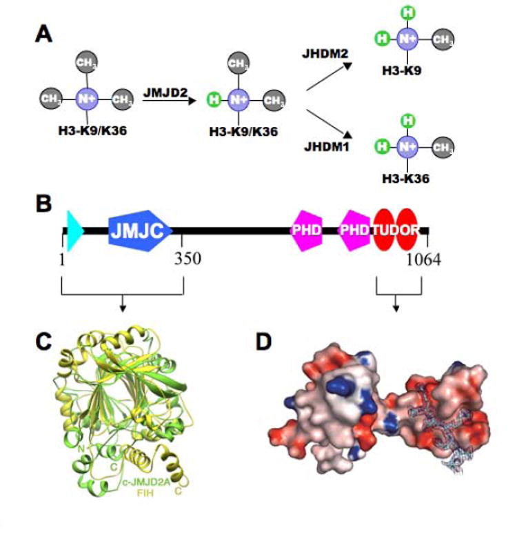
(a) JMJD2A demethyltes trimethylated H3-K9/K36 to di-methyl lysine [99], while JHDM2A and JHDM1A demethylate respectively dimethylated H3-K9 and H3-K36 [98,111]. (b) Domain organization of JMJD2A. (c) The superposition of N-terminal catalytic core of JMJD2A (colored green) and FIH (colored yellow) [112]. (d) Structure of C-terminal double tudor domain [87].
Recently, several structures of PHD fingers complexed with histone peptides have been deposited in the Protein Data Bank (Table 1), suggesting that this wide spread protein domain also participates in the recognition of methyllysine signal.
Demethylation by oxidation or hydroxylation
Methylation can function as a reversible signal, as in the case of O(xygen)-methylation, in which the side chain carboxyl groups of glutamate residues or the C-terminal carboxyl groups are reversibly methylated [90,91]. There are many examples of N(itrogen)-methylations (of arginine, lysine, glutamine, asparagine, histidine residues, and the amino group at the N terminus) in the cell. While it is not certain that all N-methylations are reversible, recent discoveries showed that a human nuclear peptidyl arginine deiminase, PAD4, antagonizes methylation on the arginine residues by converting arginine to citrulline [92-94] (Figure 5); a human nuclear amine oxidases, LSD1, functions as a histone di/mono-methyl-lysine demethylase via an oxidation reaction [95-97]; and JmjC domain-containing hydroxylase-like proteins are able to demethylate mono-, di-, or tri-methylated lysines [98,99], which was first proposed for S. pombe Epe1[100].
Figure 5. Human PAD4 (peptidylarginine deiminase 4) structure.
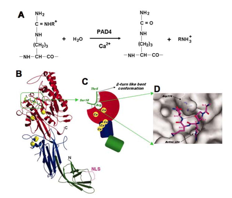
The figure panels are adopted from [94]. (a) Citrullination (or deimination) of arginine (R=H) and NG-monomethylarginine (R=CH3) residues by PAD4. (b) Ribbon representation of PAD4 in complex with peptide H3. Ca2+ ions and the histone peptide are shown as yellow balls and as a green stick model, respectively. The N-terminal subdomains 1 (residues 1–118) and 2 (residues 119–300) and the C-terminal domain (residues 301–663) are colored green, blue, and red, respectively. The nuclear localization signal (NLS) region is shown as a dotted line. (c) Schematic representation of the structure shown in panel B. Dotted lines show hydrogen bonds that form a consensus recognition motif at the molecular surface near the active site. (d) Top view of the peptide H3 together with a molecular surface representation near the active site cleft.
LSD1
LSD1 (also known as BHC110), named after protein lysine-specific-demethylase 1 [95], is found in histone modification complexes that control cell-specific gene expression. Within these complexes, REST (RE1-silencing transcription factor) corepressor CoREST enables LSD1 to demethylate nucleosomes [96,97], while BHC80 (BRAF–HDAC complex) inhibits LSD1 activity [96]. The LSD1 polypeptide chain can be divided into several structural/functional regions (Figure 6a): the N-terminal putative nuclear localization signal, followed by a SWIRM (Swi3p, Rsc8p, and Moira) domain (Figure 6b; [101])– found in several nucleosome-interacting proteins – and a monoamine oxidase domain – capable of demethylating lysines in an flavin-dependent manner [102]. From a sequence conservation standpoint, LSD1 belongs to the class of flavin-dependent mono-amine oxidases (such as human monoamino oxidase B;Figure 6c), which typically catalyze the oxidation of an amine-containing substrate using molecular oxygen as the electron acceptor [103]. The amino group of the methylated Lys is oxidized presumably to generate the corresponding imine compound, which is subsequently hydrolyzed to produce formaldehyde (Figure 6d). Substrate oxidation leads to the two-electron reduction of the protein-bound FAD cofactor, which is regenerated to its oxidized form by molecular oxygen to produce hydrogen peroxide. Biochemically, LSD1 demethylates mono- or dimethylated Lys-4 of H3 equally well, but does not demethylates trimethylated Lys-4 [104].
Figure 6. Monoamine oxidation.
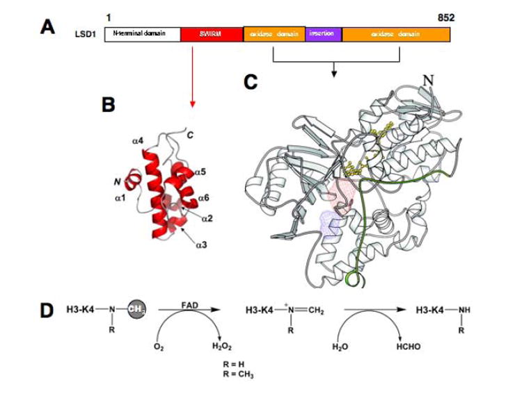
(a) Schematic representation of human LSD1 domain organization. The oxidase domain contains an atypical insertion of unknown function not found in other oxidases [95]. (b) The solution NMR structure of the SIWRM domain of LSD1. The panel is adopted from [101]. (c) Overall structure of human monoamino oxidase B (PDB 1G0S). The FAD cofactor is in yellow. The active site cavity is colored in red, whereas the entrance cavity is in blue. The panel is adopted from [104] and [103]. (d) Scheme of the demethylation reaction catalyzed by LSD1.
JmjC-containing deMTases
Studies of DNA repair in Escherichia coli demonstrated that methyl groups of 1-methyladenine (1-meA) and 3-methylcytosine (3-meC) in DNA can be removed by the AlkB family of enzymes through oxidative demethylation [105-107] (Figures 7a-b). The similarity between the chemistry of removing a methyl group from 1-meA and 3-meC and methyl-lysine (Figure 7c) prompted the proposal that the fission yeast protein Epe1 is a putative histone demethylase that could act by oxidative demethylation [100]. Epe1 modulates the stability of silent chromatin and contains a JmjC domain [108]. The Epe1 protein can be modelled onto the structure of the 2-oxoglutarate-Fe(II)-dependent dioxygenase, factor inhibiting hypoxia inducible factor (FIH), which is a protein hydroxylase that also contains a JmjC domain [109](Figure 7d). JmjC domain-containing proteins are predicted to be metalloenzymes that regulate chromatin function [110]. Using a biochemical assay based on the detection of formaldehyde, one of the predicted release products, coupled with chromatography, JHDM1 (JmjC domain-containing histone demethylase 1) and JHDM2A were found to demethylate mono- and dimethylated Lys-36 and Lys-9 of histone H3 respectively [98,111]. Another JmjC domain-contaning protein, JMJD2A, is found to reverse trimethylated H3-K9/K36 to di-but not mono-or unmethylated products [99](Figure 8a). Indeed, recently determined structure of N-terminal catalytic core JmjC-containing domain of JMJD2A is a close relative of FIH but with apparent differences outside of the jellyroll region (Figures 8b-c)[112]. Thus, chromatin-associated JmjC-domain proteins may generally be protein N(itrogen)-demethylases that can remove methyl group(s) from many protein substrates containing variety modifed residues.
Figure 7. Demethylation by hydroxylation.

(a) Mechanism of demethylation of 3-methylcytosine by AlkB. (b) Structure of E. coli AlkB with Fe(II), 2OG, and a methylated trinucleotide [107]. The sphere representing the Fe cofactor is coloured orange, whereas atoms in 2OG and dT-(1-me-dA)-dT are coloured according to atomic identity (carbon, white; oxygen, red; nitrogen, blue; and phosphorous, orange). (c) Mechanism of demethylation of histones by JmjC-domain proteins. (d) Predicted structure of Epe1 modelled on the structure of FIH [100]. The JmjC domain including the double-stranded-helix (DSBH) is shown in cyan.
In summary, with the increasing interest in protein (histone) methylation as a mechanism for gene regulation, we will undoubtedly discover other exciting roles for MTases and deMTases and the cellular processes that they direct.
Acknowledgments
Work in our laboratory is supported in part by grants from the National Institute of Health (GM49245 and GM68680). X.C. is a Georgia Research Alliance Eminent Scholar.
Footnotes
Publisher's Disclaimer: This is a PDF file of an unedited manuscript that has been accepted for publication. As a service to our customers we are providing this early version of the manuscript. The manuscript will undergo copyediting, typesetting, and review of the resulting proof before it is published in its final citable form. Please note that during the production process errors may be discovered which could affect the content, and all legal disclaimers that apply to the journal pertain.
Literature cited
- 1.Zhang Y, Reinberg D. Transcription regulation by histone methylation: interplay between different covalent modifications of the core histone tails. Genes Dev. 2001;15:2343–2360. doi: 10.1101/gad.927301. [DOI] [PubMed] [Google Scholar]
- 2.Lachner M, O’Sullivan RJ, Jenuwein T. An epigenetic road map for histone lysine methylation. J Cell Sci. 2003;116:2117–2124. doi: 10.1242/jcs.00493. [DOI] [PubMed] [Google Scholar]
- 3.Shiio Y, Eisenman RN. Histone sumoylation is associated with transcriptional repression. Proc Natl Acad Sci U S A. 2003;100:13225–13230. doi: 10.1073/pnas.1735528100. [DOI] [PMC free article] [PubMed] [Google Scholar]
- 4.Turner BM. Decoding the nucleosome. Cell. 1993;75:5–8. [PubMed] [Google Scholar]
- 5.Strahl BD, Allis CD. The language of covalent histone modifications. Nature. 2000;403:41–45. doi: 10.1038/47412. [DOI] [PubMed] [Google Scholar]
- 6.Jenuwein T, Allis CD. Translating the histone code. Science. 2001;293:1074–1080. doi: 10.1126/science.1063127. [DOI] [PubMed] [Google Scholar]
- 7.Turner BM. Cellular memory and the histone code. Cell. 2002;111:285–291. doi: 10.1016/s0092-8674(02)01080-2. [DOI] [PubMed] [Google Scholar]
- 8.Dutnall RN. Cracking the histone code: one, two, three methyls, you’re out! Mol Cell. 2003;12:3–4. doi: 10.1016/s1097-2765(03)00282-x. [DOI] [PubMed] [Google Scholar]
- 9.Wang Y, Fischle W, Cheung W, Jacobs S, Khorasanizadeh S, Allis CD. Beyond the double helix: writing and reading the histone code. Novartis Found Symp. 2004;259:3–17. discussion 17-21, 163-169. [PubMed] [Google Scholar]
- 10.de la Cruz X, Lois S, Sanchez-Molina S, Martinez-Balbas MA. Do protein motifs read the histone code? Bioessays. 2005;27:164–175. doi: 10.1002/bies.20176. [DOI] [PubMed] [Google Scholar]
- 11.Dion MF, Altschuler SJ, Wu LF, Rando OJ. Genomic characterization reveals a simple histone H4 acetylation code. Proc Natl Acad Sci U S A. 2005;102:5501–5506. doi: 10.1073/pnas.0500136102. [DOI] [PMC free article] [PubMed] [Google Scholar]
- 12.Margueron R, Trojer P, Reinberg D. The key to development: interpreting the histone code? Curr Opin Genet Dev. 2005;15:163–176. doi: 10.1016/j.gde.2005.01.005. [DOI] [PubMed] [Google Scholar]
- 13.Cosgrove MS, Wolberger C. How does the histone code work? Biochem Cell Biol. 2005;83:468–476. doi: 10.1139/o05-137. [DOI] [PubMed] [Google Scholar]
- 14.van Attikum H, Gasser SM. The histone code at DNA breaks: a guide to repair? Nat Rev Mol Cell Biol. 2005;6:757–765. doi: 10.1038/nrm1737. [DOI] [PubMed] [Google Scholar]
- 15.Fischle W, Wang Y, Allis CD. Binary switches and modification cassettes in histone biology and beyond. Nature. 2003;425:475–479. doi: 10.1038/nature02017. [DOI] [PubMed] [Google Scholar]
- 16.Fischle W, Tseng BS, Dormann HL, Ueberheide BM, Garcia BA, Shabanowitz J, Hunt DF, Funabiki H, Allis CD. Regulation of HP1-chromatin binding by histone H3 methylation and phosphorylation. Nature. 2005;438:1116–1122. doi: 10.1038/nature04219. [DOI] [PubMed] [Google Scholar]
- 17.Fischle W, Wang Y, Jacobs SA, Kim Y, Allis CD, Khorasanizadeh S. Molecular basis for the discrimination of repressive methyl-lysine marks in histone H3 by Polycomb and HP1 chromodomains. Genes Dev. 2003;17:1870–1881. doi: 10.1101/gad.1110503. [DOI] [PMC free article] [PubMed] [Google Scholar]
- 18.Bedford MT. The family of protein arginine methyltransferases. The Enzymes. 2006;24 doi: 10.1016/S1874-6047(06)80004-1. [DOI] [PubMed] [Google Scholar]
- 19.McBride AE. Diverse roles of protein arginine methyltransferases. The Enzymes. 2006;24:51–103. doi: 10.1016/S1874-6047(06)80005-3. [DOI] [PubMed] [Google Scholar]
- 20.Zhang X, Cheng X. Structure of protein arginine methyltransferases. The Enzymes. 2006;24:105–121. doi: 10.1016/S1874-6047(06)80006-5. [DOI] [PubMed] [Google Scholar]
- 21.Jenuwein T, Laible G, Dorn R, Reuter G. SET domain proteins modulate chromatin domains in eu- and heterochromatin. Cell Mol Life Sci. 1998;54:80–93. doi: 10.1007/s000180050127. [DOI] [PMC free article] [PubMed] [Google Scholar]
- 22.Rea S, Eisenhaber F, O’Carroll D, Strahl BD, Sun ZW, Schmid M, Opravil S, Mechtler K, Ponting CP, Allis CD, Jenuwein T. Regulation of chromatin structure by site-specific histone H3 methyltransferases. Nature. 2000;406:593–599. doi: 10.1038/35020506. [DOI] [PubMed] [Google Scholar]
- 23.Zhang X, Tamaru H, Khan SI, Horton JR, Keefe LJ, Selker EU, Cheng X. Structure of the Neurospora SET domain protein DIM-5, a histone H3 lysine methyltransferase. Cell. 2002;111:117–127. doi: 10.1016/s0092-8674(02)00999-6. [DOI] [PMC free article] [PubMed] [Google Scholar]
- 24.Baumbusch LO, Thorstensen T, Krauss V, Fischer A, Naumann K, Assalkhou R, Schulz I, Reuter G, Aalen RB. The Arabidopsis thaliana genome contains at least 29 active genes encoding SET domain proteins that can be assigned to four evolutionarily conserved classes. Nucleic Acids Research. 2001;29:4319–4333. doi: 10.1093/nar/29.21.4319. [DOI] [PMC free article] [PubMed] [Google Scholar]
- 25.Kouzarides T. Histone methylation in transcriptional control. Curr Opin Genet Dev. 2002;12:198–209. doi: 10.1016/s0959-437x(02)00287-3. [DOI] [PubMed] [Google Scholar]
- 26.Cheng X, Collins RE, Zhang X. Structural and sequence motifs of protein (histone) methylation enzymes. Annu Rev Biophys Biomol Struct. 2005;34:267–294. doi: 10.1146/annurev.biophys.34.040204.144452. [DOI] [PMC free article] [PubMed] [Google Scholar]
- 27.Zhang X, Yang Z, Khan SI, Horton JR, Tamaru H, Selker EU, Cheng X. Structural basis for the product specificity of histone lysine methyltransferases. Mol Cell. 2003;12:177–185. doi: 10.1016/s1097-2765(03)00224-7. [DOI] [PMC free article] [PubMed] [Google Scholar]
- 28.Min J, Zhang X, Cheng X, Grewal SI, Xu RM. Structure of the SET domain histone lysine methyltransferase Clr4. Nat Struct Biol. 2002;9:828–832. doi: 10.1038/nsb860. [DOI] [PubMed] [Google Scholar]
- 29.Jacobs SA, Harp JM, Devarakonda S, Kim Y, Rastinejad F, Khorasanizadeh S. The active site of the SET domain is constructed on a knot. Nat Struct Biol. 2002;9:833–838. doi: 10.1038/nsb861. [DOI] [PubMed] [Google Scholar]
- 30.Kwon T, Chang JH, Kwak E, Lee CW, Joachimiak A, Kim YC, Lee J, Cho Y. Mechanism of histone lysine methyl transfer revealed by the structure of SET7/9-AdoMet. Embo J. 2003;22:292–303. doi: 10.1093/emboj/cdg025. [DOI] [PMC free article] [PubMed] [Google Scholar]
- 31.Wilson JR, Jing C, Walker PA, Martin SR, Howell SA, Blackburn GM, Gamblin SJ, Xiao B. Crystal structure and functional analysis of the histone methyltransferase SET7/9. Cell. 2002;111:105–115. doi: 10.1016/s0092-8674(02)00964-9. [DOI] [PubMed] [Google Scholar]
- 32.Xiao B, Jing C, Wilson JR, Walker PA, Vasisht N, Kelly G, Howell S, Taylor IA, Blackburn GM, Gamblin SJ. Structure and catalytic mechanism of the human histone methyltransferase SET7/9. Nature. 2003;421:652–656. doi: 10.1038/nature01378. [DOI] [PubMed] [Google Scholar]
- 33.Chuikov S, Kurash JK, Wilson JR, Xiao B, Justin N, Ivanov GS, McKinney K, Tempst P, Prives C, Gamblin SJ, Barlev NA, Reinberg D. Regulation of p53 activity through lysine methylation. Nature. 2004;432:353–360. doi: 10.1038/nature03117. [DOI] [PubMed] [Google Scholar]
- 34.Couture JF, Collazo E, Hauk G, Trievel RC. Structural basis for the methylation site specificity of SET7/9. Nat Struct Mol Biol. 2006;13:140–146. doi: 10.1038/nsmb1045. [DOI] [PubMed] [Google Scholar]
- 35.Couture JF, Collazo E, Brunzelle JS, Trievel RC. Structural and functional analysis of SET8, a histone H4 Lys-20 methyltransferase. Genes Dev. 2005;19:1455–1465. doi: 10.1101/gad.1318405. [DOI] [PMC free article] [PubMed] [Google Scholar]
- 36.Xiao B, Jing C, Kelly G, Walker PA, Muskett FW, Frenkiel TA, Martin SR, Sarma K, Reinberg D, Gamblin SJ, Wilson JR. Specificity and mechanism of the histone methyltransferase Pr-Set7. Genes Dev. 2005;19:1444–1454. doi: 10.1101/gad.1315905. [DOI] [PMC free article] [PubMed] [Google Scholar]
- 37.Manzur KL, Farooq A, Zeng L, Plotnikova O, Koch AW, Sachchidanand, Zhou MM. A dimeric viral SET domain methyltransferase specific to Lys27 of histone H3. Nat Struct Biol. 2003;10:187–196. doi: 10.1038/nsb898. [DOI] [PubMed] [Google Scholar]
- 38.Trievel RC, Beach BM, Dirk LM, Houtz RL, Hurley JH. Structure and catalytic mechanism of a SET domain protein methyltransferase. Cell. 2002;111:91–103. doi: 10.1016/s0092-8674(02)01000-0. [DOI] [PubMed] [Google Scholar]
- 39.Trievel RC, Flynn EM, Houtz RL, Hurley JH. Mechanism of multiple lysine methylation by the SET domain enzyme Rubisco LSMT. Nat Struct Biol. 2003;10:545–552. doi: 10.1038/nsb946. [DOI] [PubMed] [Google Scholar]
- 40.Takusagawa F, Kamitori S. A Real Knot in Protein. J Am Chem Soc. 1996;118:8945–8946. [Google Scholar]
- 41.Michel G, Sauve V, Larocque R, Li Y, Matte A, Cygler M. The structure of the RlmB 23S rRNA methyltransferase reveals a new methyltransferase fold with a unique knot. Structure (Camb) 2002;10:1303–1315. doi: 10.1016/s0969-2126(02)00852-3. [DOI] [PubMed] [Google Scholar]
- 42.Nureki O, Shirouzu M, Hashimoto K, Ishitani R, Terada T, Tamakoshi M, Oshima T, Chijimatsu M, Takio K, Vassylyev DG, Shibata T, Inoue Y, Kuramitsu S, Yokoyama S. An enzyme with a deep trefoil knot for theactive-site architecture. Acta Crystallogr D Biol Crystallogr. 2002;58:1129–1137. doi: 10.1107/s0907444902006601. [DOI] [PubMed] [Google Scholar]
- 43.Nureki O, Watanabe K, Fukai S, Ishii R, Endo Y, Hori H, Yokoyama S. Deep knot structure for construction of active site and cofactor binding site of tRNA modification enzyme. Structure (Camb) 2004;12:593–602. doi: 10.1016/j.str.2004.03.003. [DOI] [PubMed] [Google Scholar]
- 44.Taylor WR, Xiao B, Gamblin SJ, Lin K. A knot or not a knot? SETting the record ‘straight’ on proteins. Comput Biol Chem. 2003;27:11–15. doi: 10.1016/s1476-9271(02)00099-3. [DOI] [PubMed] [Google Scholar]
- 45.Vasak M, Hasler DW. Metallothioneins: new functional and structural insights. Curr Opin Chem Biol. 2000;4:177–183. doi: 10.1016/s1367-5931(00)00082-x. [DOI] [PubMed] [Google Scholar]
- 46.Bode W, Fernandez-Catalan C, Tschesche H, Grams F, Nagase H, Maskos K. Structural properties of matrix metalloproteinases. Cell Mol Life Sci. 1999;55:639–652. doi: 10.1007/s000180050320. [DOI] [PMC free article] [PubMed] [Google Scholar]
- 47.Coussens LM, Fingleton B, Matrisian LM. Matrix metalloproteinase inhibitors and cancer: trials and tribulations. Science. 2002;295:2387–2392. doi: 10.1126/science.1067100. [DOI] [PubMed] [Google Scholar]
- 48.Bhuiyan MP, Kato T, Okauchi T, Nishino N, Maeda S, Nishino TG, Yoshida M. Chlamydocin analogs bearing carbonyl group as possible ligand toward zinc atom in histone deacetylases. Bioorg Med Chem. 2006;14:3438–3446. doi: 10.1016/j.bmc.2005.12.063. [DOI] [PubMed] [Google Scholar]
- 49.Tamaru H, Zhang X, McMillen D, Singh PB, Nakayama J, Grewal SI, Allis CD, Cheng X, Selker EU. Trimethylated lysine 9 of histone H3 is a mark for DNA methylation in Neurospora crassa. Nat Genet. 2003;34:75–79. doi: 10.1038/ng1143. [DOI] [PubMed] [Google Scholar]
- 50.Czermin B, Melfi R, McCabe D, Seitz V, Imhof A, Pirrotta V. Drosophila enhancer of Zeste/ESC complexes have a histone H3 methyltransferase activity that marks chromosomal Polycomb sites. Cell. 2002;111:185–196. doi: 10.1016/s0092-8674(02)00975-3. [DOI] [PubMed] [Google Scholar]
- 51.Santos-Rosa H, Schneider R, Bannister AJ, Sherriff J, Bernstein BE, Emre NC, Schreiber SL, Mellor J, Kouzarides T. Active genes are tri-methylated at K4 of histone H3. Nature. 2002;419:407–411. doi: 10.1038/nature01080. [DOI] [PubMed] [Google Scholar]
- 52.Tachibana M, Sugimoto K, Nozaki M, Ueda J, Ohta T, Ohki M, Fukuda M, Takeda N, Niida H, Kato H, Shinkai Y. G9a histone methyltransferase plays a dominant role in euchromatic histone H3 lysine 9 methylation and is essential for early embryogenesis. Genes Dev. 2002;16:1779–1791. doi: 10.1101/gad.989402. [DOI] [PMC free article] [PubMed] [Google Scholar]
- 53.Peters AH, Kubicek S, Mechtler K, O’Sullivan RJ, Derijck AA, Perez-Burgos L, Kohlmaier A, Opravil S, Tachibana M, Shinkai Y, Martens JH, Jenuwein T. Partitioning and plasticity of repressive histone methylation states in mammalian chromatin. Mol Cell. 2003;12:1577–1589. doi: 10.1016/s1097-2765(03)00477-5. [DOI] [PubMed] [Google Scholar]
- 54.Rice JC, Briggs SD, Ueberheide B, Barber CM, Shabanowitz J, Hunt DF, Shinkai Y, Allis CD. Histone methyltransferases direct different degrees of methylation to define distinct chromatin domains. Mol Cell. 2003;12:1591–1598. doi: 10.1016/s1097-2765(03)00479-9. [DOI] [PubMed] [Google Scholar]
- 55.Osipovich O, Milley R, Meade A, Tachibana M, Shinkai Y, Krangel MS, Oltz EM. Targeted inhibition of V(D)J recombination by a histone methyltransferase. Nat Immunol. 2004;5:309–316. doi: 10.1038/ni1042. [DOI] [PubMed] [Google Scholar]
- 56.Collins RE, Tachibana M, Tamaru H, Smith KM, Jia D, Zhang X, Selker EU, Shinkai Y, Cheng X. In vitro and in vivo analyses of a Phe/Tyr switch controlling product specificity of histone lysine methyltransferases. J Biol Chem. 2005;280:5563–5570. doi: 10.1074/jbc.M410483200. [DOI] [PMC free article] [PubMed] [Google Scholar]
- 57.Jackson JP, Johnson L, Jasencakova Z, Zhang X, PerezBurgos L, Singh PB, Cheng X, Schubert I, Jenuwein T, Jacobsen SE. Dimethylation of histone H3 lysine 9 is a critical mark for DNA methylation and gene silencing in Arabidopsis thaliana. Chromosoma. 2004;112:308–315. doi: 10.1007/s00412-004-0275-7. [DOI] [PubMed] [Google Scholar]
- 58.Adhvaryu KK, Morris SA, Strahl BD, Selker EU. Methylation of histone H3 lysine 36 is required for normal development in Neurospora crassa. Eukaryot Cell. 2005;4:1455–1464. doi: 10.1128/EC.4.8.1455-1464.2005. [DOI] [PMC free article] [PubMed] [Google Scholar]
- 59.van Dijk K, Marley KE, Jeong BR, Xu J, Hesson J, Cerny RL, Waterborg JH, Cerutti H. Monomethyl histone H3 lysine 4 as an epigenetic mark for silenced euchromatin in Chlamydomonas. Plant Cell. 2005;17:2439–2453. doi: 10.1105/tpc.105.034165. [DOI] [PMC free article] [PubMed] [Google Scholar]
- 60.Ng HH, Feng Q, Wang H, Erdjument-Bromage H, Tempst P, Zhang Y, Struhl K. Lysine methylation within the globular domain of histone H3 by Dot1 is important for telomeric silencing and Sir protein association. Genes Dev. 2002;16:1518–1527. doi: 10.1101/gad.1001502. [DOI] [PMC free article] [PubMed] [Google Scholar]
- 61.Fingerman IM, Wu CL, Wilson BD, Briggs SD. Global loss of Set1-mediated H3 Lys4 trimethylation is associated with silencing defects in Saccharomyces cerevisiae. J Biol Chem. 2005;280:28761–28765. doi: 10.1074/jbc.C500097200. [DOI] [PMC free article] [PubMed] [Google Scholar]
- 62.Schlichter A, Cairns BR. Histone trimethylation by Set1 is coordinated by the RRM, autoinhibitory, and catalytic domains. Embo J. 2005;24:1222–1231. doi: 10.1038/sj.emboj.7600607. [DOI] [PMC free article] [PubMed] [Google Scholar]
- 63.Morillon A, Karabetsou N, Nair A, Mellor J. Dynamic lysine methylation on histone H3 defines the regulatory phase of gene transcription. Mol Cell. 2005;18:723–734. doi: 10.1016/j.molcel.2005.05.009. [DOI] [PubMed] [Google Scholar]
- 64.Shahbazian MD, Zhang K, Grunstein M. Histone H2B ubiquitylation controls processive methylation but not monomethylation by Dot1 and Set1. Mol Cell. 2005;19:271–277. doi: 10.1016/j.molcel.2005.06.010. [DOI] [PubMed] [Google Scholar]
- 65.Terranova R, Agherbi H, Boned A, Meresse S, Djabali M. Histone and DNA methylation defects at Hox genes in mice expressing a SET domain-truncated form of Mll. Proc Natl Acad Sci U S A. 2006;103:6629–6634. doi: 10.1073/pnas.0507425103. [DOI] [PMC free article] [PubMed] [Google Scholar]
- 66.Kouskouti A, Scheer E, Staub A, Tora L, Talianidis I. Gene-specific modulation of TAF10 function by SET9-mediated methylation. Mol Cell. 2004;14:175–182. doi: 10.1016/s1097-2765(04)00182-0. [DOI] [PubMed] [Google Scholar]
- 67.Feng Q, Wang H, Ng HH, Erdjument-Bromage H, Tempst P, Struhl K, Zhang Y. Methylation of H3-Lysine 79 Is Mediated by a New Family of HMTases without a SET Domain. Curr Biol. 2002;12:1052–1058. doi: 10.1016/s0960-9822(02)00901-6. [DOI] [PubMed] [Google Scholar]
- 68.Lacoste N, Utley RT, Hunter JM, Poirier GG, Cote J. Disruptor of telomeric silencing-1 is a chromatin-specific histone H3 methyltransferase. J Biol Chem. 2002;277:30421–30424. doi: 10.1074/jbc.C200366200. [DOI] [PubMed] [Google Scholar]
- 69.van Leeuwen F, PR G, E D. Gottschling Dot1p Modulates Silencing in Yeast by Methylation of the Nucleosome Core. Cell. 2002;109:745–756. doi: 10.1016/s0092-8674(02)00759-6. [DOI] [PubMed] [Google Scholar]
- 70.Singer MS, Kahana A, Wolf AJ, Meisinger LL, Peterson SE, Goggin C, Mahowald M, Gottschling DE. Identification of high-copy disruptors of telomeric silencing in Saccharomyces cerevisiae. Genetics. 1998;150:613–632. doi: 10.1093/genetics/150.2.613. [DOI] [PMC free article] [PubMed] [Google Scholar]
- 71.Wysocki R, Javaheri A, Allard S, Sha F, Cote J, JiD S. Kron Role of Dot1-dependent histone H3 methylation in G1 and S phase DNA damage checkpoint functions of Rad9. Mol Cell Biol. 2005;25:8430–8443. doi: 10.1128/MCB.25.19.8430-8443.2005. [DOI] [PMC free article] [PubMed] [Google Scholar]
- 72.Giannattasio M, Lazzaro F, Plevani P, Muzi-Falconi M. The DNA damage checkpoint response requires histone H2B ubiquitination by Rad6-Bre1 and H3 methylation by Dot1. J Biol Chem. 2005;280:9879–9886. doi: 10.1074/jbc.M414453200. [DOI] [PubMed] [Google Scholar]
- 73.Okada Y, Feng Q, Lin Y, Jiang Q, Li Y, Coffield VM, Su L, Xu G, Zhang Y. hDOT1L links histone methylation to leukemogenesis. Cell. 2005;121:167–178. doi: 10.1016/j.cell.2005.02.020. [DOI] [PubMed] [Google Scholar]
- 74.Dlakic M. Chromatin silencing protein and pachytene checkpoint regulator Dot1p has a methyltransferase fold. Trends Biochem Sci. 2001;26:405–407. doi: 10.1016/s0968-0004(01)01856-4. [DOI] [PubMed] [Google Scholar]
- 75.Schubert HL, Blumenthal RM, Cheng X. Many paths to methyltransfer: a chronicle of convergence. Trends Biochem Sci. 2003;28:329–335. doi: 10.1016/S0968-0004(03)00090-2. [DOI] [PMC free article] [PubMed] [Google Scholar]
- 76.Min J, Feng Q, Li Z, Zhang Y, Xu RM. Structure of the catalytic domain of human DOT1L, a non-SET domain nucleosomal histone methyltransferase. Cell. 2003;112:711–723. doi: 10.1016/s0092-8674(03)00114-4. [DOI] [PubMed] [Google Scholar]
- 77.Sawada K, Yang Z, Horton JR, Collins RE, Zhang X, Cheng X. Structure of the conserved core of the yeast Dot1p, a nucleosomal histone H3 lysine 79 methyltransferase. J Biol Chem. 2004;279:43296–43306. doi: 10.1074/jbc.M405902200. [DOI] [PMC free article] [PubMed] [Google Scholar]
- 78.Briggs SD, Xiao T, Sun ZW, Caldwell JA, Shabanowitz J, Hunt DF, Allis CD, Strahl BD. Gene silencing: trans-histone regulatory pathway in chromatin. Nature. 2002;418:498. doi: 10.1038/nature00970. [DOI] [PubMed] [Google Scholar]
- 79.Hicke L, Schubert HL, Hill CP. Ubiquitin-binding domains. Nat Rev Mol Cell Biol. 2005;6:610–621. doi: 10.1038/nrm1701. [DOI] [PubMed] [Google Scholar]
- 80.Sun ZW, Allis CD. Ubiquitination of histone H2B regulates H3 methylation and gene silencing in yeast. Nature. 2002;418:104–108. doi: 10.1038/nature00883. [DOI] [PubMed] [Google Scholar]
- 81.Jacobs SA, Khorasanizadeh S. Structure of HP1 chromodomain bound to a lysine 9-methylated histone H3 tail. Science. 2002;295:2080–2083. doi: 10.1126/science.1069473. [DOI] [PubMed] [Google Scholar]
- 82.Min J, Zhang Y, Xu RM. Structural basis for specific binding of Polycomb chromodomain to histone H3 methylated at Lys 27. Genes Dev. 2003;17:1823–1828. doi: 10.1101/gad.269603. [DOI] [PMC free article] [PubMed] [Google Scholar]
- 83.Flanagan JF, Mi LZ, Chruszcz M, Cymborowski M, Clines KL, Kim Y, Minor W, Rastinejad F, Khorasanizadeh S. Double chromodomains cooperate to recognize the methylated histone H3 tail. Nature. 2005;438:1181–1185. doi: 10.1038/nature04290. [DOI] [PubMed] [Google Scholar]
- 84.Han Z, Guo L, Wang H, Shen Y, Deng XW, Chai J. Structural Basis for the Specific Recognition of Methylated Histone H3 Lysine 4 by the WD-40 Protein WDR5. Mol Cell. 2006;22:137–144. doi: 10.1016/j.molcel.2006.03.018. [DOI] [PubMed] [Google Scholar]
- 85.Maurer-Stroh S, Dickens NJ, Hughes-Davies L, Kouzarides T, Eisenhaber F, Ponting CP. The Tudor domain ‘Royal Family’: Tudor, plant Agenet, Chromo, PWWP and MBT domains. Trends Biochem Sci. 2003;28:69–74. doi: 10.1016/S0968-0004(03)00004-5. [DOI] [PubMed] [Google Scholar]
- 86.Kim J, Daniel J, Espejo A, Lake A, Krishna M, Xia L, Zhang Y, Bedford Tudor MT. MBT and chromo domains gauge the degree of lysine methylation. EMBO Rep. 2006;7:397–403. doi: 10.1038/sj.embor.7400625. [DOI] [PMC free article] [PubMed] [Google Scholar]
- 87.Huang Y, Fang J, Bedford MT, Zhang Y, Xu RM. Recognition of Histone H3 Lysine-4 Methylation by the Double Tudor Domain of JMJD2A. Science. 2006 doi: 10.1126/science.1125162. [DOI] [PubMed] [Google Scholar]
- 88.Huyen Y, Zgheib O, Ditullio RA, Jr, Gorgoulis VG, Zacharatos P, Petty TJ, Sheston EA, Mellert HS, Stavridi ES, Halazonetis TD. Methylated lysine 79 of histone H3 targets 53BP1 to DNA double-strand breaks. Nature. 2004;432:406–411. doi: 10.1038/nature03114. [DOI] [PubMed] [Google Scholar]
- 89.Charier G, Couprie J, Alpha-Bazin B, Meyer V, Quemeneur E, Guerois R, Callebaut I, Gilquin B, Zinn-Justin S. The Tudor tandem of 53BP1: a new structural motif involved in DNA and RG-rich peptide binding. Structure. 2004;12:1551–1562. doi: 10.1016/j.str.2004.06.014. [DOI] [PubMed] [Google Scholar]
- 90.Longin S, Goris J. Reversible methylation of protein phosphatase 2A. The Enzymes. 2006;24 doi: 10.1016/S1874-6047(06)80013-2. [DOI] [PubMed] [Google Scholar]
- 91.Antomattei FM, Weis RM. Reversible methylation of glutamate residues in the receptor proteins of bacterial sensory systems. The Enzymes. 2006;24:325–382. doi: 10.1016/S1874-6047(06)80014-4. [DOI] [PubMed] [Google Scholar]
- 92.Cuthbert GL, Daujat S, Snowden AW, Erdjument-Bromage H, Hagiwara T, Yamada M, Schneider R, Gregory PD, Tempst P, Bannister AJ, Kouzarides T. Histone deimination antagonizes arginine methylation. Cell. 2004;118:545–553. doi: 10.1016/j.cell.2004.08.020. [DOI] [PubMed] [Google Scholar]
- 93.Wang Y, Wysocka J, Sayegh J, Lee YH, Perlin JR, Leonelli L, Sonbuchner LS, McDonald CH, Cook RG, Dou Y, Roeder RG, Clarke S, Stallcup MR, Allis CD, Coonrod SA. Human PAD4 regulates histone arginine methylation levels via demethylimination. Science. 2004;306:279–283. doi: 10.1126/science.1101400. [DOI] [PubMed] [Google Scholar]
- 94.Arita K, Shimizu T, Hashimoto H, Hidaka Y, Yamada M, Sato M. Structural basis for histone N-terminal recognition by human peptidylarginine deiminase 4. Proc Natl Acad Sci U S A. 2006;103:5291–5296. doi: 10.1073/pnas.0509639103. [DOI] [PMC free article] [PubMed] [Google Scholar]
- 95.Shi Y, Lan F, Matson C, Mulligan P, Whetstine JR, Cole PA, Casero RA. Histone Demethylation Mediated by the Nuclear Amine Oxidase Homolog LSD1. Cell. 2004;119:941–953. doi: 10.1016/j.cell.2004.12.012. [DOI] [PubMed] [Google Scholar]
- 96.Shi YJ, Matson C, Lan F, Iwase S, Baba T, Shi Y. Regulation of LSD1 histone demethylase activity by its associated factors. Mol Cell. 2005;19:857–864. doi: 10.1016/j.molcel.2005.08.027. [DOI] [PubMed] [Google Scholar]
- 97.Lee MG, Wynder C, Cooch N, Shiekhattar R. An essential role for CoREST in nucleosomal histone 3 lysine 4 demethylation. Nature. 2005;437:432–435. doi: 10.1038/nature04021. [DOI] [PubMed] [Google Scholar]
- 98.Tsukada Y, Fang J, Erdjument-Bromage H, Warren ME, Borchers CH, Tempst P, Zhang Y. Histone demethylation by a family of JmjC domain-containing proteins. Nature. 2006;439:811–816. doi: 10.1038/nature04433. [DOI] [PubMed] [Google Scholar]
- 99.Whetstine JR, Nottke A, Lan F, Huarte M, Smolikov S, Chen Z, Spooner E, Li E, Zhang G, Colaiacovo M, Shi Y. Reversal of Histone Lysine Trimethylation by the JMJD2 Family of Histone Demethylases. Cell. 2006 doi: 10.1016/j.cell.2006.03.028. [DOI] [PubMed] [Google Scholar]
- 100.Trewick SC, McLaughlin PJ, Allshire RC. Methylation: lost in hydroxylation? EMBO Rep. 2005;6:315–320. doi: 10.1038/sj.embor.7400379. [DOI] [PMC free article] [PubMed] [Google Scholar]
- 101.Tochio N, Umehara T, Koshiba S, Inoue M, Yabuki T, Aoki M, Seki E, Watanabe S, Tomo Y, Hanada M, Ikari M, Sato M, Terada T, Nagase T, Ohara O, Shirouzu M, Tanaka A, Kigawa T, Yokoyama S. Solution structure of the SWIRM domain of human histone demethylase LSD1. Structure. 2006;14:457–468. doi: 10.1016/j.str.2005.12.004. [DOI] [PubMed] [Google Scholar]
- 102.Forneris F, Binda C, Vanoni MA, Mattevi A, Battaglioli E. Histone demethylation catalysed by LSD1 is a flavin-dependent oxidative process. FEBS Lett. 2005;579:2203–2207. doi: 10.1016/j.febslet.2005.03.015. [DOI] [PubMed] [Google Scholar]
- 103.Binda C, Mattevi A, Edmondson DE. Structure-function relationships in flavoenzyme-dependent amine oxidations: a comparison of polyamine oxidase and monoamine oxidase. J Biol Chem. 2002;277:23973–23976. doi: 10.1074/jbc.R200005200. [DOI] [PubMed] [Google Scholar]
- 104.Forneris F, Binda C, Vanoni MA, Battaglioli E, Mattevi A. Human histone demethylase LSD1 reads the histone code. J Biol Chem. 2005;280:41360–41365. doi: 10.1074/jbc.M509549200. [DOI] [PubMed] [Google Scholar]
- 105.Falnes PO, Johansen RF, Seeberg E. AlkB-mediated oxidative demethylation reverses DNA damage in Escherichia coli. Nature. 2002;419:178–182. doi: 10.1038/nature01048. [DOI] [PubMed] [Google Scholar]
- 106.Trewick SC, Henshaw TF, Hausinger RP, Lindahl T, Sedgwick B. Oxidative demethylation by Escherichia coli AlkB directly reverts DNA base damage. Nature. 2002;419:174–178. doi: 10.1038/nature00908. [DOI] [PubMed] [Google Scholar]
- 107.Yu B, Edstrom WC, Benach J, Hamuro Y, Weber PC, Gibney BR, Hunt JF. Crystal structures of catalytic complexes of the oxidative DNA/RNA repair enzyme AlkB. Nature. 2006;439:879–884. doi: 10.1038/nature04561. [DOI] [PubMed] [Google Scholar]
- 108.Ayoub N, Noma K, Isaac S, Kahan T, Grewal SI, Cohen A. A novel jmjC domain protein modulates heterochromatization in fission yeast. Mol Cell Biol. 2003;23:4356–4370. doi: 10.1128/MCB.23.12.4356-4370.2003. [DOI] [PMC free article] [PubMed] [Google Scholar]
- 109.Elkins JM, Hewitson KS, McNeill LA, Seibel JF, Schlemminger I, Pugh CW, Ratcliffe PJ, Schofield CJ. Structure of factor-inhibiting hypoxia-inducible factor (HIF) reveals mechanism of oxidative modification of HIF-1 alpha. J Biol Chem. 2003;278:1802–1806. doi: 10.1074/jbc.C200644200. [DOI] [PubMed] [Google Scholar]
- 110.Clissold PM, Ponting CP. JmjC: cupin metalloenzyme-like domains in jumonji, hairless and phospholipase A2beta. Trends Biochem Sci. 2001;26:7–9. doi: 10.1016/s0968-0004(00)01700-x. [DOI] [PubMed] [Google Scholar]
- 111.Yamane K, Toumazou C, Tsukada YI, Erdjument-Bromage H, Tempst P, Wong J, Zhang Y. JHDM2A, a JmjC-Containing H3K9 Demethylase, Facilitates Transcription Activation by Androgen Receptor. Cell. 2006;125:483–495. doi: 10.1016/j.cell.2006.03.027. [DOI] [PubMed] [Google Scholar]
- 112.Chen Z, Zang J, Whetstine J, Hong X, Davrazou F, Kutateladze TG, Simpson M, Mao Q, Pan CH, Dai S, Hagman J, Hansen K, Shi Y, Zhang G. Structural Insights into Histone Demethylation by JMJD2 Family Members. Cell. 2006;125:691–702. doi: 10.1016/j.cell.2006.04.024. [DOI] [PubMed] [Google Scholar]
- 113.Li H, Ilin S, Wang W, Duncan EM, Wysocka J, Allis CD, Patel DJ. Molecular basis for site-specific read-out of histone H3K4me3 by the BPTF PHD finger of NURF. Nature. 2006;442:91–95. doi: 10.1038/nature04802. [DOI] [PMC free article] [PubMed] [Google Scholar]
- 114.Pena PV, Davrazou F, Shi X, Walter KL, Verkhusha VV, Gozani O, Zhao R, Kutateladze T. Molecular mechanism of histone H3K4me3 recognition by plant homeodomain of ING2. Nature. 2006;442:100–103. doi: 10.1038/nature04814. [DOI] [PMC free article] [PubMed] [Google Scholar]