

Abstract
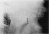
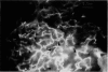
The severity of microangiopathy in patients with chronic venous insufficiency (CVI) determines the extent of the trophic disturbances of the skin. Resulting from valvular incompetence of deep and/or perforating veins and the accompanying venous outflow obstruction caused by deep venous thrombosis (DVT), the increased ambulatory venous pressure heads are transmitted retrograde into the microvasculature of the skin at the ankle region. In the present study, we have assessed the changes in the cutaneous microvasculature by dynamic fluorescence video microscopy, fluorescence microlymphography, and transcutaneous oxygen tension (tcPO2) measurements. In mild forms of CVI, capillary density, morphologic characteristics, and tcPO2 are still normal. Fluorescent light intensity is, however, significantly increased, indicating an increased transcapillary diffusion of sodium fluorescein (NaF) as a marker for enhanced leakage of the capillaries in the early stage of the disease. The pericapillary halo diameters are significantly enlarged, compared to controls (p < 0.01). In the severe stages of CVI and in patients with venous ulcers, capillary thromboses, probably caused by endothelium-blood cell interactions, may lead to a reduced capillary density. In order to enlarge the exchange surface area, the remaining skin capillaries become tortuous (capillary tufts). Parallel to the reduced capillary number, tcPO2 decreases and can be extremely low at the ulcer rim or at white atrophy spots. Fibrin cuffs are not a specific finding for venous ulceration and do not significantly impair oxygen diffusion. Fluorescence microlymphography permits visualization of the lymphatic capillaries of the superficial skin. In severe stages of CVI, the lymphatic capillary network at the medial ankle area is destroyed, and the remaining lymphatic capillary fragments have an increased permeability to FITC-dextran with a molecular weight of 150,000. These findings demonstrate a special lymphatic microangiopathy in CVI, suggesting an additional lymphatic component in the edema formation.
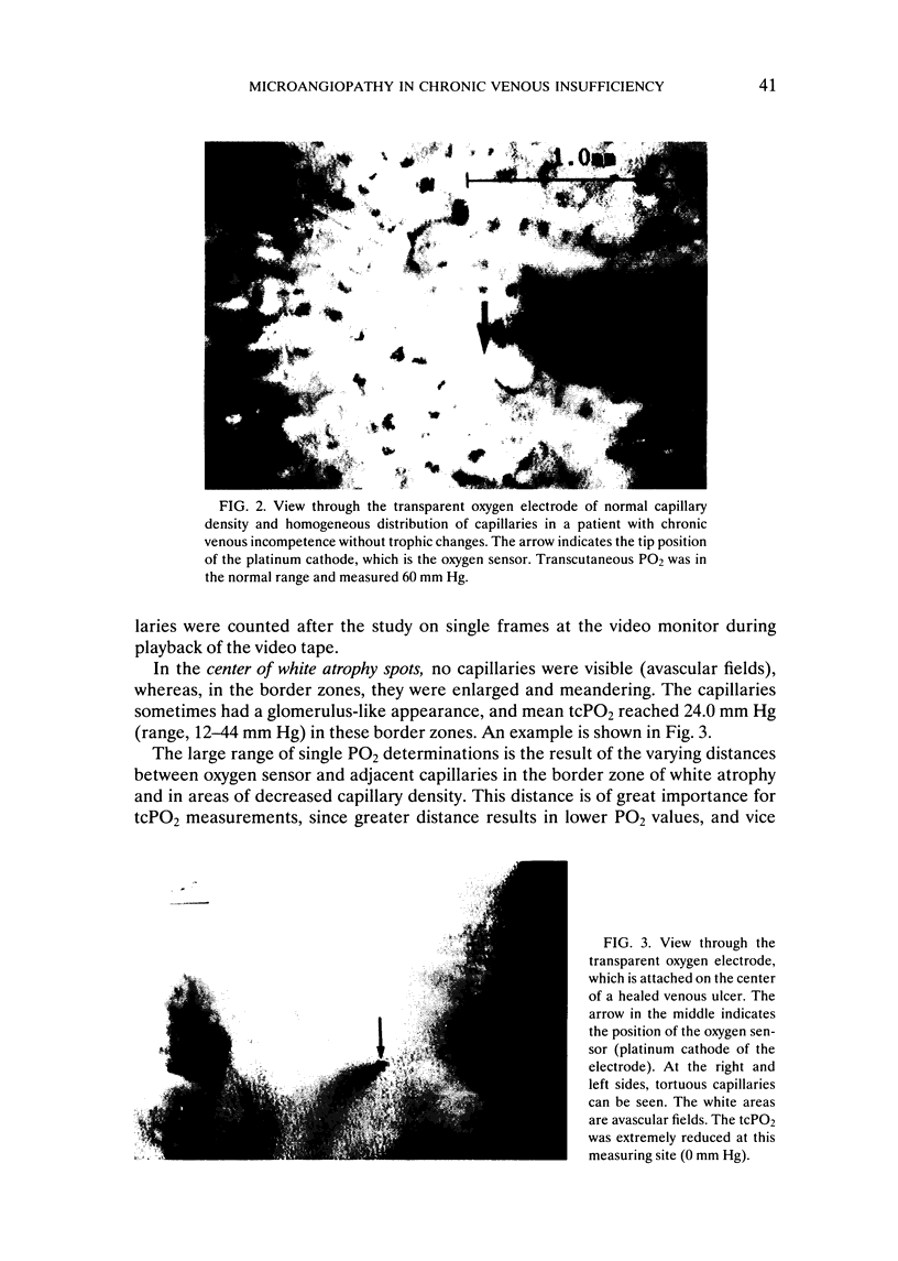
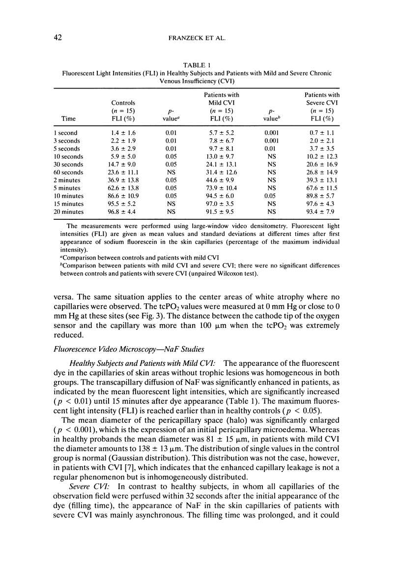
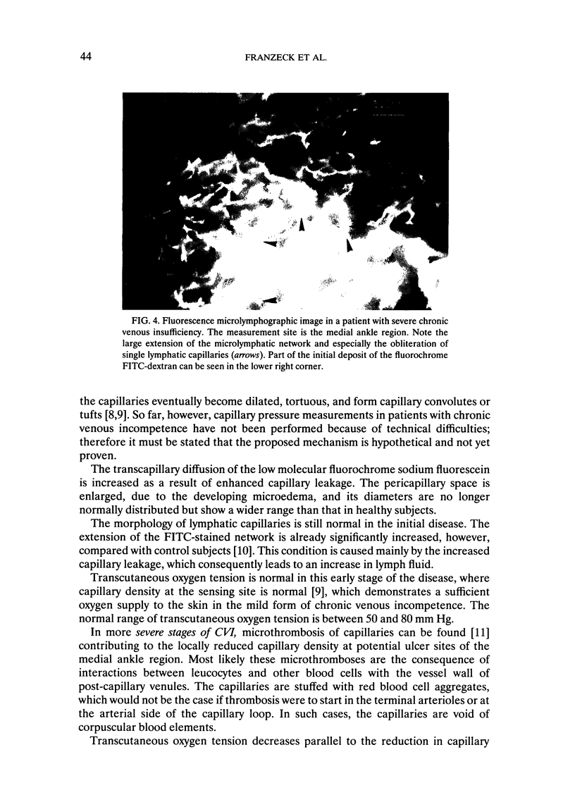
Full text
PDF
Images in this article
Selected References
These references are in PubMed. This may not be the complete list of references from this article.
- Bollinger A., Jäger K., Geser A., Sgier F., Seglias J. Transcapillary and interstitial diffusion of Na-fluorescein in chronic venous insufficiency with white atrophy. Int J Microcirc Clin Exp. 1982;1(1):5–17. [PubMed] [Google Scholar]
- Bollinger A., Jäger K., Roten A., Timeus C., Mahler F. Diffusion, pericapillary distribution and clearance of Na-fluorescein in the human nailfold. Pflugers Arch. 1979 Nov;382(2):137–143. doi: 10.1007/BF00584215. [DOI] [PubMed] [Google Scholar]
- Bollinger A., Jäger K., Sgier F., Seglias J. Fluorescence microlymphography. Circulation. 1981 Dec;64(6):1195–1200. doi: 10.1161/01.cir.64.6.1195. [DOI] [PubMed] [Google Scholar]
- Bollinger A., Leu A. J. Evidence for microvascular thrombosis obtained by intravital fluorescence videomicroscopy. Vasa. 1991;20(3):252–255. [PubMed] [Google Scholar]
- Burnand K. G., Whimster I., Naidoo A., Browse N. L. Pericapillary fibrin in the ulcer-bearing skin of the leg: the cause of lipodermatosclerosis and venous ulceration. Br Med J (Clin Res Ed) 1982 Oct 16;285(6348):1071–1072. doi: 10.1136/bmj.285.6348.1071. [DOI] [PMC free article] [PubMed] [Google Scholar]
- Franzeck U. K., Bollinger A., Huch R., Huch A. Transcutaneous oxygen tension and capillary morphologic characteristics and density in patients with chronic venous incompetence. Circulation. 1984 Nov;70(5):806–811. doi: 10.1161/01.cir.70.5.806. [DOI] [PubMed] [Google Scholar]
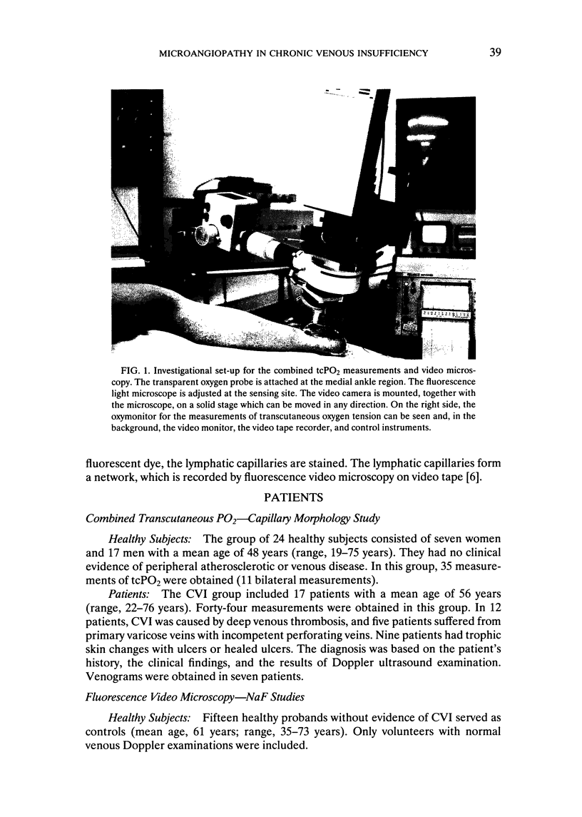
- Franzeck U. K., Isenring G., Frey J., Jäger K., Mahler F., Bollinger A. Eine Apparatur zur dynamischen intravitalen Videomikroskopie. Vasa. 1983;12(3):233–238. [PubMed] [Google Scholar]
- Franzeck U. K., Talke P., Bernstein E. F., Golbranson F. L., Fronek A. Transcutaneous PO2 measurements in health and peripheral arterial occlusive disease. Surgery. 1982 Feb;91(2):156–163. [PubMed] [Google Scholar]
- Huch A., Franzeck U. K., Huch R., Bollinger A. A transparent transcutaneous oxygen electrode for simultaneous studies of skin capillary morphology, flow dynamics and oxygenation. Int J Microcirc Clin Exp. 1983;2(2):103–108. [PubMed] [Google Scholar]
- Vanscheidt W., Stengele K., Wokalek H., Schöpf E. Perikapilläre Fibrinmanschetten--ein O2-Diffusionsblock? Vasa. 1991;20(2):142–146. [PubMed] [Google Scholar]