Abstract
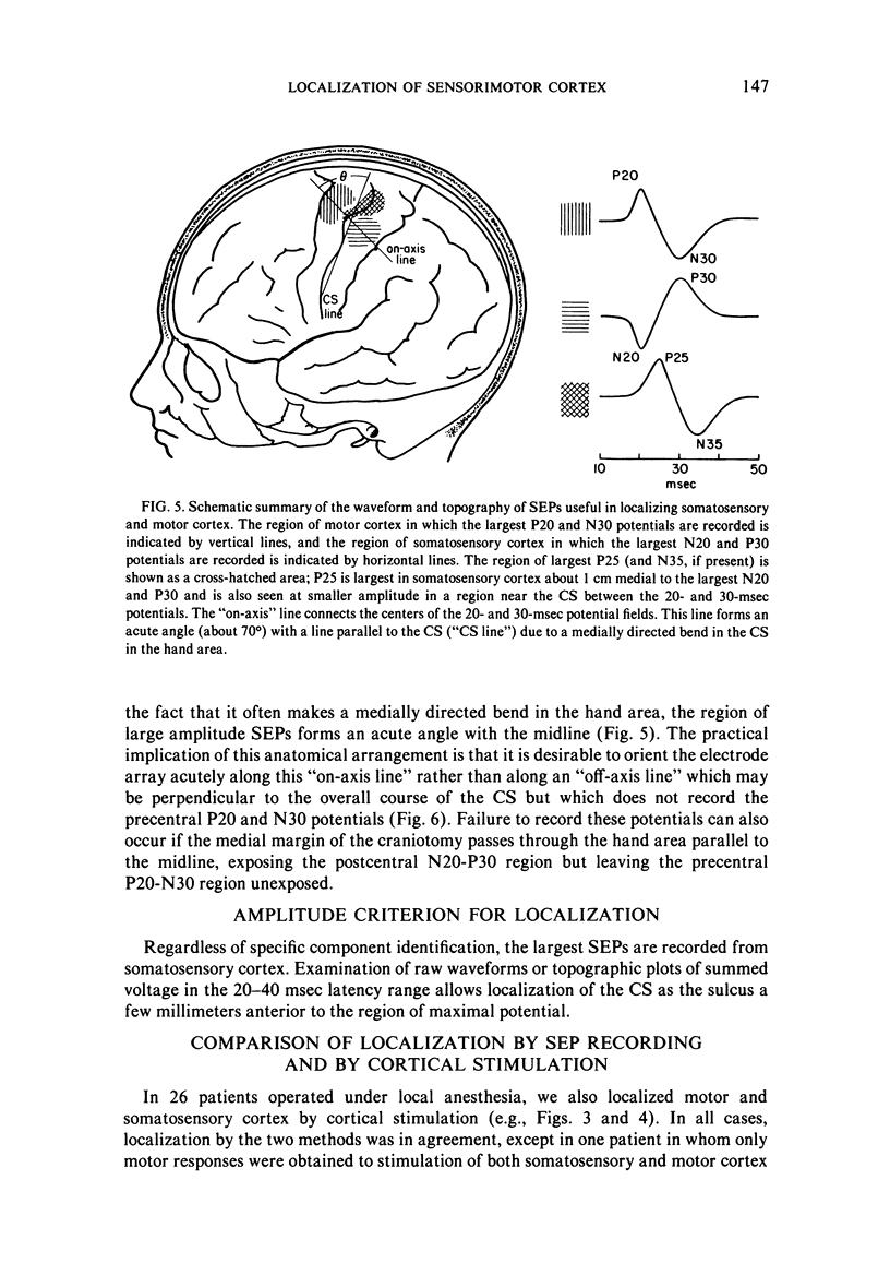
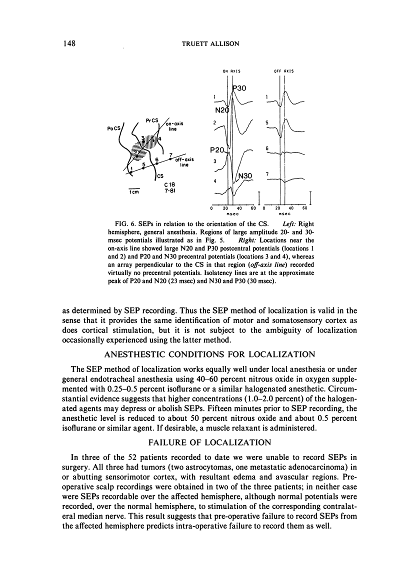
The current method of localizing somatosensory and motor cortex during neurosurgical removal of abnormal tissue is Penfield's method of cortical stimulation. While useful, this method has drawbacks, in particular the need to operate under local anesthesia. Another method of localization, described here, involves intra-operative recording of short-latency somatosensory evoked potentials to stimulation of the contralateral median nerve, from electrodes placed directly on central cortex. Proper localization involves identification of potentials which invert in polarity across the central sulcus, identification of other potentials which are largest in the medial portion of the hand area of somatosensory cortex and do not polarity invert, and determination of the region of maximal potential amplitude. This method of localization works equally well whether the patient is under local or general anesthesia, but it occasionally fails in patients with tumors abutting or invading the hand area of sensorimotor cortex.
Full text
PDF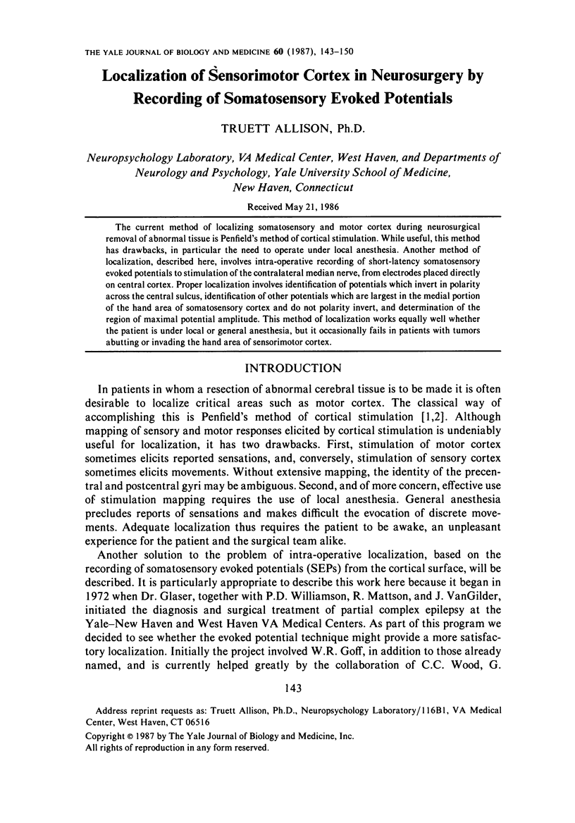
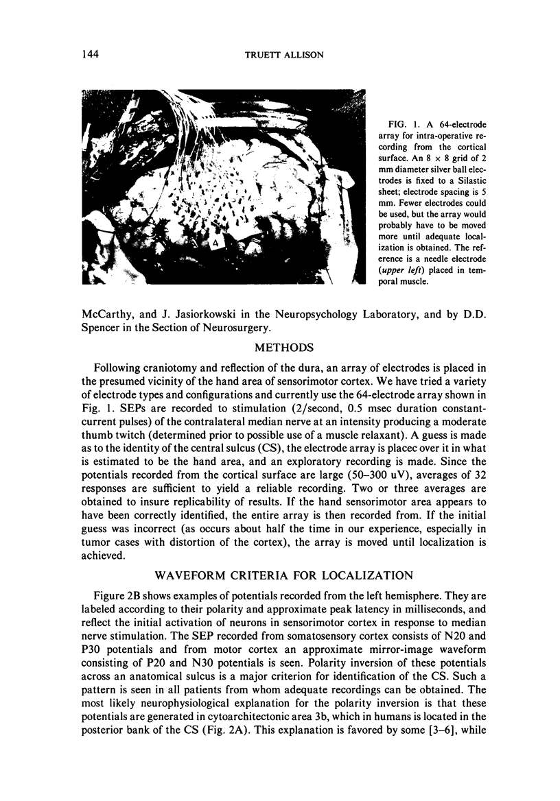
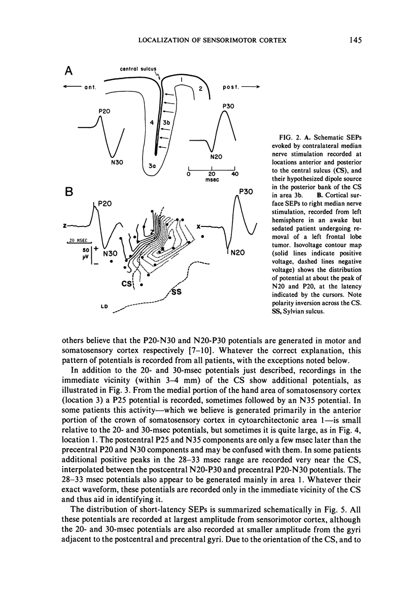
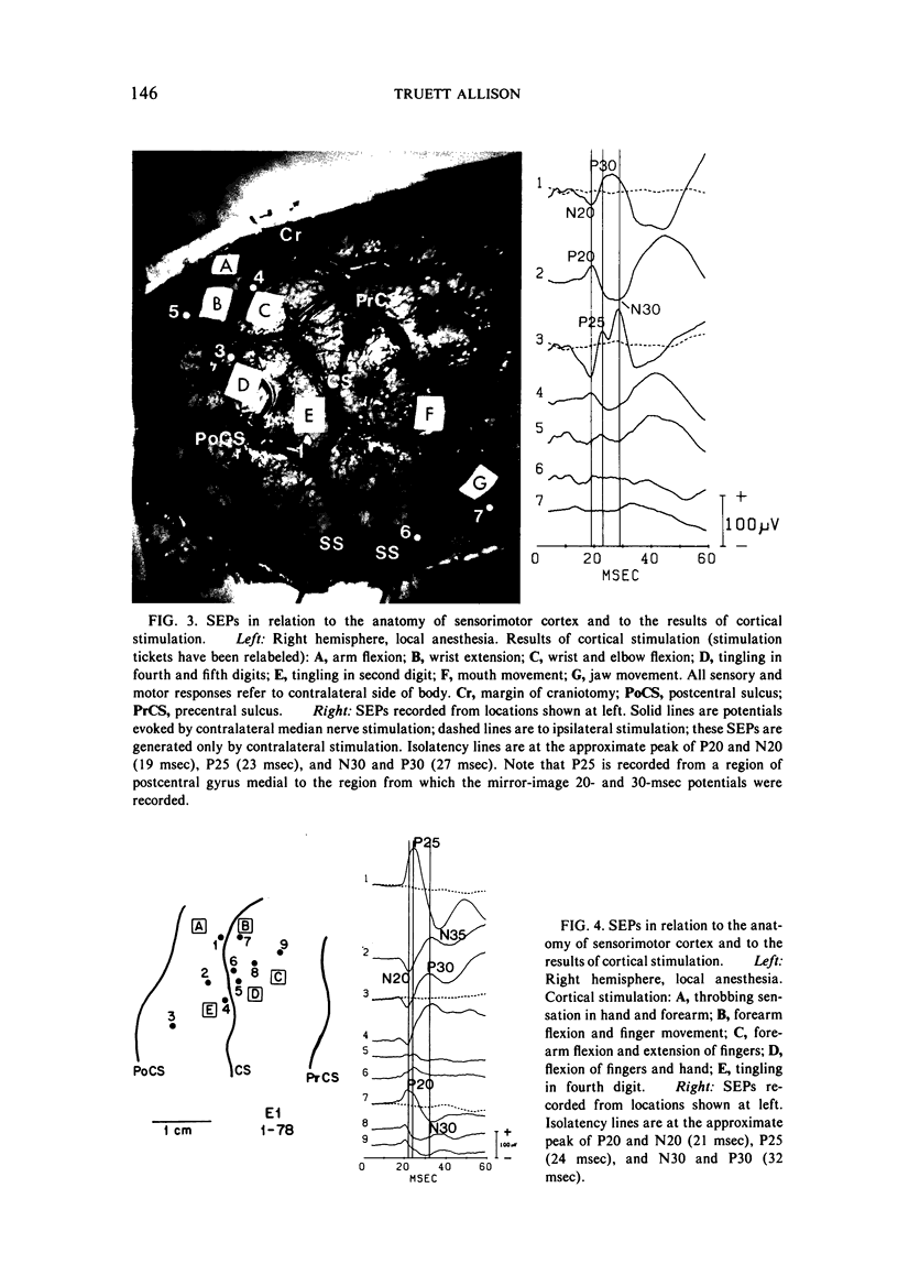
Images in this article
Selected References
These references are in PubMed. This may not be the complete list of references from this article.
- Allison T. Scalp and cortical recordings of initial somatosensory cortex activity to median nerve stimulation in man. Ann N Y Acad Sci. 1982;388:671–678. doi: 10.1111/j.1749-6632.1982.tb50833.x. [DOI] [PubMed] [Google Scholar]
- Broughton R., Rasmussen T., Branch C. Scalp and direct cortical recordings of somatosensory evoked potentials in man (circa 1967). Can J Psychol. 1981 Jun;35(2):136–158. doi: 10.1037/h0081151. [DOI] [PubMed] [Google Scholar]
- Desmedt J. E., Cheron G. Non-cephalic reference recording of early somatosensory potentials to finger stimulation in adult or aging normal man: differentiation of widespread N18 and contralateral N20 from the prerolandic P22 and N30 components. Electroencephalogr Clin Neurophysiol. 1981 Dec;52(6):553–570. doi: 10.1016/0013-4694(81)91430-9. [DOI] [PubMed] [Google Scholar]
- Goldring S., Gregorie E. M. Surgical management of epilepsy using epidural recordings to localize the seizure focus. Review of 100 cases. J Neurosurg. 1984 Mar;60(3):457–466. doi: 10.3171/jns.1984.60.3.0457. [DOI] [PubMed] [Google Scholar]
- Lueders H., Lesser R. P., Hahn J., Dinner D. S., Klem G. Cortical somatosensory evoked potentials in response to hand stimulation. J Neurosurg. 1983 Jun;58(6):885–894. doi: 10.3171/jns.1983.58.6.0885. [DOI] [PubMed] [Google Scholar]
- Maccabee P. J., Pinkhasov E. I., Cracco R. Q. Short latency somatosensory evoked potentials to median nerve stimulation: effect of low frequency filter. Electroencephalogr Clin Neurophysiol. 1983 Jan;55(1):34–44. doi: 10.1016/0013-4694(83)90144-x. [DOI] [PubMed] [Google Scholar]
- Yamada T., Kayamori R., Kimura J., Beck D. O. Topography of somatosensory evoked potentials after stimulation of the median nerve. Electroencephalogr Clin Neurophysiol. 1984 Feb;59(1):29–43. doi: 10.1016/0168-5597(84)90018-2. [DOI] [PubMed] [Google Scholar]


