Summary
In response to DNA damage in G2, mammalian cells must avoid entry into mitosis and instead initiate DNA repair. Here we show that in response to genotoxic stress in G2, the phosphatase Cdc14B translocates from the nucleolus to the nucleoplasm and induces the activation of the ubiquitin ligase APC/CCdh1, with the consequent degradation of Plk1, a prominent mitotic kinase. This process induces the stabilization of Claspin and Wee1, as the proteolysis of these two proteins requires phosphorylation by Plk1, and allows an efficient G2 checkpoint. As a by-product of APC/CCdh1 reactivation in DNA-damaged G2 cells, Claspin, which we show to be a novel substrate of APC/CCdh1 in G1, is targeted for degradation. However, this process is counteracted by the deubiquitylating enzyme Usp28 to permit Claspin-mediated activation of Chk1 in response to DNA damage. These findings define a novel pathway that is crucial for the G2 DNA damage response checkpoint.
Introduction
The Anaphase Promoting Complex or Cyclosome (APC/C) is a ubiquitin ligase that plays a crucial role in the regulation of mitosis and the G1 phase of the cell cycle (Peters, 2006). In early mitosis, APC/C is activated through binding to Cdc20, and in late M, Cdc20 is replaced by Cdh1, the second activator of APC/C. During G1, APC/CCdh1 remains active to ensure that certain positive regulators of the cell cycle do not accumulate prematurely. Then, at the G1/S transition, APC/CCdh1 is inactivated by phosphorylation to allow stabilization of its substrates and promote progression into S phase. Cdk1-cyclin A and Cdk2-cyclin A mediate the phosphorylation of Cdh1, resulting in the dissociation of Cdh1 from the APC/C core (Lukas et al., 1999; Mitra et al., 2006; Sorensen et al., 2001). Other mechanisms inhibiting APC/CCdh1 activity include Emi1 binding and degradation of both Cdh1 and Ubc10 (a ubiquitin conjugating enzyme that works with APC/C). During G2, both Cdh1 and Ubc10 reaccumulate, but APC/CCdh1 remains inactive due to CDK-dependent phosphorylation of Cdh1 and the presence of Emi1. In early mitosis, Emi1 is eliminated via the SCFβTrcp ubiquitin ligase, but the bulk of APC/CCdh1 remains inactive due to high Cdk1 activity. Ultimately, Cdh1 activation in anaphase involves Cdk1 inactivation by APC/CCdc20 and Cdh1 dephosphorylation. In yeast, this dephosphorylation is carried out by the Cdc14 phosphatase, but the mechanism in mammals remains unclear (D'Amours and Amon, 2004; Sullivan and Morgan, 2007).
Upon DNA damage, proliferating cells activate a regulatory signaling network to either arrest the cell cycle and enable DNA repair or, if the DNA damage is too extensive to be repaired, induce apoptosis (Bartek and Lukas, 2007; Harper and Elledge, 2007; Kastan and Bartek, 2004). The DNA damage response involves a number of factors that ultimately coordinate the spatiotemporal assembly of protein complexes at the site of DNA damage to initiate and maintain the checkpoint. Depending on the type of genotoxic stress, different checkpoint pathways are activated. UV and stalled replication forks activate the ATR-Chk1 pathway, whereas double-strand breaks result in the activation of both the ATM-Chk2 and the ATR-Chk1 pathways. After ATR is recruited to the site of damage, it phosphorylates and activates the effector kinase Chk1, a process requiring the mediator protein Claspin. Important downstream targets of Chk1 include p53 and Cdc25A, a transcription factor and an activator of Cdk1, respectively. Chk1-mediated phosphorylation induces the stabilization of p53 (with the consequent expression of the CDK inhibitor p21) and is required for the SCFβTrcp-mediated degradation of Cdc25A; thus, Chk1 activation results in the attenuation of Cdk1 activity, with the consequent inhibition of mitosis.
During the recovery from DNA replication and DNA damage stresses, the G2 checkpoint must be silenced. This process involves the degradation of Claspin via the SCFβTrcp ubiquitin ligase following the phosphorylation of Claspin by Plk1 (Mailand et al., 2006; Mamely et al., 2006; Peschiaroli et al., 2006). However, if DNA damage occurs during G2, SCFβTrcp-dependent ubiquitylation of Claspin is inhibited to re-establish the checkpoint. The blocking of Claspin ubiquitylation is at least partially due to the inhibition of Plk1, which occurs in response to DNA damage (Smits et al., 2000). In fact, Claspin is not phosphorylated on its degron and does not bind βTrcp in G2 cells that have been subjected to DNA damage (Peschiaroli et al., 2006). However, despite the lack of SCFβTrcp-Plk1-dependent ubiquitylation, Claspin continues to be ubiquitylated, only remaining stable due to a deubiquitylating enzyme (DUB), namely Usp28 (Zhang et al., 2006), suggesting the presence of an additional, unknown ligase, targeting Claspin after DNA damage.
In this study, we initially identified APC/CCdh1 as the ubiquitin ligase that targets Claspin in G1 and after DNA damage in G2. Since Usp28 stabilizes Claspin after genotoxic stresses, we wished to identify the proteins that are targeted by APC/CCdh1 in response to DNA damage. Furthermore, we studied the mechanisms leading to the activation of APC/CCdh1 during the DNA damage response checkpoint in G2. The results of these studies are presented herein.
Results
Claspin is a substrate of APC/CCdh1 in the G1 phase of the cell cycle
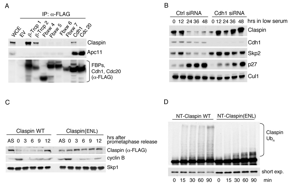
The levels of Claspin oscillate throughout the cell cycle (Mailand et al., 2006; Mamely et al., 2006; Peschiaroli et al., 2006). The highest expression levels are observed in S phase and early G2, and levels decrease thereafter, becoming almost undetectable during mitosis and the following G1 phase (Figure S1A). Levels of Claspin also decrease when cells withdraw from the cell cycle and enter quiescence (Figure S1B). Since SCFβTrcp mediates the degradation of Claspin degradation at G2/M, we asked whether this ligase is also responsible for Claspin degradation in G1. Therefore, we downregulated both βTrcp1 and βTrcp2 using an established siRNA oligo and analyzed Claspin levels in both M and G1. While silencing of βTrcp induced an accumulation of Claspin in prometaphase cells, no effect was visible in G1 cells (Figure S1C), suggesting that a ligase different from SCFβTrcp targets Claspin for degradation in G0 and G1. Thus, we screened for this G1 specific ubiquitin ligase, investigating the ability of Claspin to bind ubiquitin ligase subunits. We found that only Cdh1 was able to co-immunoprecipitate endogenous Claspin, whereas Fbxw4, Fbxw5, Fbxw6, Fbxw7, and Cdc20 (Figure 1A), as well as thirteen different F-box proteins (Peschiaroli et al., 2006), failed to bind Claspin.
Figure 1. Claspin is degraded in G0 and G1 via the APC/CCdh1 ubiquitin ligase.
(A) Cdh1 interacts with Claspinin vivo HEK293T cells were transfected with the indicated FLAG-tagged constructs or an empty vector (EV). Whole cell extracts (WCE) were immunoprecipitated (IP) with anti-FLAG resin, and immunocomplexes were probed with antibodies to the indicated proteins.
(B) T98G cells were transfected with a control (Ctrl) siRNA oligo or an siRNA oligo directed against Cdh1 mRNA. Cells were then switched to culture media containing 0.02% FBS to arrest them in G0/G1. Samples were collected at the indicated times after the beginning of the serum starvation and subjected to immunoblot analysis using antibodies to the indicated proteins. Skp2 and p27 were used as cell cycle markers.
(C) Claspin(ENL) is stable in G1. HeLa cells were infected with retroviruses expressing either FLAG-tagged wild type Claspin or FLAG-tagged Claspin(ENL) and subsequently treated for 16 hours with nocodazole to induce a mitotic block. Round, prometaphase cells were then collected by gentle shake-off and replated in fresh medium for the indicated times. Cells were harvested, and cell extracts were analyzed by immunoblotting with antibodies to the indicated proteins. Synchronization was monitored by flow cytometry and by the levels of cyclin B.
(D) Ubiquitin ligation assays of 35S-labeled, in vitro translated Claspin N-terminal fragments (amino acid 1–678) of either wild type Claspin or Claspin(ENL) were conducted in the presence of unlabeled, in vitro translated Cdh1. Samples were incubated at 30°C for the indicated times. The lower autoradiography image represents a short exposure time, and the upper image represents a long exposure time. The bracket on the right side of the top panel marks a ladder of bands corresponding to polyubiquitylated Claspin.
To investigate whether APC/CCdh1 has a role in the degradation of Claspin during G0/G1, we reduced the expression of Cdh1 in T98G cells using a previously validated siRNA oligo. T98G cells were serum starved following knockdown of Cdh1, and the expression levels of Claspin were analyzed at various time points thereafter. Downregulation of Cdh1 strongly inhibited the degradation of Claspin in cells progressively accumulating in G0/G1 (Figure 1B). Similarly, T98G cells released from a block in prometaphase retained Claspin expression throughout G1 phase upon silencing of Cdh1 (data not shown). Together, these data show a role for APC/CCdh1 in targeting Claspin for degradation during G0 and G1.
We also systematically mapped the Cdh1 binding motif of Claspin. A series of binding experiments using multiple Claspin deletion mutants narrowed the binding motif to an N-terminal region located between amino acids 79–102 (Figure S2A–D). Finally, we inserted five unique, triple-point-mutations to Alanine across Claspin amino acids 79–102, following the pattern of evolutionary conserved residues (Figure S2E), and assayed these mutants for binding to Cdh1. Both mutant #2 [Claspin(EEN)] and mutant #3 [Claspin(ENL)] failed to bind Cdh1 (Figure S2F). Thus, these studies identified the motif "EENxENL," located at residues 86–92 of Claspin, as the site mediating binding to Cdh1. Accordingly, in contrast to wild type Claspin, Claspin(ENL) was stable in HeLa cells progressing through G1 (Figure 1C).
To further support that Claspin is ubiquitylated via APC/CCdh1, we reconstituted the ubiquitylation of Claspin in vitro.The N-terminus of Claspin (amino acids 1–678) was efficiently ubiquitylated only when Cdh1 was present (Figure 1D and S3). In contrast, no Cdh1-dependent ubiquitylation of Claspin(ENL) was observed (Figure 1D). Similarly, the C-terminus of Claspin (amino acids 679–1333), lacking the Cdh1-binding domain, was not ubiquitylated despite the presence of Cdh1 (data not shown).
Thus, experiments in cell systems and in vitroshow that Cdh1 promotes the ubiquitylation and consequent degradation of Claspin in a manner that requires an intact Cdh1-interaction motif.
Upon DNA damage in G2, Usp28 protects Claspin, but not Plk1, fromAPC/CCdh1–mediated degradation
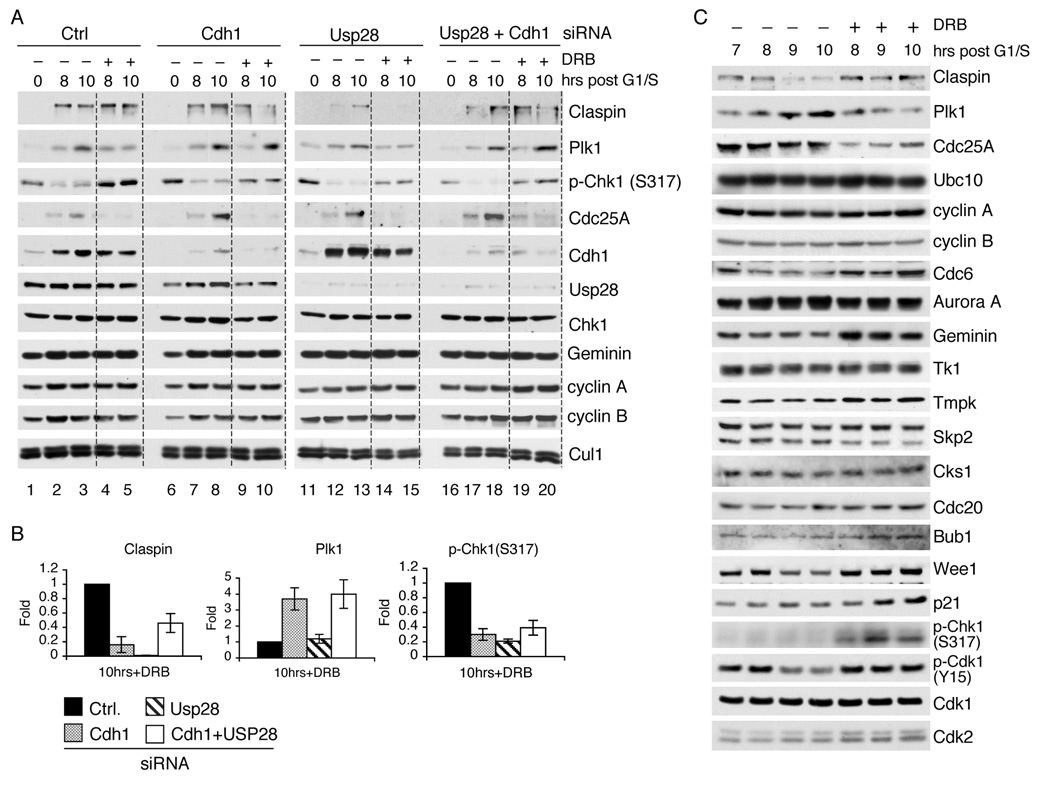
Usp28 deubiquitylates and consequently stabilizes Claspin in response to DNA damage (Zhang et al., 2006). After genotoxic stresses, the recognition of Claspin by the SCFβTrcp ubiquitin ligase is impaired due to the inhibition of Plk1 (Mailand et al., 2006; Mamely et al., 2006; Peschiaroli et al., 2006; Smits et al., 2000). Under these conditions, Claspin is continuously ubiquitylated via a ubiquitin ligase different from SCFβTrcp that had remained unidentified. Notably, a G2 phase-specific reactivation of APC/CCdh1 after DNA damage has been described in vertebrates (Sudo et al., 2001), but the reason for this reactivation is not known. We confirmed that Cdh1 re-associates with Cdc27 (an APC/C core subunit) in human G2 cells subjected to genotoxic stresses, and this APC/CCdh1 is active (Figure S4 and Figure S5). Thus, we decided to investigate whether APC/CCdh1 targets Claspin during G2 in response to DNA damage, which would explain the need for Usp28. To this end, we silenced the expression of Cdh1, Usp28, or both Cdh1 and Usp28 in U2OS cells using previously validated siRNA oligos. After transfection, cells were synchronized at G1/S and then allowed to progress through the cell cycle. Seven hours after the release from G1/S (when cells were in G2), cells were pulsed with doxorubicin for one hour to induce DNA damage and harvested at different times thereafter (Figure 2A,B). As expected, downregulation of Usp28 resulted in decreased levels of Claspin (Figure 2A, lanes 14 and 15). However, when both Usp28 and Cdh1 were silenced together, the expression of Claspin was partially restored (Figure 2A, lanes 19 and 20), indicating that Usp28 counteracts Cdh1-dependent degradation of Claspin.
Figure 2. In response to DNA damage in G2, APC/CCdh1 is reactivated to target Plk1 and Claspin (but the latter is protected by Usp28).
(A) U2OS cells were transfected with the indicated siRNA oligos and synchronized at G1/S using a double thymidine block. Cells were then released from the block to allow progression towards G2. At seven hours post release, cells were pulsed for one hour with either solvent (−) or doxorubicin (DRB) (+) and collected at the indicated times thereafter. Whole cell extracts were analyzed by immunoblotting with antibodies to the indicated proteins. Synchrony was verified by flow cytometry. To facilitate comparison, a gray line separates samples treated with doxorubicin from untreated samples.
(B) The graphs show the quantification of the levels of Claspin, Plk1, and Chk1 phosphorylated on Ser317 shown in (A) at the 10 hour timepoint averaged with two additional, independent experiments. The value given for the amount of protein present in the control sample two hours after the end of the doxorubicin pulse was set as 1 (n=3, ± SD).
(C) U2OS cells were synchronized and treated with DRB as described in (A). Samples were collected at the indicated times and processed for immunoblot analysis with antibodies to the indicated proteins.
If Claspin is protected from proteolysis by Usp28 in DNA-damaged G2 cells, what substrates are targeted for degradation by APC/CCdh1? To answer this question, we analyzed the levels of 15 G1 substrates of Cdh1 (Claspin, Plk1, Cdc25A, Ubc10, cyclin A, cyclin B, Cdc6, Aurora A, Geminin, Tk1, Tmpk, Skp2, Cks1, Cdc20, and Bub1). U2OS cells were synchronized in G2 and pulsed with doxorubicin, as described for Figure 2A. Samples were collected at different times thereafter and subjected to immunoblotting. We observed that the levels of Plk1 decreased in response to DNA damage (Figure 2C) in a manner similar to Cdc25A, whose degradation after genotoxic stresses is dependent on SCFβTrcp (Busino et al., 2003; Jin et al., 2003). The other 13 substrates of APC/CCdh1 remained unchanged or showed slight increases in their abundance (Figure 2C). The effect on Plk1 was also observed following treatment with ionizing radiation (Figure S6).
These results prompted us to investigate whether Plk1 degradation is dependent on the reactivated APC/CCdh1 complex. We found that levels of Plk1 were considerably reduced in the presence of doxorubicin at the 10-hour time point compared to the untreated sample (compare lane 3 to lane 5 in Figure 2A). In contrast, when cells were treated with Cdh1siRNA oligos, Plk1 levels remained unchanged despite the presence of DNA damage (compare lane 3 to lanes 10 and 20 in Figure 2A). Downregulation of Usp28 did not affect Plk1 levels or Plk1 half-life (Figure 2A and Figure S7), indicating that Usp28 does not oppose ubiquitylation of Plk1 as it does with Claspin.
As expected, knockdown of Cdh1 and/or Usp28 had no effect on Cdc25A (Figure 2A). Interestingly, compared to samples treated with control oligos, downregulation of Cdh1 induced a reduction in the levels of Claspin and in the activating phosphorylation of Chk1 on Ser317 (Figure 2A,B). This last result shows that APC/CCdh1 activity is necessary to sustain Claspin expression in response to genotoxic stress in G2. Since Plk1 promotes Claspin degradation, the stabilization of Plk1, which occurred in cells where Cdh1 expression was silenced, may explain why Claspin levels decrease under these conditions.
The above results show that APC/CCdh1 targets Claspin and Plk1 in response to DNA damage in G2 and that Usp28 selectively counteracts this action on Claspin. In addition, these results suggest that the APC/CCdh1-mediated degradation of Plk1 promotes Claspin stabilization and the consequent activation of Chk1.
Cdh1-dependent degradation of Plk1 is required for an efficient DNA damage-induced G2 checkpoint
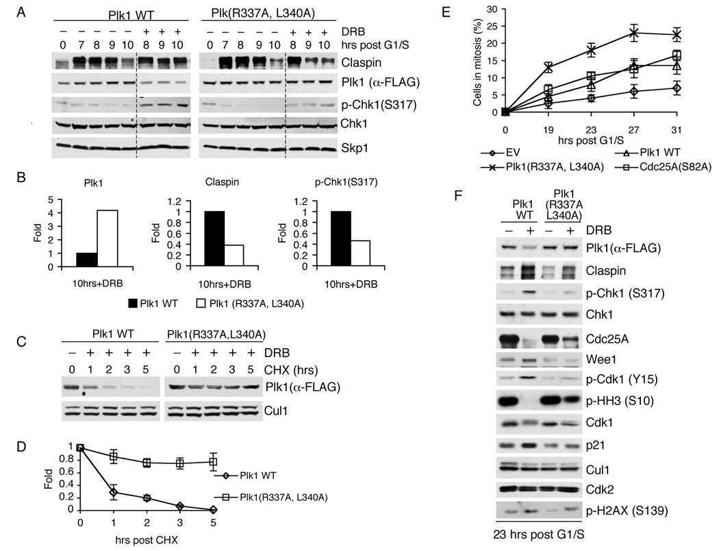
A destruction box in Plk1 is required for the APC/CCdh1-dependent degradation of Plk1 during late M and in G1 (Lindon and Pines, 2004). Based on this information, we generated a stable Plk1 mutant [Plk1(R337A,L340A)] to investigate the biological function of Plk1 degradation in response to DNA damage in G2. To this end, U2OS cells retrovirally infected with either wild type Plk1 or Plk1(R337A,L340A) were synchronized in G2, pulsed for one hour with doxorubicin, and followed for an additional three hour period. Plk1(R337A,L340A) was not degraded following treatment with doxorubicin, whereas a significant decrease in the levels of wild type Plk1 was observed (Figure 3A,B). The difference in Plk1 levels corresponded to the difference in the half-lives of wild type Plk1 versus Plk1(R337A,L340A) (Figure 3C,D). This finding shows that DNA damage-induced degradation of Plk1 in G2 is mediated by its Cdh1-binding site.
Figure 3. The DNA damage checkpoint in G2 requires Cdh1-dependent degradation of Plk1.
(A) U2OS cells retrovirally infected with the indicated Plk1 constructs were synchronized and treated with DRB as described in (2A). Cells were then collected at the indicated times and immunoblotted with antibodies to the indicated proteins. To facilitate comparison, a gray line separates samples treated with doxorubicin from untreated samples.
(B) The graphs show the quantification of the levels of Claspin, Plk1, and Chk1 phosphorylated on Ser317 shown in (A) at the 10 hour timepoint averaged with an additional, independent experiment. The value given for the amount of protein present in the control sample two hours after the end of the doxorubicin pulse was set as 1 (n=2).
(C) U2OS cells were infected, synchronized, and pulsed with doxorubicin as in (A), except that cycloheximide (CHX) was added seven hours after release from G1/S. At different times after the addition of CHX, cells were collected, and cell lysates were immunoblotted with antibodies to the indicated proteins.
(D) The graph shows the quantification of the levels of wild type Plk1 and Plk1(R337A,L340A) shown in (C) averaged with two additional, independent experiments (n=3, ± SD).
(E) U2OS cells infected with either an empty virus (EV) or retroviruses encoding wild type Plk1, Plk1(R337A,L340A), or Cdc25A(S82A) were synchronized and treated with DRB as described in (A). Thereafter, cells were incubated in fresh medium containing nocodazole to trap cells in mitosis. Samples were then collected at the indicated times, and the percentage of mitotic cells was monitored by immunodetection of Histone H3 phosphorylated on Ser10 using flow cytometry (n=3, ± SD).
(F) U2OS cells treated as in (A) were collected 23 hours after release from G1/S (16 hours after the doxorubicin pulse), and then cell lysates were immunoblotted with antibodies to the indicated proteins.
Importantly, following DNA damage, the lack of Plk1(R337A,L340A) degradation was associated with decreased levels of Claspin and reduced phosphorylation of Chk1 (Figure 3A,B), similar to what is observed when Cdh1 is silenced (Figure 2A,B). Since Plk1 initiates the SCFβTrcp-dependent degradation of Claspin following recovery from DNA damage stresses, these data argue for a role of Cdh1 in limiting Plk1 levels to preserve Claspin stability and Chk1 activation in response to DNA damage in G2.
To investigate the impact of stable Plk1 on the maintenance of the checkpoint, U2OS cells treated as in Figure 3A were incubated with nocodazole (to trap cells in mitosis) and followed for longer time points (up to 31 hours after release from G1/S,i.e. 24 hours after the pulse with doxorubicin) (Figure 3E). In addition to cells expressing wild type Plk1 or Plk1(R337A,L340A), this experiment included cells expressing a stable Cdc25A(S82A) mutant, which is known to impair the checkpoint response to DNA damage (Busino et al., 2003). Mitotic entry was determined by immunostaining for Ser10 phosphorylated Histone H3. Cells infected with an empty viral vector showed only marginal mitotic entry, indicative of an intact G2 checkpoint, while cells expressing wild type Plk1 or Cdc25A(S82A) displayed an increase in cells entering mitosis. Strikingly, mitotic entry was significantly more pronounced in cells expressing Plk1(R337A,L340A), indicating that Cdh1-mediated degradation of Plk1 is a critical step to establish and maintain the DNA damage checkpoint in G2. The relatively moderate elevation of mitotic entry observed when wild type Plk1 was expressed is likely due to the increased demand on the proteolytic machinery to degrade the excess of Plk1. Importantly, the expression of a stable Claspin mutant (lacking the βTrcp degron) prevented Plk1(R337A,L340A) from bypassing the checkpoint (Figure S8), confirming that the induced degradation of Claspin is a major mechanism for Plk1 to induce premature mitosis.
To elucidate the molecular mechanisms by which cells expressing Plk1(R337A,L340A) are able to bypass this checkpoint, cell extracts were also analyzed by immunoblotting (Figure 3F). All major events downstream from Plk1, which ultimately lead to the activation of Cdk1 and entry into mitosis, were observed in DNA-damaged cells expressing the stable Plk1 mutant but not wild type Plk1. Figure 3F shows that compared to wild type Plk1 (lane 2), Plk1(R337A,L340A) expression (lane 4) resulted in: (i) degradation of Claspin, with the consequent reduction in activating phosphorylation of Chk1; (ii) degradation of the Cdk1 inhibitor Wee1 [another SCFβTrcp substrate, whose degradation is promoted by Plk1 (Watanabe et al., 2004)]; (iii) elevated Cdc25A levels (likely due to low Chk1 activity); (iv) dephosphorylation of Cdk1 on Tyr15 (likely due to low levels of Wee1 and high Cdc25A expression); (v) lower p21 levels; and (vi) phosphorylation of Histone H3 on Ser10. Thus, cells entered mitosis despite the presence of DNA damage, visualized by the presence of H2AX phosphorylation (bottom panel in Figure 3F and S9).
Therefore, based on these observations, we concluded that Cdh1-driven degradation of Plk1 in response to DNA damage is vital to maintain an efficient G2 checkpoint.
Cdc14B relocates to the nucleus and associates with Cdh1 in G2 in a DNA-damage-dependent manner
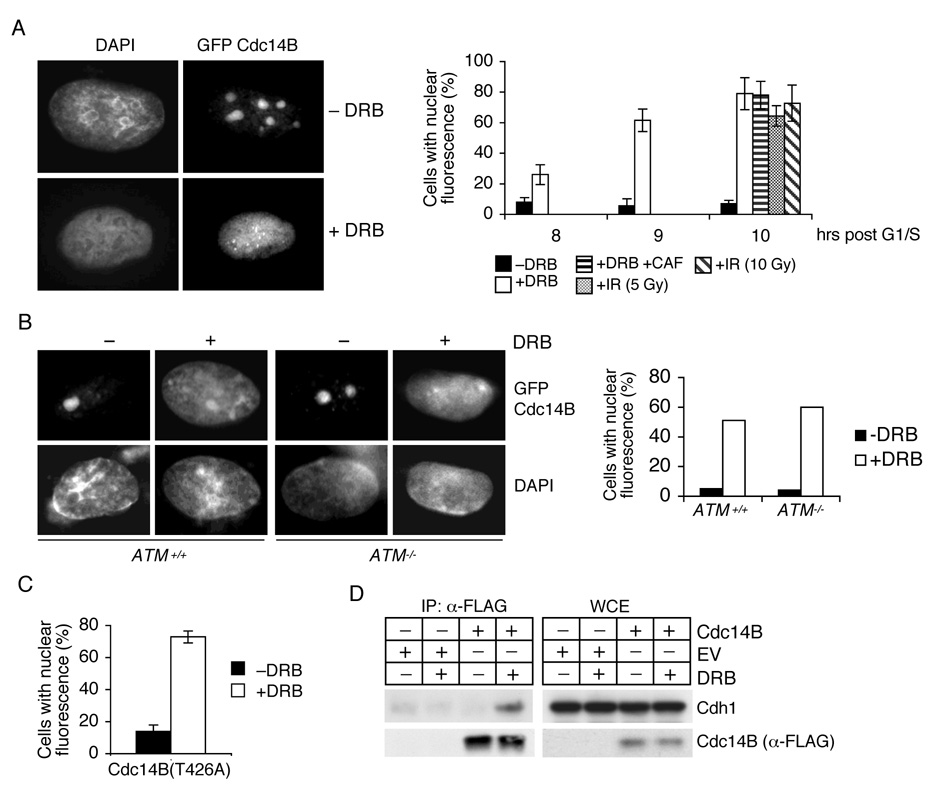
To be able to associate with and activate APC/C in late mitosis, Cdh1 must be dephosphorylated at sites that were phosphorylated by CDKs during S and G2 (Lukas et al., 1999; Mitra et al., 2006; Sorensen et al., 2001). Our data indicates that APC/CCdh1 is reactivated in G2 following DNA damage to target Plk1; therefore, we reasoned that a phosphatase may be required to remove the inhibitory phosphates from Cdh1. Studies in yeast have shown that Cdc14 dephosphorylates Cdh1 in late mitosis (D'Amours and Amon, 2004; Sullivan and Morgan, 2007). Two Cdc14 paralogs exist in mammals: Cdc14A and Cdc14B. Whereas Cdc14A is localized to the centrosomes, Cdc14B localizes to the nucleolus (Cho et al., 2005; Mailand et al., 2002). As a first step to study the possibility that one of the Cdc14 phosphatases may dephosphorylate and activate Cdh1 in G2 phase in response to a genotoxic stress, we investigated their subcellular localizations after treatment with doxorubicin or ionizing radiation. As expected, Cdc14B was initially localized to the nucleolus; however, following DNA damage, it moved to the nucleoplasm (Figure 4A). In contrast, no change in subcellular localization was observed for Cdc14A (data not shown).
Figure 4. Cdh1 associates with Cdc14B in G2 in response to DNA damage.
(A) Cdc14B moves from the nucleolus to the nucleoplasm in response to DNA damage. U2OS cells transfected with a construct expressing GFP-tagged Cdc14B were synchronized as described in (2A) and then either treated with DRB [+/−caffeine (CAF)] or subjected to ionizing radiations (IR). Cells were then collected and analyzed by direct immunofluorescence. Left panels: micrographs of representative cells showing the subcellular localization of Cdc14B. Right panel: Quantification of cells with nuclear Cdc14B fluorescence at the indicated times post pulse with doxorubicin (n=3, ± SD).
(B) Cdc14B translocation to the nucleus is independent of ATM. The experiment was performed as in (A), except that asynchronous ATM+/+ and ATM−/− fibroblasts were used (n=2).
(C) The experiment was performed as in (A), except that GFP-tagged Cdc14B(T426A) was used (n=3).
(D) Cdh1 binds to Cdc14B in a DNA damage-dependent manner. U2OS cells infected with either an empty retrovirus (EV) or viruses encoding FLAG-tagged Cdc14B were synchronized and treated with DRB as described in (A). Cells were collected two hours later, and whole cell extracts (WCE) were immunoprecipitated (IP) with anti-FLAG resin. The indicated proteins were detected by immunoblotting.
Interestingly, the nuclear localization of Cdc14B did not depend on ATM, as relocalization from the nucleolus was also observed in the presence of caffeine (Figure 4A) and in ATM-/- fibroblasts (Figure 4B). Human Cdc14B has three ATM phosphorylation consensus sites (one SQ and two TQs), two of which are not conserved in many other mammals. In contrast, the third site (Thr426) is conserved in all vertebrates. We mutated Thr426 to Ala and found that Cdc14B(T426A) continued to move to the nucleus after DNA damage (Figure 4C), confirming that ATM does not play a role in Cdc14B translocation.
Next, to test whether Cdc14B and Cdh1 physically interact in response to DNA damage in G2, we performed co-immunoprecipitation experiments in G2 U2OS cells. Remarkably, an interaction between the two proteins was only observed after treatment with doxorubicin (Figure 4D and Figure S10).
Thus, during the G2 DNA damage response, Cdc14B moves to nucleus and binds Cdh1.
Cdc14B activates APC/CCdh1 after DNA damage in G2
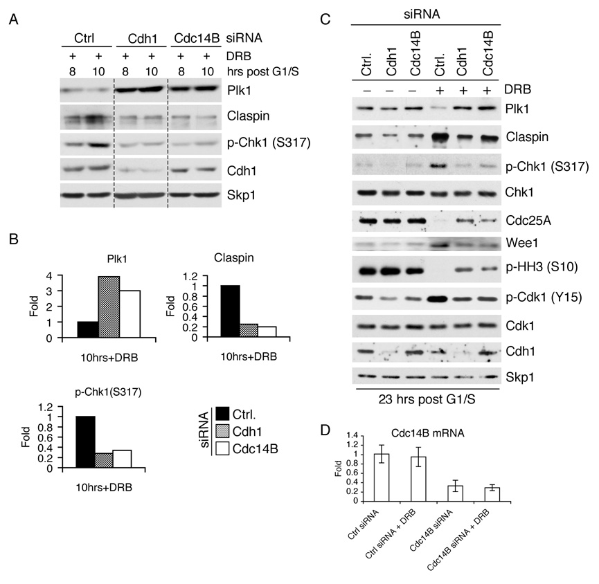
The finding that Cdc14B and Cdh1 interact in a DNA damage-dependent manner in G2 indicated that Cdc14B is involved in the reactivation of APC/CCdh1. To further test this hypothesis, we investigated the effect of silencing Cdc14B. U2OS cells were transfected with previously validated siRNA oligos directed against eitherCdc14B or Cdh1 and then synchronized. Upon reaching G2 (seven hours after release from G1/S), cells were pulsed with doxorubicin and collected at different times points thereafter (Figure 5A). Strikingly, silencing of Cdc14B resulted in the stabilization of Plk1 to an extent comparable to that obtained from silencing Cdh1 (Figure 2A and 5A,B). Moreover, reduced levels of Claspin and impaired phosphorylation of Chk1 at Ser317 were observed in the Cdc14B knockdown samples, suggesting that the elevated Plk1 levels produced an effect on downstream targets. Thus, the presence of Cdc14B appears vital for Plk1 degradation in G2 upon genotoxic stress.
Figure 5. DNA damage-dependent reactivation of APC/CCdh1 in G2 requires Cdc14B.
(A) U2OS cells transfected with the indicated siRNA oligos were synchronized and treated with DRB as described in (2A). Cells were collected at the indicated times, and cell lysates were analyzed by immunoblotting with antibodies to the indicated proteins.
(B) The graphs show the quantification of Claspin, Plk1, and Chk1 phosphorylated on Ser317 shown in (A) at the 10 hour timepoint averaged with an additional, independent experiment. The value given for the amount of protein present in the control sample two hours after the end of the doxorubicin pulse was set as 1 (n=2).
(C) U2OS cells, transfected with the indicated siRNA oligos, were treated as in (A). Twenty-three hours after release from G1/S (16 hours after the doxorubicin pulse), cells were collected, and cell lysates were analyzed by immunoblotting with antibodies to the indicated proteins.
(D)Cdc14B mRNA levels of cells used in (A) and (C) were analyzed eight hours after release from G1/S using real time PCR in triplicate measurements (± SD). The value given for the amount of Cdc14B mRNA present in the sample treated with control oligos was set as 1.
To investigate the impact of silencing Cdc14B on the maintenance of the G2 checkpoint, U2OS cells treated as in Figure 5A were followed for a longer time course (up to 23 hours after release from G1/S). Even at these late time points, Plk1 was stabilized when Cdc14B was downregulated (Figure 5C and S11). Importantly, similar to what was observed when the stable Plk1 mutant was expressed (Figure 3F), events downstream of Plk1, which lead to entry into mitosis, were observed in the Cdc14B knockdown samples (Figure 5C,D): increased degradation of Claspin and Wee1, reduction in the activating phosphorylation of Chk1, elevated Cdc25A levels, reduction in the inactivating phosphorylation of Cdk1, and phosphorylation of Histone H3 on Ser10.
Thus, Cdc14B, similar to Cdh1, is critical to establish and maintain the DNA damage checkpoint in G2. Interestingly, while downregulation of Cdc14B induced checkpoint bypass, the combination ofCdh1 and Cdc14B siRNAs had no additional effect (Figure S12), showing that both proteins function in the same pathway.
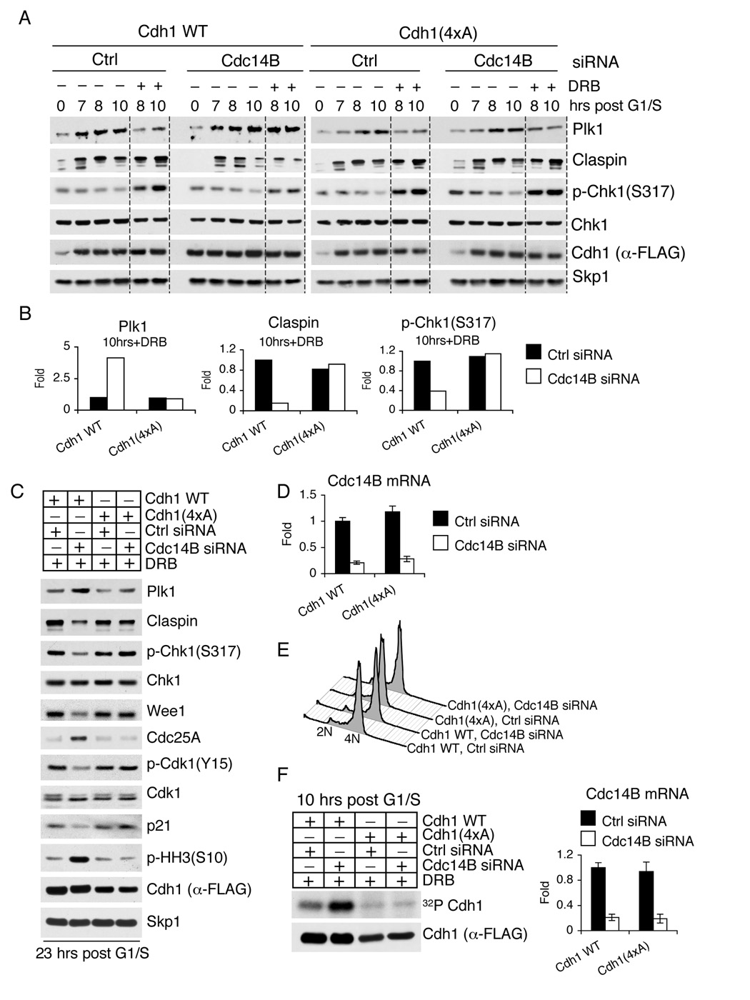
The phosphatase activity of Cdc14B towards Cdh1 is predicted to occur on sites that are phosphorylated by CDKs. The four sites phosphorylated in Cdh1 by CDKs during S and G2 have been previously described (Lukas et al., 1999; Sorensen et al., 2001). To further investigate the role of Cdc14B in activating Cdh1 in G2 upon DNA damage, U2OS cells were infected with retroviruses expressing either wild type Cdh1 or Cdh1(4xA), a constitutively active, phosphorylation-deficient Cdh1 mutant, in which the four CDK phosphorylation sites are mutated to Alanine. Subsequently, these cells were subjected to experiments identical to those shown in Figure 5. As in non-infected cells, the silencing of Cdc14B in cells expressing wild type Cdh1 resulted in the stabilization of Plk1 in response to DNA damage and in downstream events regulated by Plk1 (Figure 6A–E). In marked contrast, silencing of Cdc14B did not stabilize Plk1 in cells expressing Cdh1(4xA) (Figure 6A–E and S13). Consequently, Claspin was not degraded, and Chk1 was efficiently phosphorylated upon DNA damage. Moreover, despite the downregulation of Cdc14B, cells expressing Cdh1(4xA) displayed high levels of Wee1, inactivating phosphorylation of Cdk1, high levels of p21, and low levels of phosphorylation of Histone H3 on Ser10 (Figure 6C, compare lane 2 and 4), showing that in the presence of a constitutively active Cdh1 mutant, Cdc14B becomes dispensable for sustaining the G2 checkpoint.
Figure 6. A constitutively active Cdh1 mutant is refractory to the silencing of Cdc14B.
(A) U2OS cells were retrovirally infected with either wild type Cdh1 or Cdh1(4xA) mutant and subsequently transfected with the indicated siRNA oligos. Cells were then synchronized and treated with DRB as described in (2A), collected at the indicated times, and immunoblotted with antibodies to the indicated proteins. To facilitate comparison, a gray line separates samples treated with doxorubicin from untreated samples.
(B) The graphs show the quantification of the levels of Claspin, Plk1, and Chk1 phosphorylated on Ser317 shown in (A) at the indicated timepoint averaged with an additional, independent experiment. The value given for the amount of protein present in the control sample two hours after the end of the doxorubicin pulse was set as 1 (n=2).
(C) U2OS cells expressing wild type Cdh1 or Cdh1(4xA) were transfected with the indicated siRNA oligos and treated as in (A). Twenty-three hours after release from G1/S (16 hours after the doxorubicin pulse), cells were collected, and cell lysates were analyzed by immunoblotting with antibodies to the indicated proteins.
(D)Cdc14B mRNA levels of cells used in (A) and (C) were analyzed eight hours after release from G1/S using real time PCR in triplicate measurements (± SD). The value given for the amount of Cdc14B mRNA present in the sample expressing wild type Cdh1 and treated with control oligos was set as 1.
(E) Synchrony in G2 was ascertained by flow cytometry for cells used in (A) at the time of doxorubicin treatment. Identical synchrony in G2 was obtained for cells used in (C).
(F) The experiment was performed as in (A), except that an in vivo labeling with 32P-orthophosphate was performed during the last three hours before cells were collected. Cdh1 was then immunoprecipitated under denaturing conditions, resolved by SDS-PAGE, and visualized by autoradiography (upper panel) or immunoblotting (bottom panel). Right panel shows Cdc14B mRNA levels analyzed by real time PCR.
Importantly, after DNA damage, thein vivo phosphorylation of wild type Cdh1 - but not that of Cdh1(4xA) - increased after Cdc14B silencing (Figure 6F), indicating that in response to genotoxic stress, Cdc14B dephosphorylates Cdh1 on the four sites phosphorylated by Cdk2.
These results indicate that upon DNA damage in G2, Cdh1 is the relevant target of Cdc14B and that CDK-mediated phosphorylation of Cdh1 is the relevant constraint that is removed by Cdc14B.
The Cdc14B-Cdh1-Plk1 axis is deregulated in human tumors
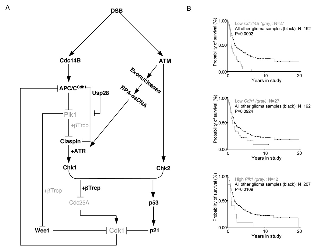
Our results show that the Cdc14B-Cdh1-Plk1 axis controls the G2 DNA damage checkpoint (see model in Figure 7A). To investigate a potential involvement of this pathway in human cancer, we searched the Oncomine database for the differential expression ofCdc14B, Cdh1, andPlk1in normal versus tumor tissues. The expression of Cdc14B(always) and Cdh1 (in most cases), was significantly decreased in many common tumors, such as breast, prostate, ovary, liver, and brain tumors (Figure S14 and Figure S15). Conversely, levels of Plk1 were consistently and significantly increased in most tumors (Figure S16). Importantly, the decrease in Cdc14B expression and the increase in Plk1 levels were significantly correlated with the grade of breast cancer. Finally, downregulation ofCdc14B and Cdh1 and/or upregulation of Plk1 correlated with survival in glioma patients (Figure 7B). These results suggest that a failure in the Cdc14B-Cdh1-Plk1 axis might impair the G2 DNA damage checkpoint, with the consequent acquisition of genomic instability.
Figure 7. The Cdc14B-Plk1-Cdh1 axis controls the DNA damage response in G2 and is deregulated in human tumors.
(A) A model of the G2 DNA damage response checkpoint. Black signifies activated forms of the respective proteins, and gray indicates inactive forms or degraded proteins. After induction of double strand breaks (DSB) in G2, ATM activates Chk2. In addition, ATM activates certain exonucleases to induce DSB resection, resulting in RPA coated, single-stranded DNA (ssDNA), which contributes to the recruitment of ATR. In parallel, Cdc14B is released from the nucleolus to the nucleoplasm, activating APC/CCdh1, which in turn targets Plk1 for proteasomal degradation. Because of the low levels of Plk1, phosphorylation of Claspin and Wee1 is reduced, preventing βTrcp-mediated degradation. Claspin is protected from APC/CCdh1-mediated degradation by Usp28. Stable Claspin promotes the ATR-mediated activation of Chk1, which, together with Chk2, targets Cdc25A (inducing its degradation) and p53 (promoting its stabilization and consequent induction of p21). As a result, Cdk1 activity is attenuated, and cells arrest in G2. Stable Wee1 contributes to this inhibition by directly phosphorylating Cdk1. The reduction in Cdk1 activity further removes the constraints on APC/CCdh1 activity.
(B) Poor patient survival correlates with low levels of Cdc14B and Cdh1 and high levels of Plk1 (gray).Rembrandt glioma database (http://rembrandt.nci.nih.gov)-generated Kaplan-Meier survival curves of 219 patients with gliomas of all histological grades grouped by gene expression levels of Cdc14B (top plot), Cdh1 (middle plot), and Plk1 (bottom plot). The associated P values are shown for each plot.
Discussion
Following genotoxic stresses, Claspin promotes ATR-mediated phosphorylation and activation of Chk1 (Harper and Elledge, 2007). In G2, during the recovery from DNA replication stress or DNA damage, Claspin is phosphorylated by Plk1 and consequently degradade via SCFβTrcp (Mailand et al., 2006; Mamely et al., 2006; Peschiaroli et al., 2006). This process contributes to turning off the checkpoint to promote the entry of cells into mitosis. SCFβTrcp-dependent ubiquitylation of Claspin remains active at least until prometaphase; however, Claspin reaccumulates only at the G1/S transition of the next cell cycle, suggesting that Claspin degradation is active during G1.
Several substrates degraded via SCF ligases in specific phases of the cell cycle are eliminated by the means of APC/C at different times. For example, p21 and Mll are degraded at G1/S via SCFSkp2 and at G2/M via APC/CCdc20 (Amador et al., 2007; Liu et al., 2007). Similarly, Cdc25A, whose degradation is mediated by SCFβTrcp during S, is eliminated in G1 via APC/CCdh1 (Busino et al., 2003; Donzelli et al., 2002; Jin et al., 2003). Herein we show that, in contrast to degradation of Claspin in G2 via SCFβTrcp, Claspin proteolysis in G1 cells is directed by APC/CCdh1 (Figure 1, Figure S1, Figure S2, and Figure S3).
APC/CCdh1 maintains the G0/G1 state by ensuring that many positive regulators of S and M do not accumulate prematurely, a function critical to genome stability (Guardavaccaro and Pagano, 2006; Peters, 2006). Claspin plays a poorly understood, positive role in DNA replication, which appears distinct from its role in checkpoint signaling. In fact, not only does expression of Claspin represent a reliable marker of cell proliferation in both human cancer and normal tissues (Tsimaratou et al., 2007), but Claspin overexpression has also been shown to increase cell proliferation (Lin et al., 2004). Our finding suggests that Claspin represents another substrate that APC/CCdh1 keeps at low levels during G1 to avoid premature entry into S.
This initial finding proved to be an invaluable portal to gain entry into the signaling network that mediates the G2 checkpoint, starting from the following observation. Upon DNA damage in G2, Claspin is no longer phosphorylated on its degron, blocking recognition by βTrcp (Peschiaroli et al., 2006). Therefore, why is Usp28 required to deubiquitylate and stabilize Claspin upon DNA damage (Zhang et al., 2006) (see model in Figure S17)? In answer to this question, we show here that APC/CCdh1 is reactivated in response to DNA damage (Figure S4 and Figure S5), thereby inducing ubiquitylation of Claspin, which is neutralized by Usp28 (Figure 2A,B).
This observation prompted a novel question. If Claspin is ubiquitylated in response to DNA damage as a byproduct of APC/CCdh1 activation but is protected from degradation by Usp28, what are the relevant substrate(s) that are degraded via APC/CCdh1 under these conditions? We surveyed 15 substrates that are known to be targeted by this ligase in G0/G1 and found that only two were downregulated: Plk1 and Cdc25A (Figure 2C). The latter is known to be degraded via SCFβTrcp after genotoxic stresses (Busino et al., 2003; Jin et al., 2003), and, in fact, its levels did not promptly increase when Cdh1 was silenced (Figure 2A). In contrast, Plk1 degradation was abolished when Cdh1 levels were lowered by siRNA (Figure 2A,B and Figure 5A–C), showing that Cdh1 promotes Plk1 degradation. The lack of Claspin phosphorylation and consequent stabilization (Peschiaroli et al., 2006) is attributed to the inactivation of Plk1 that occurs after DNA damage (Smits et al., 2000), but how Plk1 is inactivated had remained largely unknown (see model in Figure S17). We show that Plk1 is degraded via APC/CCdh1 following DNA damage. Downregulation of Cdh1 or the expression of a stable Plk1 mutant both resulted in the downregulation of Claspin in G2 DNA-damaged cells (Figure 2A,B, Figure 3A,F and Figure 5A,C). Thus, APC/CCdh1 exerts a dual control on Claspin stability: during G1, it promotes Claspin degradation, whereas after genotoxic stresses in G2, it allows Claspin stabilization by mediating the degradation of Plk1, which in turn disables Claspin proteolysis via SCFβTrcp.
Knockdown of Cdh1 or the expression of a Cdh1-insensitive mutant of Plk1 also resulted in additional defects in the following cellular responses to DNA damage: activating phosphorylation of Chk1 (that correlates with the accumulation of Claspin), Cdc25A degradation, accumulation of Wee1, inactivating phosphorylation of Cdk1, induction of p21, and low levels of phosphorylation of Histone H3 on Ser10 (Figure 2A, Figure 3A,F, Figure 5A,C, and Figure 7A). Importantly, we show that APC/CCdh1-mediated degradation of Plk1 is essential for the establishment of an efficient G2 checkpoint that prevents entry into M upon genotoxic stresses (Figure 3E,F).
The degradation of Cdc25A and induction of p21 represent two established mechanisms that contribute to the inactivation of Cdk1 in response to DNA damage. We now show that APC/CCdh1-mediated degradation of Plk1 is upstream of these events (see model in Figure 7A). Moreover, by promoting Wee1 stabilization, the degradation of Plk1 also induces a direct inhibition of Cdk1. Thus, Plk1 proteolysis via APC/CCdh1 appears to be necessary for effective inhibition of Cdk1 after genotoxic stresses.
We also investigated the molecular mechanisms leading to APC/CCdh1 reactivation in response to DNA damage. CDK-dependent phosphorylation of Cdh1 dissociates Cdh1 from APC/C to prevent its activation from G1/S until anaphase. In yeast, Cdh1 activation in anaphase involves Cdh1 dephosphorylation by the Cdc14 phosphatase, but whether this mechanism is conserved in mammals is unknown. Interestingly, the levels of Cdc14 in yeast and frog do not oscillate during the cell cycle, but the protein is sequestered in the nucleolus until mitosis (D'Amours and Amon, 2004; Kaiser et al., 2004). Likewise, the expression of human Cdc14B mRNA does not change during cell cycle progression (Whitfield et al., 2002), and Cdc14B protein is localized to the nucleolus (Cho et al., 2005). We show here that, in response to DNA damage, human Cdc14B translocates from the nucleolus to the nucleoplasm, where it physically binds and activates Cdh1 (Figure 4–Figure 6). This result explains why Cdc14B is not expressed exclusively at G2/M, like other mitotic regulators. Rather, it is sequestered in the nucleolus during interphase, allowing prompt release in response to genotoxic stresses.
Since the expression of Cdh1(4xA) makes Cdc14B dispensable upon DNA damage in G2, and silencing of Cdc14B increases the phosphorylation of wild type Cdh1 but not that of Cdh1(4xA) (Figure 6), we propose that Cdc14B activates Cdh1 by reversing the CDK-mediated phosphorylation of Cdh1.
Interestingly, Cdc14B release from the nucleolus is not dependent on ATM (whose activation is considered the most upstream signaling event after chromosomal breakage) (Figure 4A–C). The mechanism of mitotic release/activation of Cdc14 is relatively well understood in yeast, where the process is initiated by the ”early anaphase release network” and completed by the ”mitotic exit network” (D'Amours and Amon, 2004; Sullivan and Morgan, 2007). It will be important to understand whether this pathway is conserved in mammals and determine the mechanism underlying Cdc14B release from the nucleolus in response to genotoxic stresses.
In addition to Claspin, other Cdh1 substrates (e.g., cyclin A, cyclin B, and Geminin) are stable in damaged cells despite the reactivation of APC/CCdh1. Nevertheless, they are not destabilized when either Cdh1 and/or Usp28 are downregulated (Figure 2A). Thus, our findings suggest that two pools of APC/CCdh1 exist in G2. One pool (targeting cyclins and Geminin) is inactive both in the presence and absence of genotoxic stress, likely because inhibited by Emi1. (In fact, Figure S4B shows that Emi1 binding to Cdc27 is not influenced by DNA damage.) The other pool (targeting Claspin and Plk1) is inhibited by phosphorylation and is reactivated in response to DNA damage, with Claspin ubiquitylation counteracted by Usp28.
While it is simple to imagine DUBs reverting protein modification by ubiquitin (e.g., monoubiquitylation and polyubiquitylation via Lys63) and ubiquitin-like molecules that do not target proteins for degradation, it is more difficult to envision why proteins should be stabilized by deubiquitylation rather than lack of ubiquitylation. Our results show that in some cases, regulation by DUBs is necessary to counteract the activity of ubiquitin ligases on selective substrates. The specificity of DUBs, however, is poorly understood, and it is currently unknown how Usp28 targets Claspin but not Plk1. Similarly, although in response to DNA damage Usp28 is phosphorylated on two potential ATM sites (Zhang et al., 2006), it is not clear why Usp28 stabilizes Claspin in damaged G2 cells but not in G1 cells, where Usp28 levels are equally high (Figure S1A,B).
We also found that the levels of Cdc14B, Plk1, and Cdh1 are deregulated in human tumors, and this deregulation correlates with the survival of cancer patients (Figure 7B, and Figure S14–Figure S16). Future investigations will be necessary to elucidate the mechanisms by which the expression of these three cell cycle regulators are altered and to determine if their activity, in addition to changes in levels, can be deregulated by mutations. Abnormal levels or activity of these proteins is likely to disrupt orderly cell cycle progression. Furthermore, based on our findings, we propose that a failure in the Cdc14B-Cdh1-Plk1 axis induces an inefficient G2 DNA damage checkpoint, with the consequent risk of genomic instability.
In conclusion, while it is established that the major function of APC/CCdh1 is the maintenance of the G0/G1 state, our studies demonstrate that in response to DNA damage, APC/CCdh1 is reactivated to allow the elimination of the pro-mitotic kinase Plk1. Importantly, APC/CCdh1-mediated degradation of Plk1 is essential for the establishment and maintenance of an efficient G2 checkpoint. Finally, we provide evidence that APC/CCdh1 is activated by Cdc14B released from the nucleolus upon genotoxic stresses. Our findings indicate that the Cdc14B-Cdh1-Plk axis is a hub in the G2 DNA damage response in mammalian cells that is crucial for the inhibition of Cdk1 and the prevention of entry into mitosis. In contrast to the βTrcp-mediated degradation of Cdc25A and the p53-dependent induction of p21, Cdc14B, Plk1, and Cdh1 are well conserved from yeast to human, suggesting that the regulatory network described here represents an ”ancient” response to genotoxic stress that is conserved along evolution.
Experimental procedures
Cell synchronization and drug treatments
U2OS and T98G cells were synchronized as described (Dorrello et al., 2006; Peschiaroli et al., 2006).Pulse treatment of U2OS cells with doxorubicin was performed for one hour at a final concentration of 0.5 µM. To measure protein half-lives, G2 cells were incubated in the presence of 100 µg/ml cycloheximide dissolved in 100% ethanol.
Biochemical Methods
Extract preparation, immunoprecipitation, and immunoblotting have previously been described (Guardavaccaro et al., 2008; Peschiaroli et al., 2006).
Transient transfections and retrovirus-mediated gene transfer
Transient transfections of U2OS and HEK293T cells were performed using Exgene (Fermentas) or FuGENE-6 (Roche) transfection reagents, respectively. For retrovirus production, packaging GP-293 (Clontech) cells were transfected with the FuGENE-6 reagent according to the manufacturer’s instructions. Forty-eight hours after transfection, the virus-containing medium was collected and supplemented with 8 µg/ml polybrene (Sigma). Cells were then infected by replacing the cell culture medium with the viral supernatant for six hours. siRNA duplexes were transfected into subconfluent U2OS or T98G cells using HiPerfect reagent (Qiagen) according to the manufacturer’s instructions.
Cell cycle analysis
To analyze mitotic entry, cells were fixed and stained with propidium iodide and an antibody against phospho-Histone H3 (Ser10), followed by FITC-conjugated secondary antibody (Molecular Probes). The percentage of M-phase cells and cellular DNA content were determined by flow cytometry using a FACSCalibur flow-cytometer (BD Biosciences) and FlowJo software (Tree Star Inc, Stanford), as described (Bassermann et al., 2005).
Supplementary Material
Acknowledgements
We thank L. Young and A. Gaziel for their contribution; J. Lukas and J. Skaar for critically reading the manuscript; and A. Ballabeni, J. Borowiec, Z.F. Chang, S. Elledge, L. Gardner, T. Halazonetis, T. Huang, J. Lukas, C. Sorensen, and N. Watanabe for reagents. MP is grateful to T. M. Thor for continuous support. This work was supported by a fellowship from the German Research Foundation to FB, fellowships from the America Italian Cancer Foundation to LB, DG, and AP, an Emerald Foundation grant to DG, and grants from the NIH to MP.
Footnotes
Publisher's Disclaimer: This is a PDF file of an unedited manuscript that has been accepted for publication. As a service to our customers we are providing this early version of the manuscript. The manuscript will undergo copyediting, typesetting, and review of the resulting proof before it is published in its final citable form. Please note that during the production process errors may be discovered which could affect the content, and all legal disclaimers that apply to the journal pertain.
References
- Amador V, Ge S, Santamaria P, Guardavaccaro D, Pagano M. APC/C(Cdc20) controls the ubiquitin-mediated degradation of p21 in prometaphase. Mol Cell. 2007;27:462–473. doi: 10.1016/j.molcel.2007.06.013. [DOI] [PMC free article] [PubMed] [Google Scholar]
- Bartek J, Lukas J. DNA damage checkpoints: from initiation to recovery or adaptation. Curr Opin Cell Biol. 2007;19:238–245. doi: 10.1016/j.ceb.2007.02.009. [DOI] [PubMed] [Google Scholar]
- Bassermann F, von Klitzing C, Munch S, Bai R, Kawaguchi H, Morris W, Peschel C, Duyster J. NIPA defines an SCF-type mammalian E3 ligase that regulates mitotic entry. Cell. 2005;122:45–57. doi: 10.1016/j.cell.2005.04.034. [DOI] [PubMed] [Google Scholar]
- Busino L, Donzelli M, Chiesa M, Guardavaccaro D, Ganoth D, Dorrello N, Hershko A, Pagano M, Draetta GF. Degradation of Cdc25A by αTrCP during S phase and in response to DNA damage. Nature. 2003;426:87–91. doi: 10.1038/nature02082. [DOI] [PubMed] [Google Scholar]
- Cho P, Liu Y, Gomez M, Dunlap J, Tyers M, Wang Y. The dual-specificity phosphatase CDC14B bundles and stabilizes microtubules. Mol Cell Biol. 2005;25:4541–4551. doi: 10.1128/MCB.25.11.4541-4551.2005. [DOI] [PMC free article] [PubMed] [Google Scholar]
- D'Amours D, Amon A. At the interface between signaling and executing anaphase-Cdc14 and the FEAR network. Genes Dev. 2004;18:2581–2595. doi: 10.1101/gad.1247304. [DOI] [PubMed] [Google Scholar]
- Donzelli M, Squatrito M, Ganoth D, Hershko A, Pagano M, Draetta G. Dual mode of degradation of Cdc25 A phosphatase. Embo J. 2002;21:4875–4884. doi: 10.1093/emboj/cdf491. [DOI] [PMC free article] [PubMed] [Google Scholar]
- Dorrello NV, Peschiaroli A, Guardavaccaro D, Colburn NH, Sherman NE, Pagano M. S6K1- and αTRCP-mediated degradation of PDCD4 promotes protein translation and cell growth. Science. 2006;314:467–471. doi: 10.1126/science.1130276. [DOI] [PubMed] [Google Scholar]
- Guardavaccaro D, Frescas D, Dorrello N, Peschiaroli A, Multani A, Cardozo T, Lasorella A, Iavarone A, Chang S, Hernando E, Pagano M. Control of chromosome stability by the βTrCP-REST-Mad2 axis. Nature. 2008;452:365–369. doi: 10.1038/nature06641. [DOI] [PMC free article] [PubMed] [Google Scholar]
- Guardavaccaro D, Pagano M. Stabilizers and destabilizers controlling cell cycle oscillators. Mol Cell. 2006;22:1–4. doi: 10.1016/j.molcel.2006.03.017. [DOI] [PubMed] [Google Scholar]
- Harper W, Elledge SJ. The DNA damage response: ten years after. Mol Cell. 2007;28:739–745. doi: 10.1016/j.molcel.2007.11.015. [DOI] [PubMed] [Google Scholar]
- Jin J, Shirogane T, Xu L, Nalepa G, Qin J, Elledge S, Harper W. SCFβTRCP links Chk1 signaling to degradation of the Cdc25A protein phosphatase. Genes Dev. 2003;17:3062–3074. doi: 10.1101/gad.1157503. [DOI] [PMC free article] [PubMed] [Google Scholar]
- Kaiser BK, Nachury MV, Gardner BE, Jackson P. Xenopus Cdc14 α/β are localized to the nucleolus and centrosome and are required for embryonic cell division. BMC Cell Biol. 2004;5:27. doi: 10.1186/1471-2121-5-27. [DOI] [PMC free article] [PubMed] [Google Scholar]
- Kastan M, Bartek J. Cell-cycle checkpoints and cancer. Nature. 2004;432:316–323. doi: 10.1038/nature03097. [DOI] [PubMed] [Google Scholar]
- Lin SY, Li K, Stewart G, Elledge S. Human Claspin works with BRCA1 to both positively and negatively regulate cell proliferation. Proc Natl Acad Sci U S A. 2004;101:6484–6489. doi: 10.1073/pnas.0401847101. [DOI] [PMC free article] [PubMed] [Google Scholar]
- Lindon C, Pines J. Ordered proteolysis in anaphase inactivates Plk1 to contribute to proper mitotic exit in human cells. J Cell Biol. 2004;164:233–241. doi: 10.1083/jcb.200309035. [DOI] [PMC free article] [PubMed] [Google Scholar]
- Liu H, Cheng EH, Hsieh JJ. Bimodal degradation of MLL by SCFSkp2 and APCCdc20 assures cell cycle execution: a critical regulatory circuit lost in leukemogenic MLL fusions. Genes Dev. 2007;21:2385–2398. doi: 10.1101/gad.1574507. [DOI] [PMC free article] [PubMed] [Google Scholar]
- Lukas C, Sorensen C, Kramer E, Santoni E, Lindeneg C, Peters JM, Bartek J, Lukas J. Accumulation of cyclin B1 requires E2F and cyclin-A-dependent rearrangement of the anaphase-promoting complex. Nature. 1999;401:815–818. doi: 10.1038/44611. [DOI] [PubMed] [Google Scholar]
- Mailand N, Bekker-Jensen S, Bartek J, Lukas J. Destruction of Claspin by SCFβTrCP restrains Chk1 activation and facilitates recovery from genotoxic stress. Mol Cell. 2006;23:307–318. doi: 10.1016/j.molcel.2006.06.016. [DOI] [PubMed] [Google Scholar]
- Mailand N, Lukas C, Kaiser B, Jackson P, Bartek J, Lukas J. Deregulated human Cdc14A phosphatase disrupts centrosome separation and chromosome segregation. Nat Cell Biol. 2002;4:317–322. doi: 10.1038/ncb777. [DOI] [PubMed] [Google Scholar]
- Mamely I, van Vugt M, Smits V, Semple J, Lemmens B, Perrakis A, Medema R, Freire R. Polo-like kinase-1 controls proteasome-dependent degradation of Claspin during checkpoint recovery. Curr Biol. 2006;16:1950–1955. doi: 10.1016/j.cub.2006.08.026. [DOI] [PubMed] [Google Scholar]
- Mitra J, Enders G, Azizkhan J, Lengel K. Dual regulation of the anaphase promoting complex in human cells by cyclin A-Cdk2 and cyclin A-Cdk1 complexes. Cell Cycle. 2006;5:661–666. [PubMed] [Google Scholar]
- Peschiaroli A, Dorrello N, Guardavaccaro D, Venere M, Halazonetis T, Sherman N, Pagano M. SCFβTrCP-mediated degradation of Claspin regulates recovery from the DNA replication checkpoint response. Mol Cell. 2006;23:319–329. doi: 10.1016/j.molcel.2006.06.013. [DOI] [PubMed] [Google Scholar]
- Peters JM. The anaphase promoting complex/cyclosome: a machine designed to destroy. Nat Rev Mol Cell Biol. 2006;7:644–656. doi: 10.1038/nrm1988. [DOI] [PubMed] [Google Scholar]
- Smits V, Klompmaker R, Arnaud L, Rijksen G, Nigg E, Medema R. Polo-like kinase-1 is a target of the DNA damage checkpoint. Nat Cell Biol. 2000;2:672–676. doi: 10.1038/35023629. [DOI] [PubMed] [Google Scholar]
- Sorensen C, Lukas C, Kramer E, Peters JM, Bartek J, Lukas J. A conserved cyclin-binding domain determines functional interplay between anaphase-promoting complex-Cdh1 and cyclin A-Cdk2 during cell cycle progression. Mol Cell Biol. 2001;21:3692–3703. doi: 10.1128/MCB.21.11.3692-3703.2001. [DOI] [PMC free article] [PubMed] [Google Scholar]
- Sudo T, Ota Y, Kotani S, Nakao M, Takami Y, Takeda S, Saya H. Activation of Cdh1-dependent APC is required for G1 cell cycle arrest and DNA damage-induced G2 checkpoint in vertebrate cells. Embo J. 2001;20:6499–6508. doi: 10.1093/emboj/20.22.6499. [DOI] [PMC free article] [PubMed] [Google Scholar]
- Sullivan M, Morgan D. Finishing mitosis, one step at a time. Nat Rev Mol Cell Biol. 2007;8:894–903. doi: 10.1038/nrm2276. [DOI] [PubMed] [Google Scholar]
- Tsimaratou K, Kletsas D, Kastrinakis N, Tsantoulis P, Evangelou K, Sideridou M, Liontos M, Poulias I, Venere M, Salmas M, et al. Evaluation of claspin as a proliferation marker in human cancer and normal tissues. J Pathol. 2007;211:331–339. doi: 10.1002/path.2095. [DOI] [PubMed] [Google Scholar]
- Watanabe N, Arai H, Nishihara Y, Taniguchi M, Watanabe N, Hunter T, Osada H. M-phase kinases induce phospho-dependent ubiquitination of somatic Wee1 by SCFβTrCP. Proc Natl Acad Sci U S A. 2004;101:4419–4424. doi: 10.1073/pnas.0307700101. [DOI] [PMC free article] [PubMed] [Google Scholar]
- Whitfield M, Sherlock G, Saldanha A, Murray J, Ball C, Alexander K, Matese J, Perou C, Hurt M, Brown P, Botstein D. Identification of genes periodically expressed in the human cell cycle and their expression in tumors. Mol Biol Cell. 2002;13:1977–2000. doi: 10.1091/mbc.02-02-0030.. [DOI] [PMC free article] [PubMed] [Google Scholar]
- Zhang D, Zaugg K, Mak T, Elledge S. A role for the deubiquitinating enzyme USP28 in control of the DNA-damage response. Cell. 2006;126:529–542. doi: 10.1016/j.cell.2006.06.039. [DOI] [PubMed] [Google Scholar]
Associated Data
This section collects any data citations, data availability statements, or supplementary materials included in this article.


