Abstract
The Escherichia coli multidrug efflux pump protein AcrB has recently been cocrystallized with various substrates, suggesting that there is a phenylalanine-rich binding site around F178 and F615. We found that F610A was the point mutation that had the most significant impact on substrate MICs, while other targeted mutations, including conversion of phenylalanines 136, 178, 615, 617, and 628 to alanine, had smaller and more variable effects.
The Escherichia coli AcrB multidrug efflux pump is a member of the resistance-nodulation-division (RND) family and recognizes many chemically unrelated compounds, including various dyes and antibiotics (10, 11). AcrB cooperates with the membrane fusion protein AcrA and the TolC outer membrane protein.
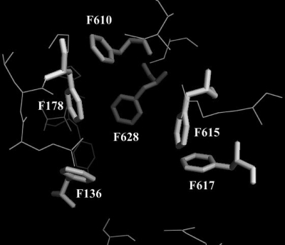
While previous crystallographic studies with crystals grown in trigonal space group R32 described a symmetric AcrB trimer, recent studies of structures derived from monoclinic crystals described an asymmetric trimer in which each protomer was suggested to correspond to a distinct functional state of a proposed three-step transport cycle reminiscent of a peristaltic pump (9, 12, 13). In this model, the protomer in its binding or tight-state conformation forms a hydrophobic pocket defined by phenylalanines 136, 178, 610, 615, 617, and 628.
Analysis of doxorubicin- and minocycline-complexed AcrB crystals suggested that these two compounds interact with different residues of the binding protomer. Minocycline seemed to interact with F178, N274, and F615, while doxorubicin seemed to interact with Q176, F615 and F617 (9). Thus, it was proposed that the extremely broad substrate spectrum of AcrB could be explained by the flexible interaction of various ligands mostly with hydrophobic phenylalanines and to a minor degree with polar residues in the binding pocket.
Support for this model also came from several mutational studies which found that substrate specificity in RND efflux pumps is determined by residues in the periplasmic domain (2-4, 7, 8). A recent study found that the V610F mutation in the E. coli RND efflux pump YhiV, which is homologous to the V612F mutation in AcrB, leads to a 16-fold increase in the linezolid MIC compared to the MIC of the YhiUV-overproducing wild-type strain (2).
However, no systematic site-directed mutagenesis study of the phenylalanine residues that form the proposed hydrophobic binding pocket in AcrB has been described previously.
In the present study we constructed and tested such phenylalanine mutants to examine the functional role of hydrophobic residues in the proposed AcrB multidrug binding site. We used as the parental strain the previously described multidrug-resistant (gyrA marR) acrB-overexpressing E. coli K-12 strain 3-AG100 that was obtained after repeated exposure to a fluoroquinolone (5).
For site-directed mutagenesis the phage λ-based homologous recombination system (Red/ET counterselection Bac modification kit; GeneBridges, Heidelberg, Germany) was used to introduce an rpsL-neo cassette into the acrB gene of strain 3-AG100 (grown in Luria-Bertani broth) and to subsequently replace the cassette with an appropriate oligonucleotide (the sequences of the PCR primers and oligonucleotides that were obtained from Thermo Electron [Ulm, Germany] are shown in Table 1). Recombination events were confirmed by PCR and nucleotide sequencing of the acrB gene using standard techniques.
TABLE 1.
Oligonucleotides and primers used for Red/ET-recombinationa
| Oligonucleotide | Sequence (5′ → 3′) |
|---|---|
| Upper-oligoI (615-628) | ATCTGACCAAAGAAAAGAACAACGTTGAGTCGGTGTTCGCCGTTAACGGCGGCCTGGTGATGATGGCGGGATCG |
| Lower-oligoI (reverse complement orientation) | TCAACTTTGTTTTCTTCGCCCGGACGATCGGCCCAGTCCTTCAAGGAAACTCAGAAGAACTCGTCAAGAAGGCG |
| Upper-oligoII (130-149) | TGGCGATGCCGTTGCTGCCGCAAGAAGTTCAGCAGCAAGGGGTGAGCGTTGGCCTGGTGATGATGGCGGGATCG |
| Lower-oligoII (reverse complement orientation) | ATGGCATCTTTCATATTCGCCGCCACGTAGTCGGAGATATCCTCCTGCGTTCAGAAGAACTCGTCAAGAAGGCG |
| Upper-oligoIII (175-194)) | TGGCGGCGAATATGAAAGATGCCATCAGCCGTACGTCGGGCGTGGGTGATGGCCTGGTGATGATGGCGGGATCG |
| Lower-oligoIII (reverse complement orientation) | TTCTGCGCTTTGATGGCGGTAATGACATCAACCGGCGTTAGCTGGAATTTTCAGAAGAACTCGTCAAGAAGGCG |
| Repair-oligo 1: rep-acrB-Phe610Ala | ACGTTGAGTCGGTGGCAGCCGTTAACGGCTTCGGCTTTGCGGGACGTGGTCAGAATACCGGTATTGCGTTCGTTTCCTTGAAGGACTGGGCCGATCGTCC |
| Repair-oligo 2: rep-acrB-Phe615Ala | ACGTTGAGTCGGTGTTCGCCGTTAACGGCGCAGGCTTTGCGGGACGTGGTCAGAATACCGGTATTGCGTTCGTTTCCTTGAAGGACTGGGCCGATCGTCC |
| Repair-oligo 3: rep-acrB-Phe617Ala | ACGTTGAGTCGGTGTTCGCCGTTAACGGCTTCGGCGCAGCGGGACGTGGTCAGAATACCGGTATTGCGTTCGTTTCCTTGAAGGACTGGGCCGATCGTCC |
| Repair-oligo 4: rep-acrB-Phe628Ala | ACGTTGAGTCGGTGTTCGCCGTTAACGGCTTCGGCTTTGCGGGACGTGGTCAGAATACCGGTATTGCGGCAGTTTCCTTGAAGGACTGGGCCGATCGTCC |
| Repair-oligo 5: rep-acrB-Phe628Phe | ACGTTGAGTCGGTGTTCGCCGTTAACGGCTTCGGCTTTGCGGGACGTGGTCAGAATACCGGTATTGCGTTTGTTTCCTTGAAGGACTGGGCCGATCGTCC |
| Repair-oligo 6: rep-acrB-Del615-617 | GAACAACGTTGAGTCGGTGTTCGCCGTTAACGGC^^^^^^^^^GCGGGACGTGGTCAGAATACCGGTATTGCGTTCGTTTCCTTGAAGGACTGGGCCGATCGTCCGGGCG |
| Repair-oligo 7: rep-acrB-Phe136Ala | GCCGCAAGAAGTTCAGCAGCAAGGGGTGAGCGTTGAGAAATCATCCAGCAGCGCACTGATGGTTGTCGGCGTTATCAACACCGATGGCACCATGACGCAGGAGGATATCTCCGACTACGTGGCGGCGA |
| Repair-oligo 8: rep-acrB-Phe178Ala | AGATGCCATCAGCCGTACGTCGGGCGTGGGTGATGTTCAGTTGGCAGGTTCACAGTACGCGATGCGTATCTGGATGAACCCGAATGAGCTGAACAAATTCCAGCTAACGCCGGTTGATGTCATTACCG |
| Forward primer for amplification of repair oligonucleotides 1-6 | ATCTGACCAAAGAAAAGAACAACGTTGAGTCGGTG |
| Reverse primer for amplification of repair oligonucleotides 1-6 | CGCCCGGACGATCGGCCCAGTCCTT |
| Check-forward primer I | CCTTCTTGCCAGATGAGGAC |
| Check-reverse primer I | GCAGTACCCAGTTCCACGAT |
| Check-forward primer II | GTGCAGATCACCCTGACCTT |
| Check-reverse primer II | CGTTCTGCGCTTTGATGG |
| Check-forward primer III | ACCATGACGCAGGAGGATA |
| Check-reverse primer III | TAAGCTGTTGGCCTTTCACC |
The upper and lower oligonucleotides include the primer sequences for amplification of the rpsL-neo cassette (indicated by italics). The 5′ parts of the oligonucleotides are homologous to the corresponding acrB regions upstream and downstream (nucleotides 1793 to 1842 and 1885 to 1934 for exchange region I, nucleotides 338 to 387 and 448 to 497 for region II, and nucleotides 473 to 522 and 583 to 632 for region III). The exchanged nucleotide triplets in the repair oligonucleotides are indicated by bold type (e.g., GTT is changed to TTT at nucleotides 1834 to 1836 in acrB). The underlined sequences in the amplification primers are the priming parts for the repair oligonucleotides, which have to be elongated. The Check-forward and Check-reverse primers are used to confirm successful exchange of the rpsL-neo cassette and to sequence the modified region of acrB (check PCR product for acrB region I, nucleotides 1685 to 2030; check PCR product for region II, nucleotides 262 to 634; check PCR product for region III, nucleotides 442 to 691).
To confirm production of the mutant AcrB protein, we performed Western blotting using standard techniques. Most of the mutants exhibited a strong immunogenic response; the only exception was an F615A/F617A/F628A triple mutant which was excluded from further study due to insufficient AcrB expression (Fig. 1).
FIG. 1.
Western blot analysis of mutant AcrB production. Total protein extracts of E. coli 3-AG100 mutants (14 μg protein) were separated by NuPAGE Novex bis-Tris (Invitrogen, California) gel electrophoresis and probed with polyclonal anti-AcrB antibodies. Lanes MW contained molecular weight markers.
We used as a positive control strain F628F, which is a pseudomutant with MICs and ethidium bromide (EtBr) and phenylalanine-arginine β-naphthylamide (PAβN) accumulation properties corresponding to those of wild-type strain 3-AG100. F628A is characterized by a silent mutation from TTC to TTT (sequence shown in Table 1) that demonstrates that the site-directed mutagenesis technique has no inherent effect.
The susceptibilities of the different mutants to various antimicrobials and dyes and to the putative efflux pump inhibitors 1-naphthylmethylpiperazine (NMP) and PAβN were characterized by determining MICs in 96-well microtiter plates as described previously (1, 2, 6) and are shown in Table 2. EtBr (external concentration, 2.5 μM) and PAβN (external concentration, 200 μM) fluorescence accumulation assays were carried out at least in duplicate for 30 min using our previously described protocol (2). Both EtBr and PAβN are excellent substrates of AcrAB-TolC and were chosen since they are structurally diverse; thus, the recognition by the AcrB binding pocket was assumed to be mediated by different residues. EtBr is a nonspecific DNA intercalator which, upon binding to its target structure, causes enhancement of fluorescence, while the intrinsically low-fluorescence compound PAβN is cleaved by esterases, yielding the highly fluorescent compound β-naphthylamine as described previously in a study using the related substrate Ala-Nap (naphthylamide) (6). The results obtained are shown in Fig. 2a and 2b. We also used an EtBr concentration of 25 μM and a PAβN concentration of 20 μM and obtained similar results (data not shown).
TABLE 2.
MICs of different pump substrates for AcrB mutants of E. coli 3-AG100a
| Mutation | MIC (μg/ml)
|
||||||||||||||||||||
|---|---|---|---|---|---|---|---|---|---|---|---|---|---|---|---|---|---|---|---|---|---|
| Oxacillin | Doxoru-bicin | Novobi-ocin | Clarithro-mycin | Erythro-mycin | Azithro-mycin | Clin-damycin | Pyronin Y | Linezolid | Mino- cyclin | EtBr | Levo-floxacin | Cipro-floxacin | Hoechst 33342 | Propidium iodide | PAβN | Chloram-phenicol | Tetra-cycline | NMP | Spectino-mycin | Genta-micin | |
| F628F wild type (pseudomutation) | >256 | >256 | 512 | 512 | 512 | 64 | 256 | 32 | 512 | 4 | >256 | 1 | 0.5 | 4 | >512 | >400 | 8 | 4 | 400 | 32 | 8 |
| acrB::rpsL-neo | 0.5 | 2 | 4 | 4 | 4 | 0.5 | 4 | 0.5 | 16 | 0.125 | 16 | 0.06 | 0.03 | 0.25 | 128 | 50 | 1 | 1 | |||
| F136A | 64 | 64 | 64 | 64 | 16 | ||||||||||||||||
| F178A | 32 | 16 | 64 | 128 | 16 | 64 | 0.5 | ||||||||||||||
| F610A | 64 | 128 | 32 | 16 | 64 | 2 | 32 | 2 | 0.25 | 128 | 0.13 | 0.06 | 256 | 100 | 2 | 1 | |||||
| F615A | 64 | 64 | 32 | 32 | |||||||||||||||||
| F617A | 128 | ||||||||||||||||||||
| Δ615-617 | 64 | 32 | 128 | 128 | 1 | ||||||||||||||||
| F628A | 128 | 128 | 64 | 16 | 8 | 1 | 1 | ||||||||||||||
Gentamicin, spectinomycin, and NMP are not substrates of AcrB and were used as controls. For the mutants only the MICs that were ≥4-fold different from the wild-type MIC (F628F pseudomutant) are shown. The substrates and control substances are in order (from left to right) based on the differences in the MICs between the wild type (pseudomutant F628F) and the inactivation mutant (acrB::rpsL-neo).
FIG. 2.
Increases in EtBr (a) and PAβN (b) fluorescence in AcrB phenylalanine mutants compared to pseudomutant AcrB strain F628F. Fluorescence was recorded for 30 min after addition of 2.5 μM EtBr or 200 μM PAβN. The values are means of at least duplicate experiments. RFU, relative fluorescence units.
The complete disruption of acrB by the rpsL-neo cassette led to a highly drug-susceptible phenotype and dramatic increases in EtBr and PAβN accumulation. The positive control pseudomutant F628F displayed no changes in the MIC assays or in the dye accumulation assays. Novobiocin was the only drug whose MIC was consistently markedly reduced for every single mutant. The F136A, F178A, F615A, F617A, and F628A mutations had very variable effects on substrate MICs. In addition to the susceptibility of the mutants to novobiocin, the MICs of oxacillin and the macrolides tested were also reduced in all but the F617A mutant. F178A markedly increased the susceptibility to linezolid, and the F628A mutation was found to reduce the MICs of Hoechst 33342, pyronine Y, and minocycline more than 4-fold and to increase EtBr and PAβN accumulation ∼2-fold after 30 min.
The crystallographic structure of the asymmetric AcrB trimer suggests that the main interactions between substrates and protein are due to an ensemble of phenylalanines mediating hydrophobic interactions, which might explain the extremely broad substrate specificity (Fig. 3). Surprisingly, although the AcrB cocrystallization with doxorubicin and minocycline suggested that there is a strong interaction of these substrates with F178 and F615, the F615A and F178A mutations had no measurable impact on the MICs of these two substrates. Deletion of amino acids 615 to 617 was associated with minor changes in the susceptibility to minocycline and some changes in the MICs of macrolides and oxacillin.
FIG. 3.
AcrB binding pocket based on the “tight” monomer 2GIF structure coordinates (12). Phenylalanines are indicated by sticks. The image was generated using the molecular visualization software PyMol (http://pymol.sourceforge.net).
The lack of correlation between the changes in susceptibility to doxorubicin and minocycline and the model derived from cocrystallization might have been due to redundancy of phenylalanines in the binding pocket. Mutating or deleting only one or even two phenylalanines might only lead to a (slight) reorientation of the substrate and use of other phenylalanines as hydrophobic interaction partners without generally compromising substrate capture. However, bulkier substrates, like the macrolides or novobiocin, might be unable to adapt properly in the altered environment of the pocket and might be affected more by a single mutation.
To test this hypothesis, we generated the F615A/F617A/F628A triple phenylalanine mutant; however, since we obtained only a weak Western blot band (Fig. 1), suggesting that the level of expression of the mutant AcrB protein was low, we did not include this mutant in further analyses.
The F610A mutant, however, displayed dramatically enhanced susceptibility to almost all AcrB substrates tested (but not to aminoglycosides and NMP, which are not AcrB substrates), although the absolute changes varied considerably for different substrates. In contrast, the F610A mutation increased EtBr accumulation only moderately and did not affect PAβN accumulation. This difference might have been due to the different time windows between the MIC and fluorescence experiments. The dramatic impact on substrate MICs indicates that the F610 residue has a special role in the substrate extrusion process, although the exact mechanism remains unclear. The other targeted mutations, including conversion of phenylalanines 136, 178, 615, 617, and 628 to alanine, generally had smaller effects on substrate susceptibility and presumably efflux function and binding, and the effects were variable depending on the substrate.
Acknowledgments
This study was supported by BMBF grant 01KI9951.
Footnotes
Published ahead of print on 10 October 2008.
REFERENCES
- 1.Bohnert, J. A., and W. V. Kern. 2005. Selected arylpiperazines are capable of reversing multidrug resistance in Escherichia coli overexpressing RND efflux pumps. Antimicrob. Agents Chemother. 49849-852. [DOI] [PMC free article] [PubMed] [Google Scholar]
- 2.Bohnert, J. A., S. Schuster, E. Fähnrich, R. Trittler, and W. V. Kern. 2007. Altered spectrum of multidrug resistance associated with a single point mutation in the Escherichia coli RND-type MDR efflux pump YhiV (MdtF). J. Antimicrob. Chemother. 591216-1222. [DOI] [PubMed] [Google Scholar]
- 3.Elkins, C. A., and H. Nikaido. 2002. Substrate specificity of the RND-type multidrug efflux pumps AcrB and AcrD of Escherichia coli is determined predominantly by two large periplasmic loops. J. Bacteriol. 1846490-6498. [DOI] [PMC free article] [PubMed] [Google Scholar]
- 4.Hearn, E. M., M. R. Gray, and J. M. Foght. 2006. Mutations in the central cavity and periplasmic domain affect efflux activity of the resistance-nodulation-division pump EmhB from Pseudomonas fluorescens cLP6a. J. Bacteriol. 188115-123. [DOI] [PMC free article] [PubMed] [Google Scholar]
- 5.Jellen-Ritter, A. S., and W. V. Kern. 2001. Enhanced expression of the multidrug efflux pumps AcrAB and AcrEF associated with insertion element transposition in Escherichia coli mutants selected with a fluoroquinolone. Antimicrob. Agents Chemother. 451467-1472. [DOI] [PMC free article] [PubMed] [Google Scholar]
- 6.Lomovskaya, O., M. S. Warren, A. Lee, J. Galazzo, R. Fronko, M. Lee, J. Blais, D. Cho, S. Chamberland, T. Renau, R. Leger, S. Hecker, W. Watkins, K. Hoshino, H. Ishida, and V. J. Lee. 2001. Identification and characterization of inhibitors of multidrug resistance efflux pumps in Pseudomonas aeruginosa: novel agents for combination therapy. Antimicrob. Agents Chemother. 45105-116. [DOI] [PMC free article] [PubMed] [Google Scholar]
- 7.Mao, W., M. S. Warren, D. S. Black, T. Satou, T. Murata, T. Nishino, N. Gotoh, and O. Lomovskaya. 2002. On the mechanism of substrate specificity by resistance nodulation division (RND)-type multidrug resistance pumps: the large periplasmic loops of MexD from Pseudomonas aeruginosa are involved in substrate recognition. Mol. Microbiol. 46889-901. [DOI] [PubMed] [Google Scholar]
- 8.Middlemiss, J. K., and K. Poole. 2004. Differential impact of MexB mutations on substrate selectivity of the MexAB-OprM multidrug efflux pump of Pseudomonas aeruginosa. J. Bacteriol. 1861258-1269. [DOI] [PMC free article] [PubMed] [Google Scholar]
- 9.Murakami, S., R. Nakashima, E. Yamashita, T. Matsumoto, and A. Yamaguchi. 2006. Crystal structures of a multidrug transporter reveal a functionally rotating mechanism. Nature 443173-179. [DOI] [PubMed] [Google Scholar]
- 10.Nikaido, H. 1998. Antibiotic resistance caused by gram-negative multidrug efflux pumps. Clin. Infect. Dis. 27(Suppl. 1)S32-S41. [DOI] [PubMed] [Google Scholar]
- 11.Piddock, L. J. 2006. Clinically relevant chromosomally encoded multidrug resistance efflux pumps in bacteria. Clin. Microbiol. Rev. 19382-402. [DOI] [PMC free article] [PubMed] [Google Scholar]
- 12.Seeger, M. A., A. Schiefner, T. Eicher, F. Verrey, K. Diederichs, and K. M. Pos. 2006. Structural asymmetry of AcrB trimer suggests a peristaltic pump mechanism. Science 3131295-1298. [DOI] [PubMed] [Google Scholar]
- 13.Sennhauser, G., P. Amstutz, C. Briand, O. Storchenegger, and M. G. Grutter. 2007. Drug export pathway of multidrug exporter AcrB revealed by DARPin inhibitors. PLoS Biol. 5e7. [DOI] [PMC free article] [PubMed] [Google Scholar]


