

Abstract
Marek's disease virus (MDV), a herpesvirus that causes a lymphoproliferative disorder in chickens, encodes a number of microRNAs derived primarily from two locations in the MDV genome. One cluster of microRNA genes flanks the meq oncogene, and a second cluster is found within the latency-associated transcript (LAT) region. The sequences of MDV microRNAs from a collection of field and reference strains with various levels of virulence were compared and found to be highly conserved. However, microRNAs from the meq cluster were detected at higher levels in lymphomas caused by a form of the virus designated very virulent plus (vv+; strain 615K, also known as T. King) than in those caused by a less virulent (very virulent [vv]) form (RB1B). For example, levels of mdv1-miR-M4, which shares a seed sequence with miR-155, a microRNA implicated in B-cell lymphoma, were threefold higher and levels of mdv1-miR-M2*/3p were more than sixfold higher in vv+ MDV-induced tumors than in vv MDV-induced tumors. In contrast, levels of the microRNAs from the LAT cluster were equivalent in tumors produced by vv and vv+ strains. Additionally, mdv1-miR-M4 is the MDV microRNA most highly expressed in tumors, where it accounts for 72% of all MDV microRNAs, as determined by deep sequencing. These data suggest that the meq cluster microRNAs play an important role in the pathogenicity of MDV.
Marek's disease virus (MDV) is an alphaherpesvirus that causes a highly contagious, lymphoproliferative disorder of chickens known as Marek's disease. Similar to those of other herpesviruses, the life cycle of MDV includes both lytic and latent stages. Initially, a productive infection occurs in epithelial cells, followed by B cells and feather follicles. Latency occurs in T lymphocytes, and following a second wave of cytolytic infection primarily in lymphoid organs, MDV transforms some T cells and produces lymphomas. MDV serotype 1 (MDV1) isolates can be grouped into four pathotypes designated mild, virulent (v), very virulent (vv), and very virulent plus (vv+). These pathotypes can all produce disease, and the nomenclature reflects increasing levels of virulence based on mortality in flocks, lesion frequency, and protection by existing vaccines (43). The sequences of the variable regions of representative strains have been determined previously (38), but the genetic cause of increased virulence has not been established. Studies over the past several decades since commercial vaccination was introduced suggest that MDV is evolving toward greater virulence (27, 43).
In MDV-induced lymphomas, a limited number of viral genes are expressed, and genes mapping to the repeat regions are thought to be key to oncogenesis (39). The meq gene, which is located in the repeat regions and encodes a bZIP transcription factor, plays a major role in transformation and is the primary candidate oncogene for MDV (20, 21). Latency-associated transcripts (LATs) are also expressed in tumors and lymphoblastoid cells and during latency (25). These LATs map in an antisense orientation relative to the ICP4 gene in the repeats flanking the unique short region and consist of a long (10-kb) transcript, at least one small RNA, and several splice variants. No protein product originating from these transcripts has been identified.
MicroRNAs are small, noncoding RNAs that regulate gene expression by base pairing with the 3′ untranslated region of mRNAs and targeting the mRNAs for translational repression or degradation. All metazoan genomes encode microRNAs, and these molecules have been shown to be involved in important biological processes such as development and differentiation, apoptosis, and the production of cancer (2, 3, 6). A number of studies point to a role of microRNAs in the regulation of the immune system (18, 22). Of particular interest is miR-155, which has been demonstrated previously to be important in regulating both B- and T-cell-dependent immune functions (28, 34, 41). Viruses that replicate in eukaryotic nuclei, including herpesviruses, encode microRNAs, and it has been suggested that these microRNAs play roles in interference with host immune responses and in the regulation of cell growth and apoptosis (for reviews, see references 10 and 30). One Kaposi's sarcoma herpesvirus (KSHV) microRNA (K12-11) shares a seed sequence with miR-155, and candidate targets that may contribute to KSHV-induced tumor formation have been validated experimentally (14, 37). Viral genes can also be targets for viral microRNAs. In cytomegalovirus (15, 16, 26) and herpes simplex virus type 1 (42), viral microRNAs have been shown to target immediate-early genes and may play an important role in the establishment and maintenance of latency.
Recent studies indicate that viral microRNAs are quite specific for each virus and are highly conserved among related strains, suggesting that sequence conservation is important for virus survival. For example, KSHV microRNAs are distinct and highly conserved across a large collection of different strains (24). Moreover, a set of conserved microRNAs in the related lymphocryptoviruses Epstein-Barr virus and rhesus lymphocryptovirus has been identified previously (7, 24). Both oncogenic MDV (MDV1) and nononcogenic MDV (MDV2) also encode microRNAs (4, 5, 46, 47). While there is no conservation of sequence between the MDV1 and MDV2 microRNAs, they are derived from similar positions in the viral genome (46).
In the present study, we have extended our previous work on the identification of MDV microRNAs (4, 5) by analyzing microRNA sequences from a collection of reference and field strains that cover the continuum of MDV pathotypes. We found that the sequences of the MDV-encoded microRNAs were highly conserved but that some microRNAs, notably those encoded upstream of the meq oncogene, were more highly expressed in tumors that had been induced by 615K (also known as T. King), an extremely pathogenic (vv+) strain. This group of highly expressed microRNAs included mdv1-miR-M4, which shares a seed sequence with miR-155. Deep sequencing of small RNAs from MDV (RB1B strain)-induced tumors indicated that mdv1-miR-M4 was more highly expressed than other MDV microRNAs. We speculate that high levels of expression of this microRNA may be related to the increased pathogenicity of the vv+ strain.
MATERIALS AND METHODS
Virus propagation and DNA sequencing.
DNA from v, vv, and vv+ reference strains and field isolates was obtained from Richard Witter (Avian Disease and Oncology Laboratory, USDA-ARS, East Lansing, MI) and John K. Rosenberger (Aviserve, LLC, Newark, DE) and has been described previously (36). DNA was amplified with primers (IDT, Coralville, IA) flanking the meq and the LAT microRNA gene clusters and the microRNA coding sequence in UL40, as well as the putative promoter region of the meq microRNA gene cluster (primers are listed in Table 1). Amplified PCR products were evaluated by agarose gel electrophoresis, reaction mixtures showing single bands were purified using a QIAquick PCR purification kit (Qiagen, Germantown, MD), and the purified products were sequenced directly by the University of Delaware Sequencing Center. Any product producing an ambiguous read was subcloned into a TA vector (pCR2.1Topo; Invitrogen, Carlsbaad, CA), and several colonies were selected for sequence confirmation.
TABLE 1.
Sequences of PCR primers used to amplify MDV fragments
| Primer name | Sequence (5′→3′) | Coordinatesa |
|---|---|---|
| meqmir-F2 | TTGTATGTGTGTGAGCAGTC | 133501-133520 |
| meqmir-R2 | AGACGTTCTACGATGGTTTT | 134449-134468 |
| LATmir-F | GACAGGAGTTCGGAATAAAC | 142150-142169 |
| LATmir-R | AGAGATTTCGAGTTGCCAAC | 142658-142677 |
| meqmirpromoter-F | TCATTGGGTATTGAAGGTAT | 132581-132600 |
| meqmirpromoter-R | TACCACCACAAACAGTAGAC | 133661-133680 |
| MDVRNR-F | GGCCCTCCGTCACATGAGAT | 96358-96377 |
| MDVRNR-R | CGTTTGACACGCTTCCAGAG | 97356-97375 |
| mirM1mirM12-F | TAATTCGGTGGTGCTGATTAGG | 136453-136474 |
| mirM1mirM12-R | ATGTCGTACGAGCCTCGTTC | 136937-136956 |
Numbering is based on the sequence with accession no. AF243438.
Infection of chickens, RNA preparation, and Northern blotting.
Fertile broiler eggs were obtained from a commercial hatchery and incubated at the University of Delaware, and hatched chickens were inoculated intraabdominally at 1 day of age with 5 × 10⁵ peripheral blood lymphocytes previously obtained from T. King-infected chickens. Two-day-old SPAFAS chickens were infected intraabdominally with chicken embryo fibroblasts (CEF) infected with RB1B (3,000 PFU/chicken). Splenic tumors were excised from chickens 7 to 8 weeks postinfection. DNA and RNA were prepared using Trizol, and viral loads were quantified using quantitative PCR analysis of meq and ICP4 genes (1). Gels were loaded with equal amounts of RNA (as determined by the A260) from samples with equivalent viral DNA loads and electrophoresed, and the RNA was hybridized to 32P-labeled antisense oligonucleotide probes as described previously (4). The intensities of the bands were determined using a Typhoon phosphorimager, and signals in each lane were normalized to that of U6 RNA prior to the determination of relative expression levels.
RT-PCR and quantitative RT-PCR.
Relative mRNA levels for meq, ICP4, and the GAPDH (glyceraldehyde-3-phosphate dehydrogenase) gene in samples used for Northern blots (as described above) were measured using protocols and primers described previously (45). All samples were treated with DNase, and no amplified product was detected in the no-reverse transcriptase (no-RT) controls.
To determine if the microRNAs encoded upstream of meq were located on the same transcript, total RNA (100 ng) from MSB1 cells was DNase treated, cDNA was generated using a gene-specific primer just downstream of the sequence encoding mdv1-miR-M4* (5′-AGACGTTCTACGATGGTTTT-3′), and then PCR amplification of that cDNA was performed using the same reverse primer with a forward primer just upstream of the sequence encoding mdv1-miR-M9-5p (5′-AAGGTAATCATTCCCGGATA-3′). This procedure resulted in the expected band of 1,288 bp. There was no amplification seen in the no-RT controls or the no-template controls.
Deep sequencing.
Total RNA from MSB1 cells, an MDV-transformed cell line, was prepared, and splenic tumors from a chicken infected with the RB1B strain of MDV were isolated and submitted to Illumina/Solexa (Hayward, CA) for the preparation of libraries (23) and sequencing. Sequences with high base quality scores were trimmed of the 3′ adapter sequences and compared to the chicken and viral genomes for exact matches. For the tumor samples, a total of 3,827,378 million signatures were obtained; of these, 1,325,729 matched the chicken genome and 13,209 matched the MDV genome. For MSB1 cells, 2,602,161 signatures were obtained, and of these, 1,119,779 matched the chicken genome and 245,475 matched the MDV genome.
RESULTS
Analysis of sequence heterogeneity in MDV microRNAs.
Regions of the MDV genome encoding microRNAs were amplified from various MDV reference strains and field isolates and sequenced. We previously identified mdv1-miR-M2, mdv1-miR-M3, mdv1-miR-M4, mdv1-miR-M5, and mdv1-miR-M12 from one region (4, 5), and two additional microRNAs (mdv1-miR-M9-5p and mdv1-miR-M9-3p) encoded further upstream were reported by Yao et al. (47) and confirmed by our deep-sequencing results (described below). All microRNAs are numbered according to the present miRBase release, and for microRNAs mapping to the repeat regions of the genome, the numbering used herein is based on the internal repeat flanking the unique short region. A schematic of these regions is shown in Fig. 1. Sequence variations found in a 1,887-nucleotide (nt) region spanning the cluster of microRNA genes upstream of the meq gene (nt 132581 to 134468) are shown in Table 2. With the exception of the sequences of mdv1-miR-M9-5p and mdv1-miR-M9-3p, the sequences of both arms of the microRNAs from this region were completely conserved among all strains examined and were identical to those of Md5 (GenBank accession no. AF243438), which was used as the reference strain. Two field isolates had a sequence variation (C→T) at position 133384 in mdv1-miR-M9-5p, and mixed bases were found in strain 543 at positions 133392 (G/A) and 133419 (T/C) in mdv1-miR-M9-3p. There were some variations at position 133826, which is located between mdv1-miR-M5 and mdv1-miR-M12, and at position 134327, which is located between mdv1-miR-M2* and mdv1-miR-M4. However, none of these sequence variations correlated with pathotypes. The region upstream of the meq microRNA gene cluster showed more sequence variation than the region encoding the actual microRNAs. One variation, at position 133206 (G→T), was present in all but one vv+ strain. RT-PCR results indicated that all the microRNAs encoded upstream of meq are present in one primary transcript (data not shown), but attempts to locate the transcriptional start site by the random amplification of cDNA ends or to approximate the transcript size by Northern blotting were unsuccessful, likely due to the low abundance and/or instability of the primary transcript. However, it appears that the 133206 (G→T) vv+ sequence variation is within the primary transcript or the promoter region of the upstream meq microRNA genes.
FIG. 1.

Schematic diagram of MDV microRNAs corresponding to the regions flanking the meq oncogene (A) and the LAT region (B). Nucleotide numbering and MDV gene names are according to the data for Md5 (GenBank accession number AF243438); microRNA naming is according to miRBase (http://microrna.sanger.ac.uk/sequences/index.shtml). Small blue arrowheads indicate the locations of the MDV microRNAs, large orange arrows indicate mRNA transcripts, and the open reading frames are hatched. The green arrow indicates the a-like sequence. Black arrowheads indicate the locations of primers used to amplify DNA from MDV isolates. MSR, MDV small RNA.
TABLE 2.
Summary of sequence variations in the meq microRNA region in various MDV strains
| Pathotype or isolate group | Strain | Nucleotide(s) or microRNA corresponding to genomic positiona:
|
||||||||||||||||||
|---|---|---|---|---|---|---|---|---|---|---|---|---|---|---|---|---|---|---|---|---|
| 132631 | 132637 | 132659 | 132671 | 132740 | 132888 | 133062 | 133098 | 133206 | 133231 | 133232 | 133311 | 133384 | 133392 | 133419 | 133444 | 133484 | 133826 | 134327 | ||
| v | CU2 | NDe | ND | ND | T/C | |||||||||||||||
| 571 | ND | ND | ND | ND | ND | T | ||||||||||||||
| 573 | ND | ND | ND | ND | T | |||||||||||||||
| 617 | ND | ND | T/C | |||||||||||||||||
| GAb | ||||||||||||||||||||
| vv | Md5c | G | C | T | C | G | T | G | A | G | A | T | T | C | A | C | C | A | A | C |
| RB1B | A/G | C/G | C/T | T/C | ||||||||||||||||
| 549A | C | ND | ND | |||||||||||||||||
| 595 | ND | T/C | ||||||||||||||||||
| 543 | T | G/A | T/C | |||||||||||||||||
| 543P | ND | ND | C | |||||||||||||||||
| vv+ | 648 | T | T/C | |||||||||||||||||
| 615K | ND | ND | ND | T | ND | T | ||||||||||||||
| 686 | T | A/G | T | |||||||||||||||||
| 660 | T | T/C | T/C | |||||||||||||||||
| MK | T | T/C | ||||||||||||||||||
| CD | T | T/A | C/T | G/A | T | |||||||||||||||
| Field isolates | 06-011 | T | ND | ND | ||||||||||||||||
| 06-012 | C/T | ND | ND | |||||||||||||||||
| 06-013 | A/G | G/A | T | T | ||||||||||||||||
| 06-014 | ND | ND | ND | |||||||||||||||||
| 06-015 | ND | ND | T | |||||||||||||||||
| 06-016 | ND | ND | ND | ND | T | |||||||||||||||
| mdv1-miR-M9-5pd | mdv1-miR-M9-5pd | mdv1-miR-M9-3pd | ||||||||||||||||||
The genomic position indicates the location in one of the repeats; there are two copies in the MDV genome.
The sequence of GA (accession no. AF147806) was obtained from GenBank.
Md5 (accession no. AF243438) is the reference strain.
mdv-miR-M9-5p spans 133374 to 133395; mdv-miR-M9-3p spans 133414 to 133435.
ND, not determined. Empty cells indicate that the nucleotide is the same as that in the reference strain.
A 528-bp region (nt 142150 to 142677) containing microRNAs from the LAT region also showed very little sequence variation (Table 3), and similar to results for the meq microRNAs, none of the polymorphisms were in the mature microRNAs or were associated with particular pathotypes. Sequence variations in the vv+ strain 615K/T. King compared to the sequences of Md5 and RB1B (3) were identified at position 142340 (T→C) in mdv1-miR-M6* and positions 142514 (C→T) and 142526 (C→T) in mdv1-miR-M7*.
TABLE 3.
Summary of sequence variations in the LAT microRNA region in various MDV strains
| Pathotype or isolate group | Strain | Nucleotide or microRNA corresponding to genomic positionb:
|
||
|---|---|---|---|---|
| 142340 | 142514 | 142526 | ||
| v | CU2 | T | ||
| BC1 | T | |||
| 571 | T | |||
| 573 | T | |||
| 617 | T | T | ||
| GAa | ||||
| vv | Md5 | T | C | C |
| 549 | ||||
| 595 | ||||
| 543 | ||||
| RL | ||||
| vv+ | 615K | C | T | T |
| 648A | ||||
| 660 | ||||
| 686 | ||||
| CD | C | T | T | |
| MK | ||||
| RLP2 | ||||
| RB1Ba | ||||
| Field isolates | 06-011 | C | T | T |
| 06-012 | C | ND | ND | |
| 06-013 | C | T | T | |
| 06-014 | C | ND | ND | |
| 06-015 | C | T | T | |
| 06-016 | C | ND | ND | |
| 06-017 | C | T | T | |
| 06-029 | C | T | T | |
| 06-031 | C | T | T | |
| 455A | ND | T | ND | |
| AV | T | T | ||
| mdv-miR-M6* | mdv-miR-M7* | mdv-miR-M7* | ||
The sequences of GA (accession no. AF147806) and RB1B (accession no. DQ534554.1) were obtained from GenBank.
ND, not determined. Empty cells indicate that the nucleotide is the same as that in the reference strain (Md5).
Amplification of the region downstream of meq revealed a single sequence variation at position 136912 (A→G) downstream of mdv1-miR-M1 in one vv strain (595), but the sequences of both the mdv1-miR-M1 and mdv1-miR-M32 microRNAs were identical to the Md5 sequences. The antisense microRNA relative to the ribonucleotide reductase small subunit (mdv1-miR-M31) was conserved in all strains, although a silent mutation (G→A) was found nearby, at position 97182, in the vv strain 549D.
Profile of MDV microRNAs in MDV-induced tumors.
We previously identified MDV microRNAs expressed in CEF, where the virus establishes a lytic infection (4). To determine if the microRNA expression profile was different in tumors where MDV established a latent infection, we performed deep sequencing of small RNAs expressed in a splenic tumor from a chicken exposed to MDV (RB1B strain). Based on sequencing frequencies (which are generally considered to correlate with relative concentrations in samples), mdv1-miR-M4 levels were the highest among those of the MDV microRNAs and accounted for 72% of the sequences matching the MDV genome (Table 4). In addition, we found that the 3p arm of mdv1-miR-M2 was more highly expressed than the 5p arm, a pattern that was opposite to that observed in sequencing small RNAs from MDV-infected CEF (4). A similar alteration in strand selection was noted for mdv1-miR-M8 as well. MDV microRNAs in MSB1 cells, which are MDV-transformed lymphoblastoid cells, also showed this preference, with mdv1-miR-M5, mdv1-miR-M2*/3p, and mdv1-miR-M8*/5p being the predominant MDV microRNAs in MSB1 cells.
TABLE 4.
Sequencing frequencies for MDV microRNAs
| MicroRNA | Sequence | % of MDV matches in:
|
||
|---|---|---|---|---|
| Tumor samples | MSB1 cells | CEFa | ||
| MicroRNAs encoded upstream of meq | ||||
| mdv1-miR-M12-3p | TTGCATAATACGGAGGGTTCTG | 2.3 | 5.0 | 0.3 |
| mdv1-miR-M12-5p | AGGCCCTCCGTATAATGTAAATGT | 0.2 | 0.0 | NDc |
| mdv1-miR-M2/5p | GTTGTATTCTGCCCGGTAGTCCG | 1.6 | 1.1 | 18.9 |
| mdv1-miR-M2*/3p | CGGACTGCCGCAGAATAGCTT | 6.9 | 21.5 | 1.6 |
| mdv1-miR-M3-5p | ATGAAAATGTGAAACCTCTCCCGC | 3.9 | 2.8 | 1.3 |
| mdv1-miR-M3-3p | TGGGGGGTTCACATTTTTAAGT | <0.1 | 0.0 | ND |
| mdv1-miR-M4 | TTAATGCTGTATCGGAACCCTTCG | 72.0 | 7.6 | 20.4 |
| mdv1-miR-M4* | AATGGTTCTGACAGCATGACC | 0.1 | 0.4 | 0.6 |
| mdv1-miR-M5/3p | TGTGTATCGTGGTCGTCTACTGT | 6.3 | 33.0 | 6.1 |
| mdv1-miR-M5*/5p | CGTATGCGATCACATTGACACG | <0.1 | 0.0 | 1.2 |
| mdv1-miR-M9-5p | TTTTCTCCTTCCCCCCGGAGTTC | 0.8 | 0.1 | ND |
| mdv1-miR-M9-3p | AAACTCCGAGGGCAGGAAAAAG | 1.0 | 0.1 | ND |
| MicroRNAs encoded downstream of meq | ||||
| mdv1-miR-M1 | TGCTTGTTCACTGTGCGGCATT | 1.0 | 3.6 | 30.1 |
| mdv1-miR-M1* | ATGCTGCGCATGAAAGAGCGA | ND | 0.0 | ND |
| mdv1-miR-M32 | TGCTACAGTCGTGAGCAGATCAA | <0.1 | 0.1 | 1.0 |
| MicroRNAs encoded in the LAT region | ||||
| mdv1-miR-M6*/5p | TGTTGTTCCGTAGTGTTCTCG | <0.1 | 0.1 | 3.8 |
| mdv1-miR-M6/3p | GAGATCCCTGCGAAATGACAGT | 0.1 | 0.1 | 8.5 |
| mdv1-miR-M7*/5p | TGTTATCTCGGGGAGATCCCGAT | ND | 0.0b | ND |
| mdv1-miR-M7/3p | TCGAGATCTCTACGAGATTACAG | ND | 0.0 | 11.0 |
| mdv1-miR-M8*/5p | TATTGTTCTGTGGTTGGTTTCGA | 3.5 | 23.5 | 1.1 |
| mdv1-miR-M8/3p | GTGACCTCTACGGAACAATAGT | 0.1 | 0.9 | 4.9 |
| mdv1-miR-M10-5p | GCGTTGTCTCGTAGAGGTCCAG | 0.1 | 0.0 | 0.2 |
| mdv1-miR-M10*/3p | TCGAAATCTCTACGAGATAACAGTT | ND | 0.0 | 0.4 |
CEF library data were extracted from a previous study (4) and were obtained by sequencing with the 454 system, while the tumor and MSB1 libraries were sequenced more deeply by Illumina.
For the MSB1 cells, 0.3% of the reads matched a sequence variant of mdv1-miR-M7*/5p containing the C→T mutation at position 142526.
ND, not determined.
MDV microRNAs are differentially expressed in tumors produced by vv and vv+ strains.
We noted previously (4) that mdv1-miR-M6 and mdv1-miR-M7 are poorly processed in tumors and CEF infected with the RB1B strain. To determine if there was a relationship between processing and the sequence variation noted in mdv1-miR-M6* and mdv1-miR-M7* between RB1B (vv) and 615K (vv+) strains, MDV microRNA levels were measured by Northern blot analysis of RNAs prepared from tumors obtained from birds infected with either the RB1B (vv) or 615K (vv+) strain. As shown in Fig. 2, microRNAs corresponding to the LAT region (mdv1-miR-M6, mdv1-miR-M7, and mdv1-miR-M8) were expressed at similar levels in tumors induced by the two different strains, and no difference in precursor versus mature forms was noted (data not shown). In contrast, microRNAs from the regions flanking the meq gene (mdv1-miR-M1 to mdv1-miR-M5 and mdv1-miR-M12) were more abundant in tumors from birds infected with the vv+ strain 615K than in those from birds infected with the vv strain RB1B. mdv1-miR-M2, mdv1-miR-M4, mdv1-miR-M5, and mdv1-miR-M12, which are encoded upstream of meq, were found at approximately three- to sixfold higher levels in 615K-induced tumors than in RB1B-induced tumors, while the levels of mdv1-miR-M3 (encoded upstream of meq) and mdv1-miR-M1 (encoded downstream of meq) were less than twofold higher.
FIG. 2.

Northern blot analysis of MDV microRNAs in tumors produced by vv (RB1B) and vv+ (615K) strains of MDV. Representative samples from different chickens (numbered 1 and 2 for each virus) are shown. Similar relative differences were noted for other samples with lower viral loads. U6 hybridization served as a control for RNA loading. Differences in signal intensities for different microRNAs are not an indication of relative expression since exposure time, probe-specific activities, and hybridization properties all vary.
Consistent with our deep-sequencing results, the MDV microRNAs originally identified as passenger strands of mdv1-miR-M2 and mdv1-miR-M8 (labeled with “*”) were readily detected on Northern blots of tumor RNA, and consistent with the expression of other microRNAs encoded upstream of meq, mdv1-miR-M2*/3p was found at higher levels in vv+ strain-induced tumors than in vv strain-induced tumors and mdv1-miR-M8*/5p was present at similar levels in all samples. We also noted that there was a local sequence duplication spanning the vicinity of mdv1-miR-M7 and mdv1-miR-M10; the sequence of mdv1-miR-M7*/5p was identical to that of mdv1-miR-M10-5p over 15 of 23 nt, and the sequence of mdv1-miR-M7-3p was identical to that of mdv1-miR-M10*/3p over 21 of 23 nt. The two bands detected in the Northern blots with antisense mdv1-miR-M7 probes likely represent cross hybridization with mdv1-miR-M10.
The meq region of the MDV genome is transcriptionally active in tumors; however, quantitative RT-PCR revealed little difference in meq mRNA levels between tumors induced by vv and vv+ strains: when normalized for GAPDH mRNA levels by the ΔΔCT method, meq and ICP4 mRNA levels were 1.4- and 1.2-fold higher in T. King (vv+) tumors than in RB1B (vv) tumors. Thus, it seems that the higher levels of the meq microRNAs are not due simply to a higher level of general transcription in this area of the genome in tumors induced by the vv+ strain.
MDV microRNA target analysis.
The functions of the MDV microRNAs are not yet known. To determine if MDV microRNAs have cellular homologs or are members of known microRNA families, we compared the seed sequences (nt 2 to 8) of all MDV microRNAs to those of all known microRNAs and found that two MDV microRNAs have limited sequence identity to known chicken microRNAs (Fig. 3). mdv1-miR-M4 contains the same seed as miR-155, a cellular microRNA that plays a role in immune function development, and the KHSV ortholog of miR-155, kshv-miR-K12-11 (14, 37). The homology among these miR-155 homologs is limited to the seed sequence. mdv1-miR-M32 shares a seed sequence with miR-221, which targets p27Kip1 protein, a key inhibitory regulator of the cell cycle (13), and the downregulation of this protein promotes the growth and proliferation of cancer cells (12).
FIG. 3.
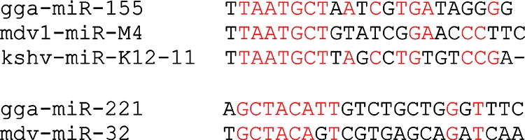
MDV microRNA homologs. MDV microRNAs are aligned with chicken (gga) and KSHV (kshv) microRNAs. Conserved residues are in red.
In order to facilitate the identification of putative target genes for all MDV microRNAs, we have created a database for the 3′ untranslated region of chicken and MDV cDNAs and implemented several available target prediction software programs (11, 31) to identify potential targets. The predicted chicken cDNA targets are too numerous to list and are best viewed online (http://mdvmicrorna.dbi.udel.edu/). Several MDV genes are candidate targets for MDV microRNAs (http://128.175.80.65/mdvHits.html). For example, the gene encoding the 14-kDa lytic phase protein is a candidate target for mdv1-miR-M1. We also noted that the viral lipase gene is a reasonable candidate for mdv1-miR-M2, and mdv-miR-M6 may target the viral CXC chemokine gene. The MDV meq microRNAs are in an antisense orientation relative to the RLORF8 transcript, which encodes a protein of unknown function. The LAT microRNAs are in an antisense orientation relative to the ICP4 transcript but appear to be downstream of the mapped end of the transcript (44). If readthrough extends the ICP4 transcript, LAT microRNAs may act as small interfering RNAs to block ICP4 translation.
DISCUSSION
In this study, we have shown that the sequences of the MDV microRNAs from a continuum of pathotypes are highly conserved. The strains examined span the disease spectrum from mild phenotypes, against which vaccination fully protects, to highly virulent forms that rapidly cause lymphoid lesions and atrophy and can result in early mortality (27, 43). Other than mdv1-miR-M9, no mature microRNAs were found to have sequence variations, and sequence variation in two of the passenger strands (mdv1-miR-M6* and mdv1-miR-M7*) did not appear to affect the levels of the mature microRNAs or the processing of the precursors. Thus, as has been found for other viral systems, the microRNAs appear to be under tight selective pressure that conserves their functions. Several sequence variations in the regions flanking the microRNAs were found; for example, the variation at position 133206 (G→T) is located upstream of all meq microRNAs and is likely to be present within the primary transcript encoding the meq microRNAs or within the promoter for this transcript. This sequence variation generally correlates with viral phenotype in that all but one vv+ strain have a T in this position and all other strains have a G. We have shown that meq microRNAs are present at higher levels in tumors induced by 615K (vv+) than in those induced by RB1B (vv). Although we do not know if the sequence variation has a direct impact on expression levels, we speculate that vv+ MDV may have evolved a mechanism for the overexpression of microRNAs that are advantageous to the virus and that, although neither the molecular mechanism for higher expression nor the targets of these microRNAs are yet known, the meq microRNAs may play a key role in the pathogenesis of MDV.
It is particularly intriguing that mdv1-miR-M4 in the meq cluster is the most abundant MDV microRNA in tumors, as determined by deep sequencing of splenic tumors from MDV (RB1B)-infected chickens. mdv1-miR-M4 shares its seed sequence with miR-155 and KHSV miR-K12-11. miR-155, which is encoded by the bic gene, has been shown previously to be important in normal lymphocyte maturation and differentiation (34), and overexpression produces tumors in both mice and chickens (9, 40). The BACH1/Brip1 gene has been identified as a target for both miR-155 and KSHV miR-K12-11, and a potential binding site in the chicken BACH1 gene is also conserved (14, 37). BACH1 is a basic leucine zipper type transcription factor that can function as an activator or a repressor and has been implicated in a number of cellular functions such as the regulation of the hypoxic response (32) and DNA damage repair in combination with BRAC1 (8). A range of additional targets involved in myeloid hyperplasia and hematopoiesis have been identified by O'Connell et al. (28). The downregulation of microRNAs in tumors is commonly observed, although certain microRNAs can contribute to tumorigenesis (18). We found that cellular miR-155 is downregulated in MDV-induced tumors relative to the levels in normal spleen tissue (our unpublished results available on our website [http://mpss.udel.edu/gga/]). It is conceivable that mdv1-miR-M4 acts to restore miR-155 activity in tumors and helps to maintain the cellular phenotype important for MDV latency and oncogenicity. MDV tumor cells, as well as lymphoblastoid cells derived from MDV-induced tumors, display an activated CD4+ phenotype and are not fully dedifferentiated (29). It is possible that mdv1-miR-M4 compensates for just a subset of miR-155 functions since only the seed sequence is shared.
The LAT-derived MDV1 microRNAs are particularly interesting since the LATs map in an antisense orientation relative to the ICP4 immediate-early gene. It has been proposed previously that herpesvirus microRNAs downregulate the expression of immediate-early proteins and therefore function in the establishment and maintenance of latency (16, 26, 42). In the case of MDV2, one microRNA (MDV2-miR-M17) (46) is in an antisense orientation relative to the coding region of MDV2 ICP4 and may potentially target MDV2 ICP4 mRNA for degradation in a manner similar to the mechanism that small interfering RNAs use to effect mRNA degradation. However, the MDV1 situation is somewhat different. For MDV1, eight microRNAs map in an antisense orientation in relation to and immediately downstream of the major transcription termination site for MDV1 ICP4 (4). It is conceivable that these microRNAs may function to suppress ICP4 expression under conditions that result in the leaky termination of ICP4 transcription; however, any role that this scenario may play in the balance between lytic and latent infection is not clear. It should be noted, however, that the positioning of microRNAs in an antisense orientation relative to and downstream of the ICP4 transcription termination site appears to be MDV1 specific and was not found for MDV2, which is nononcogenic.
Deep sequencing of MDV-induced tumor small RNAs also revealed the sequences of the passenger strands of several MDV microRNAs and confirmed two new microRNAs in the meq cluster, which were also discovered in MSB1 cells (47). In addition, strand selection for mdv1-miR-M2 and mdv1-miR-M8 in tumors appeared to be different from that in CEF (4) and MSB1 cells (47) described previously. Traditionally, the thermodynamic stability of the strands in the stem-loop structure determines which strand is selected as the microRNA, or guide strand and which is selected as the passenger strand (17, 19, 35). However, Ro et al. (33) have shown that both strands can accumulate in some tissues even in the absence of thermodynamic equivalences and, in addition, that differential strand selection in different tissues can occur. The selection of the opposite strand by the RNA-induced silencing complex will likely target a different set of mRNAs and adds to the complexity of regulation by these molecules.
To facilitate the identification of MDV microRNA targets, we have established a database for the 3′ untranslated region of chicken mRNAs and implemented current target prediction software to identify candidates. A bewildering list of potential targets was produced upon analysis of the chicken transcriptome, and only a few high-scoring viral candidates were detected. A number of cellular pathways are proposed to be regulated by viral microRNAs, including those involved in viral replication, cell proliferation, apoptosis, and stress adaptation. Each pathway reveals many candidates, and functional analysis of the pathways perturbed by the individual MDV microRNAs, in combination with our database, will facilitate the discovery of the targets.
Acknowledgments
This work was supported in part by USDA NRI and IFAFS grants.
Footnotes
Published ahead of print on 8 October 2008.
REFERENCES
- 1.Baigent, S. J., L. J. Petherbridge, K. Howes, L. P. Smith, R. J. W. Currie, and V. K. Nair. 2005. Absolute quantitation of Marek's disease virus genome copy number in chicken feather and lymphocyte samples using real-time PCR. J. Virol. Methods 12353-64. [DOI] [PubMed] [Google Scholar]
- 2.Bartel, D. P. 2004. MicroRNAs: genomics, biogenesis, mechanism, and function. Cell 116281-297. [DOI] [PubMed] [Google Scholar]
- 3.Bartel, D. P., and C.-Z. Chen. 2004. Micromanagers of gene expression: the potentially widespread influence of metazoan microRNAs. Nat. Rev. Genet. 5396-400. [DOI] [PubMed] [Google Scholar]
- 4.Burnside, J., E. Bernberg, A. Anderson, C. Lu, B. C. Meyers, P. J. Green, N. Jain, G. Isaacs, and R. W. Morgan. 2006. Marek's disease virus microRNAs map to meq and the latency-associated transcript. J. Virol. 808778-8786. [DOI] [PMC free article] [PubMed] [Google Scholar]
- 5.Burnside, J., M. Ouyang, A. Anderson, E. Bernberg, C. Lu, B. C. Meyers, P. J. Green, M. Markis, G. Isaacs, E. Huang, and R. W. Morgan. 2008. Deep sequencing of chicken microRNAs. BMC Genomics 9185. [DOI] [PMC free article] [PubMed] [Google Scholar]
- 6.Bushati, N., and S. M. Cohen. 2007. MicroRNA functions. Annu. Rev. Cell Dev. Biol. 23175-205. [DOI] [PubMed] [Google Scholar]
- 7.Cai, X., A. Schafer, S. Lu, J. P. Bilello, R. C. Desrosiers, R. Edwards, N. Raab-Traub, and B. R. Cullen. 2006. Epstein-Barr virus microRNAs are evolutionarily conserved and differentially expressed. PLoS Pathog. 2e23. [DOI] [PMC free article] [PubMed] [Google Scholar]
- 8.Cantor, S., D. Bell, S. Ganesan, E. Kass, R. Drapkin, S. Grossman, D. Wahrer, D. Sgroi, W. Lane, D. Haber, and D. Livingston. 2001. BACH1, a novel helicase-like protein, interacts directly with BRCA1 and contributes to its DNA repair function. Cell 105149-160. [DOI] [PubMed] [Google Scholar]
- 9.Costinean, S., N. Zanesi, Y. Pekarsky, E. Tili, S. Volinia, N. Heerema, and C. M. Croce. 2006. Pre-B cell proliferation and lymphoblastic leukemia/high-grade lymphoma in E mu-miR155 transgenic mice. Proc. Natl. Acad. Sci. USA 1037024-7029. [DOI] [PMC free article] [PubMed] [Google Scholar]
- 10.Cullen, B. 2006. Viruses and microRNAs. Nat. Genet. 38525-530. [DOI] [PubMed] [Google Scholar]
- 11.Enright, A., B. John, U. Gaul, T. Tuschl, C. Sander, and D. Marks. 2003. MicroRNA targets in Drosophila. Genome Biol. 5R1. [DOI] [PMC free article] [PubMed] [Google Scholar]
- 12.Fornari, F., L. Gramantieri, M. Ferracin, A. Veronese, S. Sabbioni, G. A. Calin, G. L. Grazi, C. Giovannini, C. M. Croce, L. Bolondi, and M. Negrini. 2 June 2008, posting date. MiR-221 controls CDKN1C//p57 and CDKN1B//p27 expression in human hepatocellular carcinoma. Oncogene 275651-5661. [DOI] [PubMed] [Google Scholar]
- 13.Galardi, S., N. Mercatelli, E. Giorda, S. Massalini, G. V. Frajese, S. A. Ciafre, and M. G. Farace. 2007. miR-221 and miR-222 expression affects the proliferation potential of human prostate carcinoma cell lines by targeting p27Kip1. J. Biol. Chem. 28223716-23724. [DOI] [PubMed] [Google Scholar]
- 14.Gottwein, E., N. Mukherjee, C. Sachse, C. Frenzel, W. H. Majoros, J.-T. A. Chi, R. Braich, M. Manoharan, J. Soutschek, U. Ohler, and B. R. Cullen. 2007. A viral microRNA functions as an orthologue of cellular miR-155. Nature 4501096-1099. [DOI] [PMC free article] [PubMed] [Google Scholar]
- 15.Grey, F., H. Meyers, E. A. White, D. H. Spector, and J. Nelson. 2007. A human cytomegalovirus-encoded microRNA regulates expression of multiple viral genes involved in replication. PLoS Pathog. 31593-1602. [DOI] [PMC free article] [PubMed] [Google Scholar]
- 16.Grey, F., and J. Nelson. 2008. Identification and function of human cytomegalovirus microRNAs. J. Clin. Virol. 41186-191. [DOI] [PMC free article] [PubMed] [Google Scholar]
- 17.Han, J., Y. Lee, K. Yeom, J. Nam, I. Heo, J. Rhee, S. Sohn, Y. Cho, B. Zhang, and V. Kim. 2006. Molecular basis for the recognition of primary microRNAs by the Drosha-DGCR8 complex. Cell 125887-901. [DOI] [PubMed] [Google Scholar]
- 18.Kent, O. A., and J. T. Mendell. 2006. A small piece in the cancer puzzle: microRNAs as tumor suppressors and oncogenes. Oncogene 256188-6196. [DOI] [PubMed] [Google Scholar]
- 19.Khvorova, A., A. Reynolds, and S. D. Jayasena. 2003. Functional siRNAs and miRNAs exhibit strand bias. Cell 115209-216. [DOI] [PubMed] [Google Scholar]
- 20.Levy, A. M., O. Gilad, L. Xia, Y. Izumiya, J. Choi, A. Tsalenko, Z. Yakhini, R. Witter, L. Lee, C. J. Cardona, and H.-J. Kung. 2005. Marek's disease virus Meq transforms chicken cells via the v-Jun transcriptional cascade: a converging transforming pathway for avian oncoviruses. Proc. Natl. Acad. Sci. USA 10214831-14836. [DOI] [PMC free article] [PubMed] [Google Scholar]
- 21.Liu, J.-L., Y. Ye, L. F. Lee, and H.-J. Kung. 1998. Transforming potential of the herpesvirus oncoprotein MEQ: morphological transformation, serum-independent growth, and inhibition of apoptosis. J. Virol. 72388-395. [DOI] [PMC free article] [PubMed] [Google Scholar]
- 22.Lodish, H. F., B. Zhou, G. Liu, and C.-Z. Chen. 2008. Micromanagement of the immune system by microRNAs. Nat. Rev. Immunol. 8120-130. [DOI] [PubMed] [Google Scholar]
- 23.Lu, C., B. C. Meyers, and P. J. Green. 2007. Construction of small RNA cDNA libraries for deep sequencing. Methods 43110-117. [DOI] [PubMed] [Google Scholar]
- 24.Marshall, V., T. Parks, R. Bagni, C. Wang, M. Samols, J. Hu, K. Wyvil, K. Aleman, R. Little, R. Yarchoan, R. Renne, and D. Whitby. 2007. Conservation of virally encoded microRNAs in Kaposi sarcoma-associated herpesvirus in primary effusion lymphoma cell lines and in patients with Kaposi sarcoma or multicentric Castleman disease. J. Infect. Dis. 195645-659. [DOI] [PubMed] [Google Scholar]
- 25.Morgan, R. W., Q. Xie, J. L. Cantello, A. Miles, E. Bernberg, J. Kent, and A. S. Anderson. 2001. Marek's disease virus latency, p. 223-243. In K. Hirai (ed.), Current topics in microbiology and immunology, vol. 255. Marek's disease. Springer, New York, NY. [PubMed] [Google Scholar]
- 26.Murphy, E., J. Vanicek, H. Robins, T. Shenk, and A. J. Levine. 2008. Suppression of immediate-early viral gene expression by herpesvirus-coded microRNAs: implications for latency. Proc. Natl. Acad. Sci. USA 1055453-5458. [DOI] [PMC free article] [PubMed] [Google Scholar]
- 27.Nair, V. 2005. Evolution of Marek's disease—a paradigm for incessant race between the pathogen and the host. Vet. J. 170175-183. [DOI] [PubMed] [Google Scholar]
- 28.O'Connell, R. M., D. S. Rao, A. A. Chaudhuri, M. P. Boldin, K. D. Taganov, J. Nicoll, R. L. Paquette, and D. Baltimore. 2008. Sustained expression of microRNA-155 in hematopoietic stem cells causes a myeloproliferative disorder. J. Exp. Med. 205585-594. [DOI] [PMC free article] [PubMed] [Google Scholar]
- 29.Parcells, M. S., R. L. Dienglewicz, A. S. Anderson, and R. W. Morgan. 1999. Recombinant Marek's disease virus (MDV)-derived lymphoblastoid cell lines: regulation of a marker gene within the context of the MDV genome. J. Virol. 731362-1373. [DOI] [PMC free article] [PubMed] [Google Scholar]
- 30.Pfeffer, S., and O. Voinnet. 2006. Viruses, microRNAs and cancer. Oncogene 256211-6219. [DOI] [PubMed] [Google Scholar]
- 31.Rehmsmeier, M., P. Steffen, M. Höchsmann, and R. Giegerich. 2004. Fast and effective prediction of microRNA/target duplexes. RNA 101507-1517. [DOI] [PMC free article] [PubMed] [Google Scholar]
- 32.Reichard, J. F., M. A. Sartor, and A. Puga. 2008. BACH is a specific repressor of HMOX1 that is inactivated by arsenite. J. Biol. Chem. 28322363-22370. [DOI] [PMC free article] [PubMed] [Google Scholar]
- 33.Ro, S., C. Park, D. Young, K. M. Sanders, and W. Yan. 2007. Tissue-dependent paired expression of miRNAs. Nucleic Acids Res. 355944-5953. [DOI] [PMC free article] [PubMed] [Google Scholar]
- 34.Rodriguez, A., E. Vigorito, S. Clare, M. V. Warren, P. Couttet, D. R. Soond, S. van Dongen, R. J. Grocock, P. P. Das, E. A. Miska, D. Vetrie, K. Okkenhaug, A. J. Enright, G. Dougan, M. Turner, and A. Bradley. 2007. Requirement of bic/microRNA-155 for normal immune function. Science 316608-611. [DOI] [PMC free article] [PubMed] [Google Scholar]
- 35.Schwarz, D., G. Hutvágner, T. Du, Z. Xu, N. Aronin, and P. Zamore. 2003. Asymmetry in the assembly of the RNAi enzyme complex. Cell 115199-208. [DOI] [PubMed] [Google Scholar]
- 36.Shamblin, C. E., N. Greene, V. Arumugaswami, R. L. Dienglewicz, and M. S. Parcells. 2004. Comparative analysis of Marek's disease virus (MDV) glycoprotein-, lytic antigen pp38- and transformation antigen Meq-encoding genes: association of meq mutations with MDVs of high virulence. Vet. Microbiol. 102147-167. [DOI] [PubMed] [Google Scholar]
- 37.Skalsky, R. L., M. A. Samols, K. B. Plaisance, I. W. Boss, A. Riva, M. C. Lopez, H. V. Baker, and R. Renne. 2007. Kaposi's sarcoma-associated herpesvirus encodes an ortholog of miR-155. J. Virol. 8112836-12845. [DOI] [PMC free article] [PubMed] [Google Scholar]
- 38.Spatz, S. J., and R. F. Silva. 2007. Sequence determination of variable regions within the genomes of gallid herpesvirus-2 pathotypes. Arch. Virol. 1521665-1678. [DOI] [PubMed] [Google Scholar]
- 39.Sugaya, K., G. Bradley, M. Nonoyama, and A. Tanaka. 1990. Latent transcripts of Marek's disease virus are clustered in the short and long repeat regions. J. Virol. 645773-5782. [DOI] [PMC free article] [PubMed] [Google Scholar]
- 40.Tam, W., S. H. Hughes, W. S. Hayward, and P. Besmer. 2002. Avian bic, a gene isolated from a common retroviral site in avian leukosis virus-induced lymphomas that encodes a noncoding RNA, cooperates with c-myc in lymphomagenesis and erythroleukemogenesis. J. Virol. 764275-4286. [DOI] [PMC free article] [PubMed] [Google Scholar]
- 41.Thai, T.-H., D. P. Calado, S. Casola, K. M. Ansel, C. Xiao, Y. Xue, A. Murphy, D. Frendewey, D. Valenzuela, J. L. Kutok, M. Schmidt-Supprian, N. Rajewsky, G. Yancopoulos, A. Rao, and K. Rajewsky. 2007. Regulation of the germinal center response by microRNA-155. Science 316604-608. [DOI] [PubMed] [Google Scholar]
- 42.Umbach, J. L., M. F. Kramer, I. Jurak, H. W. Karnowski, D. M. Coen, and B. R. Cullen. 2008. MicroRNAs expressed by herpes simplex virus 1 during latent infection regulate viral mRNAs. Nature 454780-783. [DOI] [PMC free article] [PubMed] [Google Scholar]
- 43.Witter, R. L. 1997. Increased virulence of Marek's disease virus field isolates. Avian Dis. 41149-163. [PubMed] [Google Scholar]
- 44.Xie, Q. 1998. Identification of genes important for the maintenance of Marek's disease virus transformation and further characterization of the MDV ICP4 gene. University of Delaware, Newark, DE.
- 45.Xu, H., Y. Yao, Y. Zhao, L. P. Smith, S. J. Baigent, and V. K. Nair. 2008. Analysis of the expression profiles of Marek's disease virus-encoded microRNAs by real-time quantitative PCR. J. Virol. Methods 149201-208. [DOI] [PubMed] [Google Scholar]
- 46.Yao, Y., Y. Zhao, H. Xu, L. P. Smith, C. H. Lawrie, A. Sewer, M. Zavolan, and V. Nair. 2007. Marek's disease virus type 2 (MDV-2)-encoded microRNAs show no sequence conservation with those encoded by MDV-1. J. Virol. 817164-7170. [DOI] [PMC free article] [PubMed] [Google Scholar]
- 47.Yao, Y., Y. Zhao, H. Xu, L. P. Smith, C. H. Lawrie, M. Watson, and V. Nair. 2008. MicroRNA profile of Marek's disease virus-transformed T-cell line MSB-1: predominance of virus-encoded microRNAs. J. Virol. 824007-4015. [DOI] [PMC free article] [PubMed] [Google Scholar]