Abstract
Case report
A fetus with rhombencephalosynapsis and prenatally diagnosed tetrasomy 9p is reported. Chromosomal analysis from amniocyte culture revealed non-mosaic supernumerary chromosome identified as isochromosome 9p (9p24→q13::q13→p24). Ultrasound scan revealed intrauterine growth retardation, renal anomalies, cardiac anomalies, ventriculomegaly, and agenesis of cerebellar vermis with fusion of the cerebellar hemispheres.
Conclusion
Although most cases of cerebellar vermis agenesis in tetrasomy 9p are described with cystic malformation such as Dandy-Walker anomaly, our case indicates that this chromosomal disorder should be taken into account in fetuses with the development of cystic and non-cystic malformations of cerebellar vermis and posterior fossa.
Keywords: Tetrasomy 9p, Rhombencephalonsynapsis, Polymalformations, Prenatal diagnosis, ARTs.
Introduction
Rhombencephalosynapsis (RES) is a rare congenital abnormality characterized by vermian agenesis and fusion of the cerebellar hemispheres [1]. Most of the reported cases are associated with brain anomalies such as hydrocephalus or ventriculomegaly, or other anomalies involving corpus callosum and midline. Pathogenesis and genetic basis of such anomaly are unknown, and chromosomal studies were uneventful in all cases but one with interstitial deletion of chromosome 2q [1].
The hypoplasia of the cerebellar vermis and large infratentorial cyst due to a diverticular expansion of the forth ventricle is defined as Dandy-Walker malformation (DWM). DWM has been described in association with tetrasomy 9p (2)(OMIM 220200).
Tetrasomy 9p is an uncommon chromosomal syndrome which was first described by Ghymers et al. in 1973 [3], and the first case of prenatal diagnosis was reported in 1991 [4].
To date, about 40 cases of tetrasomy 9p due to a supernumerary isochromosome 9p have been reported, in association with a variability in morphologic expression, though the phenotypic differences in tetrasomy 9p patients seem to be the result of the degree of mosaicism [5].
We report on a polymalformed fetus with RES and non-mosaic tetrasomy 9p in a fetus conceived from intracytoplasmic sperm injection (ICSI) pregnancy.
Case report
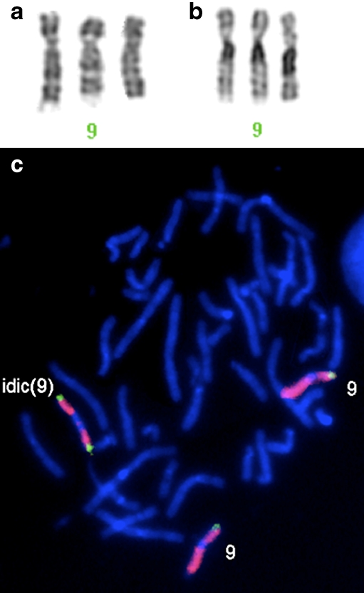
A healthy 37-year-old gravida one woman was referred for second level ultrasound scan at 19 weeks of gestation because of oligohydramnios and suspected DWM. The pregnancy was conceived from ICSI due to maternal factor (absence of tubal patency and endometriosis). The pregnancy was uneventful except for a first scan at 12 weeks’ gestation demonstrating an enlarged nuchal translucency. At the time of presentation the scan demonstrated a complex pattern of fetal anomalies characterized by intrauterine growth retardation, micromelia, brachydactyly, rocker-bottom feet, horse-shoe kidney, atrio-ventricular septal defect, truncus arteriosus, hypertelorism, bilateral cleft lip and palate with pre-maxillary protrusion, cystic hygroma, pre-auricular tag, cupped ears, cerebral hemorrhage, bilateral borderline ventriculomegaly, abnormal cerebellum with mild hypoplastic cerebellum, absence of vermis with fusion of the cerebellar hemispheres posterior fossa cyst and absence of cystic dilatation of fourth ventricle (Fig. 1a). She underwent amniocentesis and the cytogenetic analysis revealed a non-mosaic supernumerary marker chromosome (Fig. 2a) which showed two large heterochromatin blocks at C-banding (Fig. 2b). Fluorescence in situ hybridization (FISH) using chromosome 9 specific painting and subtelomeric p and q probes showed that the supernumerary marker chromosome was an isochromosome consisting of two copies of the entire short arm and the heterochromatic region of the long arm of a chromosome 9 (Fig. 2c). The karyotype was redefined as 47,XY,+idic(9)(p24→q13::q13→p24).
Fig. 1.
a Ultrasound scan demonstrating abnormal cerebellum. b Post-mortem evaluation of posterior fossa and cerebellum
Fig. 2.
a Partial karyotypes of chromosomes 9 at G-banding, b and at C-banding. c FISH analysis using subtelomere 9p probe (green) shows two 9p signals on der[9] in addition to a signal on normal chromosome 9. Chromosome 9 painting probe (red) is also shown
Parental karyotypes were normal. In order to establish parental origin of chromosome 9p tetrasomy, using C-banding no 9q heterochromatin polymorphism was observed in both parents. Parents were unavailable for DNA microsatellite analysis.
After extensive counseling the parents opted for termination of pregnancy.
At post-mortem examinations the US anomalies were confirmed. The cerebellum confirmed a RES anomaly (Fig. 1b). Additional anomalies included absence of right pectoralis muscle, downward slanting mouth, broad prominent nasal bridge with excess skin, and right abnormal pulmonary lobes, consisting in absence of division into right upper and middle lobe.
Discussion
RES is a rare central nervous system malformation that is characterized by vermian agenesis and cerebellar hemispheres fusion [1]. Additional brain anomalies are reported with RES, in one case described as associated with chromosome 2q deletion [2].
Enlarged nuchal translucency is a well-known sonographic sign related to many of the features described in the present case such as cystic hygroma and congenital heart disease.
However, Lespinasse et al. [6] described a fetus with enlarged nuchal translucency with normal standard karyotype presenting at birth a developmental delay and RES with submicrosopic unbalanced subtelomeric translocation t(2p; 10p).
Tetrasomy 9p was first described by Ghymers et al. in 1973 [3]. Since then few cases have been reported and a review of previously reported features described the variability of appearance in fetuses and newborns affected by tetrasomy 9p mosaic and non-mosaic [7]. In many cases, the syndrome is immediately recognized due to a characteristic facial appearance with ocular, nasal, oral and ear anomalies. Central nervous system malformations, skeletal anomalies, congenital heart defects, renal anomalies, cystic hygroma, and small for gestational age growth are frequently present [7].
Fetuses with tetrasomy 9p that includes the region 9pter-p11.1 are likely to present a DWM and ventriculomegaly on prenatal ultrasound [8].
So far this is the first report of RES in a fetus with tetrasomy 9p deriving from assisted reproduction technologies (ARTs).
Furthermore, albeit comparison of fetuses naturally conceived and IVF pregnancies, ICSI pregnancies describes an higher risk for chromosomal anomalies [9–10], this is the first description of tetrasomy 9p in an ART pregnancy. Although pregnant women after ART are generally older than women with spontaneously conceived pregnancies, at present no clear correlation between tetrasomy 9p occurrence and advanced maternal age is reported[11].
Our case presents classical features of tetrasomy 9p, that, albeit peculiar, are commonly described in trisomy 9 and in trisomy 13 fetuses [12]. Absence of cerebellar vermis is well-described in fetuses with either trisomy 13 and 9, albeit described in all reports as cystic malformation like DWM [2].
The present case is unique in the fact that, even though RES is a cerebellar malformation different from DWM [1], the karyotype in our case is overlapping with that reported as associated to DWM [2]. Furthermore the fetus is deriving from intracytoplasmic sperm injection (ICSI), being the first case described in an ART pregnancy.
Evaluation of this case delineates a clear phenotype for this syndrome. Prenatal ultrasound findings commonly overlap with those seen in trisomy 13. However, the diagnosis of tetrasomy 9p should be taken into account in all cases of prenatal suspect of trisomy 13 with normal results at rapid FISH analysis from uncultured amniocytes, considering the long term culture the most reliable result.
However, albeit deriving from different embryonic development, our case is confirming that a dosage effect of genes located on 9pter-9q12 may be associated with abnormal neural migration and therefore with the development of cystic [13] and non-cystic malformations of cerebellar vermis and posterior fossa [14].
We conclude that after sonographic demonstration of cerebellar vermis anomalies, particularly in combination with other fetal anomalies, prenatal diagnosis for standard fetal karyotyping is strongly suggested.
Footnotes
Capsule Tetrasomy 9p in a polymalformed fetus with rhombencephalonsynapsis is described in a pregnancy conceived by ICSI.
References
- 1.Truwit CL, Barkovich AJ, Shanahan R, Maroldo TV. MR imaging of rhombencephalosinapsis: report of three cases ond review of the literature. AJNR Am J Neuroradiol. 1991;12:957–65. [PMC free article] [PubMed]
- 2.Melaragno MI, Brunoni D, da Silva Patricia FR, Corbani M, Mustacchi Z, et al. A patient with tetrasomy 9p. Dandy-Walker cyst and Hirchsprung disease. Ann Genet. 1992;35:79–84. [PubMed]
- 3.Ghymers D, Hemann B, Distèche C, Frederic J. Tetrasomie partielle du chromosome 9 à l’ètat mosaique, chez un enfant porteur de malformations multiples. Hum Genet. 1973;20:273–82. doi:10.1007/BF00385740. [DOI] [PubMed]
- 4.Shaefer GB, Domek DB, Morgan MA, Muneer RS, Johnson SF. Tetrasomy of the short arm of chromosome 9: prenatal diagnosis and further delineation of the phenotype. Am J Med Genet. 1991;38:612–5. doi:10.1002/ajmg.1320380422. [DOI] [PubMed]
- 5.Tonk VS. Moving towards a syndrome: a review of 20 cases and a new case of non-mosaic tetrasomy 9p with long-term survival. Clin Genet. 1997;52:23–9. [DOI] [PubMed]
- 6.Lespinasse J, Testard H, Nugues F, Till M, Cordier MP, Althuser M, et al. A submicroscopic unbalanced subtelomeric translocation t(2p;10q) identified by fluorescence in situ hybridization: fetus with increased nuchal translucency and normal standard karyotype with later growth and developmental delay, rhombencephalosynapsis (RES). Ann Genet. 2004;47:405–17. doi:10.1016/j.anngen.2004.07.005. [DOI] [PubMed]
- 7.Dhandha S, Hogge WA, Surti U, McPherson E. Three cases of tetrasomy 9p. Am J Med Genet. 2002;113:375–80. doi:10.1002/ajmg.b.10826. [DOI] [PubMed]
- 8.Chen CP, Chang TY, Shih JC, Lin SP, Lin CJ, Wang W, et al. Prenatal diagnosis of dandy-Walker malformation and ventriculomegaly associated with partial trisomy 9p and distal 12p deletion. Prenat Diagn. 2002;12:1063–5. doi:10.1002/pd.459. [DOI] [PubMed]
- 9.Van Steirteghem A, Bonduelle M, Devroey P, Liebaers I. Follow-up of children born after ICSI. Hum Reprod Update. 2002;8:111–6. doi:10.1093/humupd/8.2.111. [DOI] [PubMed]
- 10.Gjerris AC, Loft A, Pinborg A, Christiansen M, Tabor A. Prenatal testing among women pregnant after assisted reproductive techniques in Denmark 1995–2000: a national cohort study. Hum Reprod. 2008;23:1545–52. doi:10.1093/humrep/den103. [DOI] [PubMed]
- 11.de Azevedo Moreira LM, Magalhanes Freitas L, Ferreira Gusmano FA, Riegel M. New case of non-mosaic Tetrasomy 9p in a severely polymalformed newborn girl. Birth Defects Res. 2003;67:985–8. doi:10.1002/bdra.10126, Part A. [DOI] [PubMed]
- 12.Feingold M, Atkins L. A case of trisomy 9. J Med Genet. 1973;10:184–7. [DOI] [PMC free article] [PubMed]
- 13.Federico A, Tomassetti P, Zollino M, Diomedi M, Dotti MT, De Stefano N, Gualdi GF, Neri G, Gigli GL. Association of trisomy 9p and band heterotopia. Neurology. 1999;2:430–2. [DOI] [PubMed]
- 14.Utsunomiya H, Takano K, Ogasawara T, Hashimoto T, Fukushima T, Okazaki M. Rhombencephalosynapsis: cerebellar embryogenesis. AJNR Am J Neuroradiol. 1998;19:547–9. [PMC free article] [PubMed]


