Abstract
Following TCR stimulation, T cells utilize the hematopoietic specific adhesion and degranulation promoting adapter protein (ADAP) to control both integrin adhesive function and NF-κB transcription factor activation. We have investigated the molecular basis by which ADAP controls these events in primary murine ADAP-/- T cells. Naïve DO11.10/ADAP-/- T cells show impaired adhesion to OVAp-bearing antigen presenting cells (APCs) that is restored following reconstitution with wild-type ADAP. Mutational analysis demonstrates that the central proline-rich domain and the C-terminal domain of ADAP are required for rescue of T:APC conjugate formation. The ADAP proline-rich domain is sufficient to bind and stabilize the expression of SKAP55, which is otherwise absent from ADAP-/- T cells. Interestingly, forced expression of SKAP55 in the absence of ADAP is insufficient to drive T:APC conjugate formation, demonstrating that both ADAP and SKAP55 are required for optimal LFA-1 function. In addition, the ADAP proline rich domain is required for optimal antigen-induced activation of CD69, CD25, and Bcl-xL, but is not required for assembly of the CARMA1/Bcl10/MALT1 signaling complex and subsequent TCR-dependent NF-κB activity. Our results indicate that ADAP is used downstream of TCR engagement to delineate two distinct molecular programs, in which the ADAP/SKAP55 module is required for control of T:APC conjugate formation and functions independently of ADAP/CARMA1 mediated NF-κB activation.
Introduction
Naïve T cells respond to antigen engagement of the TCR by rapidly increasing the functional activity of their integrin adhesion receptors, and by initiating molecular programs that promote proliferation and the gain of effector function. Cytosolic adapter proteins and enzymes control the initiation and transmission of these TCR-specific signals (1-3). One key adapter, the linker for activated T cells (LAT)4, is rapidly tyrosine phosphorylated following TCR stimulation and provides docking sites for the src homology 2 domain-containing leukocyte-specific phosphoprotein of 76 kD (SLP-76) and phospholipase C (PLC-γ1) (4). These proteins are critical for proximal signals generated by the TCR, as loss of LAT or SLP-76 expression severely disrupts T cell development (5-7). In contrast, disruption of proteins downstream of the LAT signalosome results in more selective defects in T cell activation.
Adhesion and degranulation promoting adapter protein (ADAP; formerly known as Fyb or SLAP-130) is expressed by T cells and other hematopoietic lineages except B cells. ADAP was originally identified by its TCR-dependent tyrosine phosphorylation and interaction with SLP-76 and the tyrosine kinase Fyn (8, 9). Like other adapters, ADAP contains a number of protein-protein and protein-lipid interaction domains (1, 10, 11). The C-terminus of ADAP contains several tyrosine residues that are involved in TCR dependent binding to SLP-76 and Fyn (12-14), as well as FPPP domains that have been implicated in interaction with the Ena/Vasp family of actin regulatory proteins (15). A central proline-rich domain between aa 326-426 of murine ADAP constitutively binds the SH3 domain of another adapter, SKAP55 (16-18). Immediately adjacent to this domain, aa 426-541 in ADAP contains an E/K rich domain and part of an atypical helical SH3 domain (hSH3-N) that comprise a binding site for the adapter CARMA1 that is critical for formation of the CARMA1/Bcl10/Malt1 signaling complex and NF-κB activation (19). This hSH3-N domain and a highly homologous hSH3-C located at the extreme C-terminus of ADAP bind to phospholipids in vitro (20, 21). Finally, the N-terminal ∼300 aa of ADAP binds HIP-55 (22), although the significance of this interaction is unclear.
Although initial overexpression studies reported both positive and negative roles for ADAP in T cell activation (8, 9, 12, 14), the production of ADAP-/- mice demonstrated that ADAP positively regulates T cell activation (23, 24). While ADAP-/- T cells mice show normal proximal TCR dependent responses including Ca++ mobilization and ERK phosphorylation, they exhibit impaired TCR and antigen-dependent integrin-mediated adhesion and subsequent T cell activation and survival (23-25). ADAP or SKAP55 overexpression enhances LFA-1 dependent T cell interactions with antigen-presenting cells (APCs) in a manner dependent on the SKAP55 SH3 domain (26). Similarly, T cells from SKAP55 knockdown (27) or from SKAP55-/- mice (28) exhibit defects in LFA-1 function comparable to ADAP-/- mice. The ADAP proline-rich and SKAP55 SH3 domains are critical for TCR dependent membrane targeting of Rap1 (29), a small GTPase that is important for T cell integrin activation and T:APC conjugate formation (30-32). In addition, SKAP55 constitutively associates with RIAM, which binds to the active form of Rap1 (29, 33). Thus, an ADAP/SKAP55/RIAM/Rap1 signaling arm appears to be required for control of T cell integrin activation. However, the identification of precise functions for ADAP has been complicated by the observation that stable SKAP55 expression requires a constitutive SH3 domain-mediated interaction of SKAP55 with ADAP (17, 34). The role of the ADAP/SKAP55 interaction in NF-κB activation, and the function of SKAP55 independent of ADAP expression, has not been investigated.
In the present study, we used adenovirus to express ADAP and ADAP mutant constructs in resting naïve T cells from ADAP-/- mice expressing the hCAR adenovirus receptor (35, 36) and the ovalbumin323-339 (OVAp) specific DO11.10 transgenic TCR (25, 37). Using naïve mouse T lymphocytes, we show that a small region of the ADAP proline-rich domain is required for rescue of SKAP55 expression by ADAP-/- T cells, and for optimal antigen-dependent T:APC conjugate formation and downstream T cell activation. However, SKAP55 expression alone in the absence of ADAP is not sufficient to restore T:APC conjugate formation. Furthermore, we demonstrate that formation of the ADAP/SKAP55 complex is not required for control of NF-κB activation and that SKAP55 is not found in the CARMA1/Bcl10 complex, suggesting differential control of integrin and NF-κB pathways by ADAP.
Materials and Methods
Mice
DO11.10/ADAP+/+ and ADAP-/- mice on the Balb/c background have been previously described (25) and were crossed to the hCAR transgenic mice (35) (provided by Dr. C. Weaver, University of Alabama-Birmingham). Mice were housed in specific pathogen-free facilities at the University of Minnesota, and were used between 8 and 12 weeks of age. All experimental protocols involving the use of mice were approved by the Institutional Animal Care and Use Committee at the University of Minnesota.
Cells
The Jurkat E6-1 human T cell line was obtained from American Type Culture Collection (Manassas, VA) and maintained at 37C in RPMI-1640 supplemented with fetal calf serum (Atlanta Biologicals, Atlanta, GA), L-glutamine and penicillin/streptomycin and grown in 5% CO2.
Antibodies and Reagents
The DO11.10 TCR was detected with FITC- or biotin-conjugated mAb KJ1-26 (Caltag). Anti-Thy1.1 APC or PE, anti-B220-PE-Cy5.5, anti-CD69-FITC, and anti-CD25-APC were from eBioscience (San Diego). Sheep anti-murine ADAP has been previously described (38). The following antibodies were purchased: rabbit anti-SKAP55 (Upstate, Lake Placid, NY), rabbit anti-CARMA1 (Alexis Biochemicals, San Diego, CA), goat anti-HA agarose (Bethyl Laboratories, Montgomery, TX), mouse anti-HA (Clone 16B12) (Covance), mouse anti-Bcl10 (331.3) and mouse anti-NF-κB p65 (F-9) (Santa Cruz Biotechnology, Santa Cruz, CA). Alexa 488 conjugated donkey anti-sheep and goat anti-rabbit IgG were used for intracellular staining detection. For immunoblotting, Alexa Fluor 680 (Molecular Probes) goat anti-mouse and goat anti-rabbit, and IRDye 800-conjugated goat anti-rabbit, goat anti-mouse and donkey anti-sheep IgG (Rockland Immunochemicals, Gilbertsville, PA) were used.
Recombinant DNA
The pENTR-HA-ADAP construct for adenovirus production encodes for full-length murine ADAP (130 kDa isoform) and is derived from pENTR-UP-IT (19). ADAP mutants were generated as previously described (19) using the QuickChange® II XL Site-Directed Mutagenesis kit (Stratagene) by introducing a stop codon at amino acid position 426 (mutants Δ426–819) or by using primers flanking both sides of the deleted site (mutants Δ1-327, Δ326-426, Δ338-358, and Δ426-541). FLAG-tagged SKAP55 was obtained from Dr. B. Schraven (Otto-von-Guericke University, Magdeburg, Germany) and was subcloned into pENTR-UP-IT by blunt ligation. GFP-Rap1 was generated by inserting Rap1 in frame in pEGFP-C1 (Clontech) and subcloning the GFP-Rap1 cassette into the EcoRV site of pENTR-UP-IT. All constructs were verified by DNA sequencing at the University of Minnesota Microsequencing Facility.
Adenovirus production and transduction
Adenovirus expression plasmids were generated as previously described (19) using Gateway recombination between pAD/PL-DEST (Invitrogen) and either control pENTR-UP-IT or pENTR-HA-ADAP. Adenovirus was produced in AD-293 packaging cells (Stratagene), and viral particles were purified and titered as described (35, 39, 40). Freshly isolated, resting ADAP+/+ and ADAP-/- lymph node T cells expressing the hCAR receptor were transduced with control, ADAP, or SKAP55 adenoviruses as described (35) and incubated at 37C for 3 days in complete T cell medium containing 5 ng/ml mouse IL-7 (R&D Systems, Minneapolis, MN). Preliminary experiments were performed to optimize the length of incubation required to achieve peak ADAP or SKAP55 expression. Expression of HA-ADAP or FLAG-SKAP55 was confirmed in all experiments by intracellular flow cytometry for ADAP (38), HA, or SKAP55. Transduction of Jurkat T cells was performed similarly to the primary T cells (19). Briefly, 10 x 106 Jurkat cells at a density of 50 x 106/ml in DMEM containing 10mM HEPES (pH 7.4) were incubated with 500 x 106 infectious units of adenovirus (MOI 50) for 30min at 37C. Cells were washed and cultured in T cell medium at 0.5-1 x 10⁶ cells/ml for 2d. Flow cytometric analysis (data not shown) indicated that 80-90% of cells in these cultures were transduced and expressed the indicated ADAP construct.
Conjugate and activation assays
Flow cytometry-based conjugate assays were performed as previously described (25). Briefly, control wild-type (WT) and ADAP-/- DO11.10/hCAR bulk LN T cells were transduced with adenovirus as described above. Fresh non-transgenic Balb/c splenocytes were labeled with Cell Tracker Orange (Molecular Probes) and then left unpulsed or pre-pulsed for 30 minutes at 37C with OVAp (Ovalbumin aa323-339, Invitrogen) at the indicated concentrations. Transduced KJ1-26+ Thy1.1+ T cells were then combined at a 1:4 ratio with the peptide-loaded splenocytes, pelleted in a 96-well round-bottom plate, incubated for 10 min at 37C, mixed for 20 seconds in a plate shaker, fixed with 1% paraformaldehyde (PFA), and stained for flow cytometry. Conjugates were defined as KJ1-26+Thy1.1+ T cell events co-staining with Cell Tracker Orange and B220. In vitro activation was performed similar to the conjugate assays except 0.1 X 10⁶ KJ1-26+Thy1.1+ T cells were seeded into 96-well flat-bottomed plates and activated by the addition of 0.2 X 10⁶ unstained, peptide-loaded splenocytes. Cells were harvested at the indicated times and KJ1-26+Thy1.1+ T cells were stained for CD25, CD69, and Bcl-xL as previously described (25).
Immunoprecipitation and immunoblotting
Immunoprecipitation and immunoblotting for Bcl10, Carma1, and endogenous ADAP was performed as previously described (19, 41). Briefly, 10X106 cells per condition in PBS containing 0.5% BSA were stimulated with PMA (50ng/ml), lysed with an equal volume of 2X lysis buffer (2% NP-40, 100 mM Tris-HCl pH 7.6, 300 mM NaCl, 4 mM EDTA, 2 mM sodium vanadate, 10 μg/ml leupeptin, 10 μg/ml aprotonin, 2 mM PMSF), and then cleared by centrifugation at 12,000 rcf. For immunoprecipitation, individual antibodies were adsorbed to protein A-sepharose for 2 hr at 4°C, washed with 0.2 M NaBO4 (pH 9.0), resuspended in 20 mM dimethyl pimelimedate and incubated for 30 min at RT, washed with 0.2 M ethanolamine (pH 8.0) and incubated for 2 hr in 0.2 M ethanolamine. The cleared lysates were then incubated overnight at 4C with the crosslinked bead/antibody complexes. The immune complexes were washed twice with 1X lysis buffer containing 0.1% Triton-X 100 and separated by SDS-PAGE. After Western transfer of the proteins, PVDF membrane was blocked with 0.2% casein for 1hr, primary antibody was incubated overnight in PBS/0.2% Casein/0.2% Tween-20, washed with PBS containing 0.2% Tween-20, incubated for 1hr in secondary antibody in PBS/0.2% Casein /0.2% Tween-20, and finally washed with PBS/0.2% Tween-20/0.02% SDS. The membrane was imaged with on Odyssey Infrared Imager (LI-COR Biosciences). Co-immunoprecipitation of SKAP55 with HA-ADAP constructs was performed by lysing cells as described above, incubating the soluble lysate with anti-HA agarose for 3h at 4C, and immunoblotting for ADAP, HA, or SKAP55 as described above.
NF-κB nuclear translocation
Lymph node T cells isolated from ADAP+/+ or ADAP-/- mice were transduced and cultured as described above. At the time of harvest, cells were washed into PBS/0.2% BSA and stimulated with 10 μg/ml anti-CD3 (2C11) and 1 μg/ml anti-CD28. Cells were then fixed in 2% PFA, stained with anti-Thy1.1-PE, anti-p65 FITC, and 7-AAD (eBioscience), and then analyzed for NF-κB nuclear translocation using the ImageStream® 100 multispectral imaging flow cytometer (Amnis Corporation, Seattle, WA) as described (42). A minimum of 10,000 cells were collected and analyzed under each experimental condition. NF-κB p65 nuclear translocation was specifically assessed in cells expressing similar levels of Thy1.1.
Bead conjugates
5 μm latex beads (Interfacial Dynamics, Portland, OR) were coated with anti-CD3 (2C11) in PBS for 3h at 37C, blocked with 3% BSA, and pelleted briefly at a 1:1 ratio with transduced T cells. After 2min, the pellets were gently resuspended and incubated at 37C for an additional 10 min. The T cell:bead complexes were then fixed with PFA and processed for anti-SKAP55 intracellular staining or analyzed directly for GFP-Rap1 using the ImageStream cytometer as described above. Amnis Ideas 3.0 software was used to quantify the percentage of GFP-Rap1 or SKAP55 at the T cell:bead contact. First, a mask encompassing the bead area was dilated to include the adjacent T cell surface. Then the percentage of recruited protein was defined as the intensity of GFP or SKAP55 within this mask, divided by the total intensity within the entire T cell:bead event (X 100).
Statistical analysis
Measures of statistical significance were determined with GraphPad Prism 5.0 software using an unpaired Students T test. As indicated, ***, P<0.001; **, P<0.01; *, P<0.05; ns, not significant.
Results
Rescue of T:APC conjugate formation in naïve ADAP-/- T cells by expression of WT ADAP
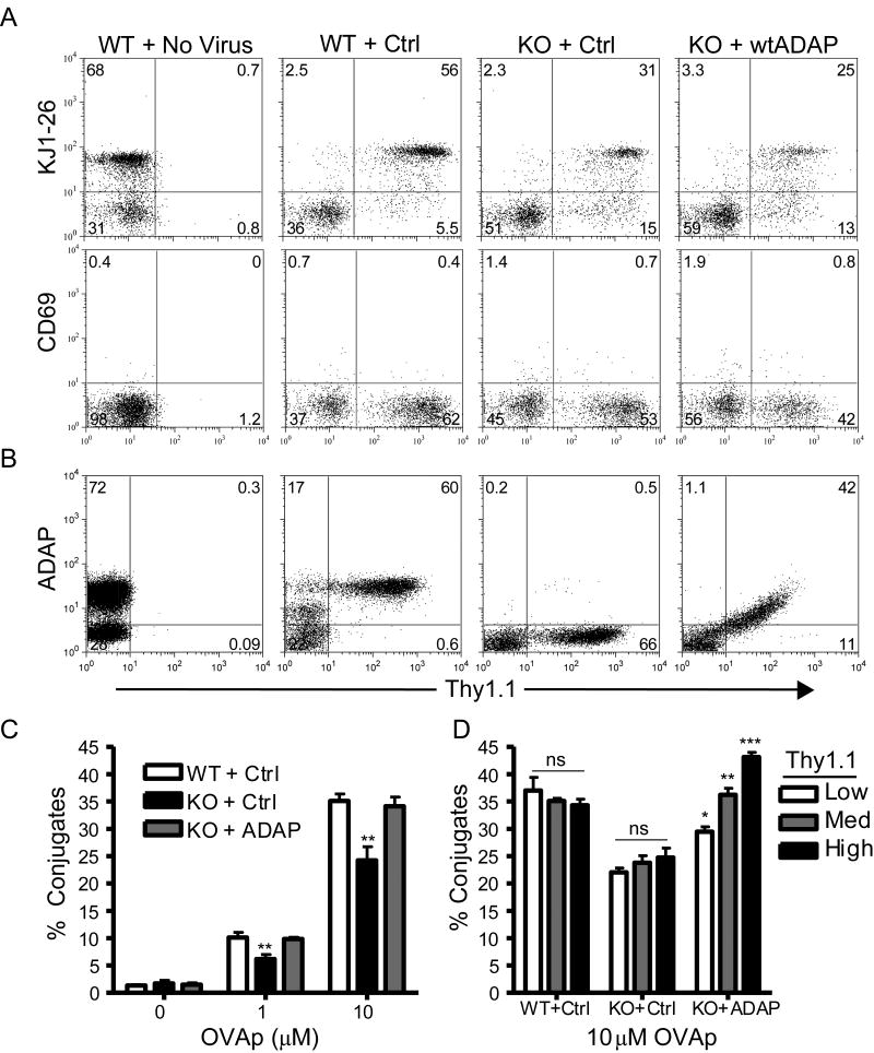
To investigate the ADAP functional domains that control integrin activation in naïve primary murine T cells, we analyzed antigen-dependent T:APC conjugate formation by DO11.10/ADAP-/- T cells expressing either WT ADAP or ADAP deletion mutants. We expressed the hCAR transgene with a T cell specific promoter (35) on the DO11.10/ADAP-/- background (25) to permit efficient adenovirus-mediated transduction of T cells from bulk lymph node preparations (data not shown) (35). Recombinant adenoviruses encoding the wt murine ADAP gene and an IRES-driven Thy1.1 (CD90.1) cell surface reporter (19) were used to reconstitute ADAP expression in freshly harvested, resting hCAR+/D011.10/ADAP-/- T cells (Fig. 1). Thy1.1 expression was observed following adenovirus transduction (Fig 1A, no virus versus control (ctrl; Thy1.1 only) virus). Both ADAP+/+ and ADAP-/- cells expressed similar levels of the DO11.10 TCR following incubation with adenovirus, as judged by equivalent intensity of the KJ1-26 staining (Fig. 1A, upper panels). There was no upregulation of expression of either CD69 (Fig. 1A, lower panels) or CD25 (data not shown, see also Fig. 7B) in Thy1.1-expressing cells across these treatment groups, indicating that the cells were not being activated while resting in culture following adenovirus transduction. Intracellular staining with an anti-ADAP antibody (Fig. 1B) indicated the presence and absence of endogenous ADAP in the majority of Thy1.1+ ADAP+/+ and ADAP-/- cells, respectively. Flow cytometric analysis of ADAP-/- cells transduced with wtADAP demonstrates that Thy1.1 expression correlated with rescue of ADAP expression to levels approaching, but not exceeding, that of endogenous ADAP expression (Fig 1B, right panel).
Figure 1. Rescue of antigen-dependent T:APC conjugate formation in ADAP-/- T cells following reconstitution of wtADAP.
Freshly harvested, naïve lymphocytes from transgenic DO11.10/hCAR/ADAP+/+ (WT) or ADAP-/- (KO) mice were transduced with adenovirus encoding Thy1.1 alone (Ctrl), Thy1.1 plus wild-type murine ADAP (wtADAP), or mock transduced (No Virus) and cultured ex vivo for 3d as described in Materials and Methods. (A) Cells were stained for Thy1.1 and either the DO11.10 TCR (KJ-126) or CD69 and analyzed by flow cytometry. (B) Cells were fixed and intracellular staining was performed with a sheep anti-ADAP antibody. (C) T:APC conjugate formation between KJ-126+/Thy1.1+ T cells and Balb/c splenocytes pre-pulsed with the indicated concentrations of OVAp (left panel) was performed as described in Materials and Methods. The same KJ-126+/Thy1.1+ population of T cells was gated into three subpopulations expressing either low, medium, or high levels of Thy1.1, and T:APC conjugate efficiency with 10μM OVAp was determined (right panel). Results are representative of at least three independent experiments performed.
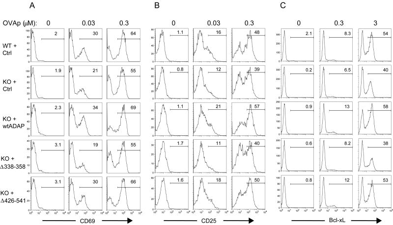
Figure 7. The ADAP proline rich domain is required for antigen-dependent T cell activation.
Naïve DO11.10/hCAR/ADAP+/+ (WT) or ADAP-/- (KO) lymphocytes expressing the indicated ADAP constructs were stimulated with fresh Balb/C splenocytes pulsed with the indicated doses of OVAp as described in Materials and Methods. (A-B) Cells were stimulated for 18h, stained for KJ-126, Thy1.1, CD69, and CD25 as indicated, fixed, and analyzed by flow cytometry. (C) Cells were stimulated for 48h with OVAp, stained for KJ-126 and Thy1.1, fixed, permeablized with saponin, stained for anti-Bcl-xL, and analyzed by flow cytometry. (A-C) The percentage of CD69+, CD25+, or Bcl-xL+ cells within the KJ1-26+Thy1.1+ gate is shown on each histogram. Results are shown for a single representative experiment and are representative of at least four independent experiments performed for CD69 and CD25 and three experiments for Bcl-xL.
DO11.10/ADAP-/- T cells show a ∼30-50% impairment in LFA-1 dependent T cell conjugate formation with OVAp-pulsed APCs at all antigen doses examined (19, 25). A similar defect in T:APC conjugate formation was observed following OVAp stimulation in this system. Background levels of T:APC conjugates in the absence of OVAp were low (<2%; Fig. 1C, 0 μM OVAp). Following incubation with APCs loaded with 10 μM OVAp, ∼35% of control adenovirus-transduced ADAP+/+ T cells interacted with APCs. In contrast, this conjugate efficiency was reduced to ∼25% for control ADAP-/- T cells (Fig. 1C). Importantly, reconstitution of ADAP-/- T cells with WT ADAP restored conjugate formation to a level similar to that found in ADAP+/ + cells (Fig. 1C) (19). Because ADAP detection by intracellular staining was sensitive enough to detect differences in ADAP expression based on the transduced level of Thy1.1 staining (43), we analyzed conjugate formation in ADAP-/- T cells gated for increasing ranges of Thy1.1 expression and found that the magnitude of this rescue depended on the level of ADAP reconstitution (Fig. 1D). For example, KJ1-26+ cells falling within the lower 1/3 of Thy1.1 expression showed only modest rescue of adhesion, while cells within the medium or high Thy1.1 gate showed highly significant increases in adhesion compared to the ADAP-/- vector control (Fig. 1D). Subsequent analyses are presented using the top ∼1/3 of Thy1.1+ (Thy1.1 High) cells expressing the wtADAP construct, with this gate being applied identically to all samples in the experiment to ensure consistency between the constructs used.
The ADAP C-terminus and proline rich domains are required for efficient T:APC conjugate formation
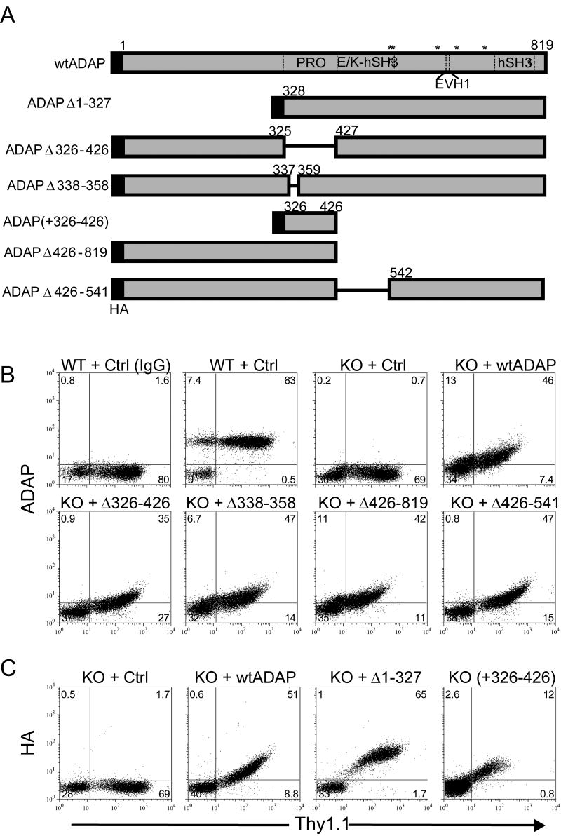
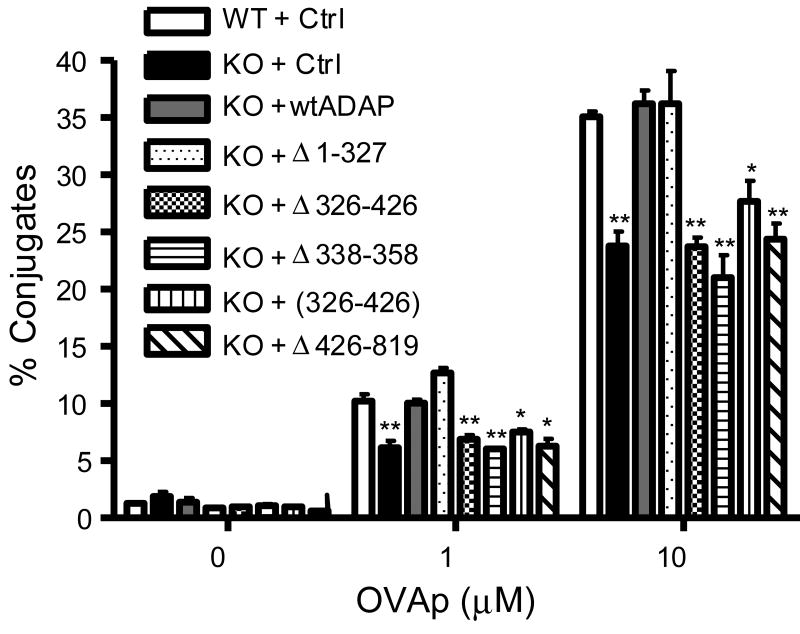
We next prepared a panel of ADAP mutants to identify the molecular domains in ADAP that are required for control of T:APC conjugate formation (Fig. 2A). The expression of constructs containing the N terminus of ADAP was demonstrated by intracellular staining with an anti-ADAP antibody that recognizes the N-terminus (Fig. 2B). The ADAPΔ1-327 and ADAP (+326-426) constructs, which lack the N-terminus, were detected by staining with an anti-HA antibody (Fig. 2C). As in Fig. 1B, Thy1.1 expression correlated with increased expression of all the ADAP constructs we examined. Wild-type ADAP and an ADAP mutant removing the first 327 aa (ADAPΔ1-327) both rescued conjugate formation (Figs. 1C and 3), indicating that the N-terminus of ADAP is not essential for this response. However, deletion of the proline rich domain of ADAP (Δ326-426) completely abolished rescue of conjugate formation, down to levels of control virus alone. Similarly, a restricted deletion within this proline rich domain (Δ338-358) also failed to rescue T:APC adhesion, indicating that this 20aa segment between aa338-358 of ADAP is critical for control of integrin function (Fig. 3). However, expression of the proline rich domain alone (+326-426) failed to rescue conjugate formation, indicating the requirement for a second domain in ADAP (Fig. 3). Indeed, removal of the C-terminal ∼400aa of ADAP (Δ426-819) also restricted rescue of conjugate formation, indicating that the C-terminus of ADAP is also required for efficient conjugate formation.
Figure 2. Schematic and expression of ADAP mutant constructs.
(A) Scale diagram of ADAP expression constructs used in this study. Amino acid numbering is given for the murine ADAP p130 isoform. Abbreviations: PRO, proline-rich domain; E/K, glutamic acid and lysine-rich domain; hSH3, N-terminal (N) or C-terminal (C) helical SH3 domain; EVH1, Ena/Vasp homology domain. Asterisks (*) are used to denote the position of tyrosines 547, 549, 584, 615, 687 found in phosphorylation consensus motifs. (B) Freshly harvested naïve DO11.10/hCAR/ADAP+/+ (WT) or ADAP-/- (KO) lymphocytes were transduced with adenoviruses encoding the indicated constructs or Thy1.1 control adenovirus (ctrl) and fixed and stained with an anti-Thy1.1 antibody and either anti-ADAP antibody or non-immune sheep serum (IgG) and analyzed by flow cytometry. (C) Same as in (B) except the indicated constructs were stained with an anti-hemaglutinin (HA) antibody. Expression profiles are representative of at least three independent analyses performed for each construct shown.
Figure 3. The ADAP proline rich domain is required for T:APC conjugate formation.
Naïve DO11.10/hCAR/ADAP-/- (KO) or ADAP+/+ (WT) lymphocytes expressing the indicated ADAP constructs were analyzed for T:APC conjugate formation between KJ-126+/Thy1.1+ T cells and Balb/c splenocytes pre-pulsed with the indicated concentrations of OVAp. Results are shown for a single representative experiment and are representative of at least four independent experiments performed for each construct.
The ADAP proline rich domain controls SKAP55 expression in naïve T cells
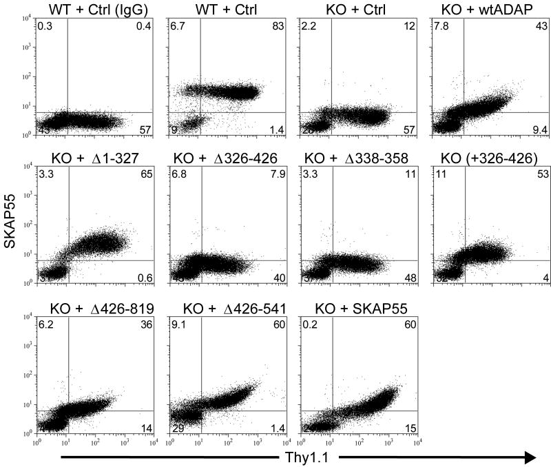
Analysis of ADAP-deficient Jurkat T cells (34) and ADAP-/- murine T cells (44) has demonstrated that SKAP55 expression is also severely impaired in the absence of ADAP, due to caspase and/or proteosome-mediated destabilization of free SKAP55 protein when it cannot interact with ADAP. We have confirmed this finding using intracellular flow cytometry of WT or ADAP-/- lymphocytes using an anti-SKAP55 antibody (Fig. 4) and Western blotting (data not shown). Wild-type T cells infected with control Thy1.1 adenovirus showed robust SKAP55 levels, while ADAP-/- T cells infected with the same control adenovirus demonstrated very low expression, consistently just above the baseline signal derived from control immunoglobulin (Fig. 4). Similar results were observed using a monoclonal anti-SKAP55 antibody for flow cytometry and western blotting, and in freshly isolated, non adenovirus-transduced lymphocytes (data not shown). Adenoviral-mediated reconstitution of ADAP expression allowed SKAP55 levels to accumulate (Fig. 4), with peak expression observed after ∼3 days of infection (data not shown). This effect was positively correlated with the level of Thy1.1 expression, indicating that SKAP55 accumulation mirrors the level of ADAP reconstitution (See Fig. 1D). Analysis of the ADAP domains required for SKAP55 stability demonstrated that neither the N-terminus (aa1-327) nor the C-terminus (aa426-819) of ADAP are essential. In contrast, removal of the central proline-rich domain of ADAP (Δ326-426) or the restricted deletion (Δ338-358) within this region precluded accumulation of SKAP55 (Fig. 4). Expression of the proline-rich domain alone (+326-426) was sufficient to stabilize SKAP55, even though this domain does not rescue conjugate formation (Fig. 4).
Figure 4. The ADAP proline rich domain controls SKAP55 expression in ADAP-/- T cells.
Naïve DO11.10/hCAR/ADAP-/- (KO) or ADAP+/+ (WT) lymphocytes expressing the indicated ADAP constructs, wild-type SKAP55, or the control adenovirus (Ctrl) were fixed and intracellular staining was performed with rabbit anti-SKAP55 antibody or control rabbit immunoglobulin (IgG) and analyzed by flow cytometry. Results are shown for a single representative experiment and are representative of at least four independent experiments performed for each construct.
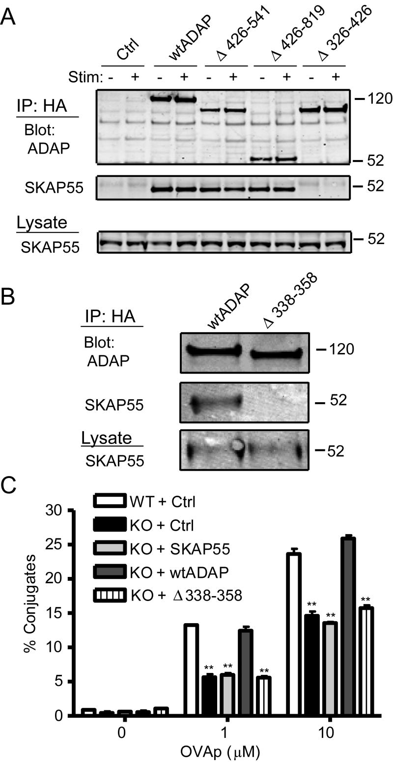
We next performed co-immunoprecipitation analysis to confirm that the interaction of ADAP with SKAP55 is dependent on the ADAP proline-rich domain. Jurkat T cells were transduced with the control virus, or HA-tagged wtADAP, Δ426-541, Δ426-819, or Δ326-426, and stimulated with anti-TCR mAb OKT3 or left unstimulated. Following lysis and anti-HA immunoprecipitation, SKAP55 constitutively interacted with all constructs except the proline-rich domain mutant (Δ326-426) (Fig. 5A) and with the restricted proline deletion (Δ338-358, data not shown). To assess the dependence on this proline-rich domain in primary murine T cells, we transduced ADAP+/+ T cells, which maintain endogenous SKAP55, with either HA-tagged wtADAP, or HA-tagged ADAPΔ338-358. Input whole-cell lysates from each sample contained equivalent levels of SKAP55, and the epitope-tagged ADAP construct was pulled down equally following anti-HA immunoprecipitation (Fig. 5B). Importantly, SKAP55 was detected in the HA immunoprecipitate from the wtADAP-transduced sample but not from the ADAPΔ338-358 sample (Fig. 5B). Thus, the ADAP proline-rich motif is critical for association with and stability of SKAP55 in primary murine T cells.
Figure 5. SKAP55 expression in ADAP-/- T cells fails to rescue T:APC conjugate formation.
(A) Jurkat T cells were transduced with control adenovirus (Ctrl) or with the indicated HA-tagged ADAP constructs. After 2d, 106 cells were left unstimulated or stimulated for 5 min with anti-TCR mAb OKT3, lysed, and immunoprecipitated with agarose-conjugated anti-HA and western blots for ADAP and SKAP55 performed. The relative molecular mass of size standards is shown on the right. Similar results were observed in four independent experiments. (B) Naïve hCAR/ADAP+/+ lymphocytes expressing endogenous levels of SKAP55 were transduced with either full-length murine ADAP (wtADAP) or ADAP lacking the restricted proline rich domain (Δ338-358). Cells were lysed as described in Materials and Methods, immunprecipitated with agarose-conjugated anti-HA antibodies, and Western blots were performed with anti-HA or anti-SKAP55 antibodies. (C) Naïve DO11.10/hCAR/ADAP-/- (KO) or ADAP+/+ (WT) lymphocytes expressing the indicated constructs were analyzed for T:APC conjugate formation between KJ-126+/Thy1.1+ T cells and Balb/c splenocytes pre-pulsed with the indicated concentrations of OVAp. Results are shown for a single representative experiment and are representative of at least four independent experiments performed for each construct.
SKAP55 expression is not sufficient for T:APC conjugate formation
Since cells expressing the ADAP proline-rich domain mutants lack normal levels of SKAP55, it remains possible that stable SKAP55 expression alone might be sufficient for T:APC conjugate formation. To directly test this model, we infected ADAP-/- T cells with adenovirus expressing SKAP55. Following transduction with the virus, SKAP55 was detected by intracellular flow cytometry (see Fig. 4, final panel) at levels approaching that of endogenous SKAP55 found in ADAP+/+ cells. This level of SKAP55 is at or above the level of SKAP55 that accumulated following expression of WT ADAP. Furthermore, the exogenously supplied SKAP55 was able to interact with ADAP when expressed in ADAP+/+ T cells (data not shown). However, SKAP55 expression in DO11.10/ADAP-/- T cells failed to rescue T:APC conjugate formation (Fig. 5C). This result suggests that SKAP55 is not sufficient for this function and that both ADAP and SKAP55 are required for control of antigen-receptor dependent integrin function in naïve T cells.
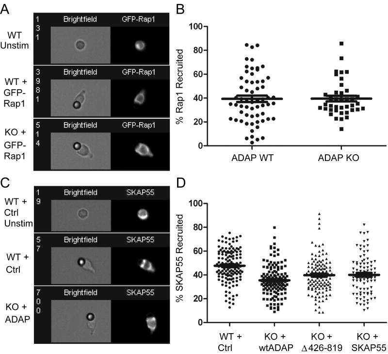
We additionally tested the dependence on ADAP for recruitment of Rap1 and SKAP55 to TCR-coated beads, as a measure of the membrane recruitment potential of SKAP55 and Rap1. ADAP has been has been shown to be important for TCR-induced membrane recruitment of Rap1 (29, and R.B.M and Y.S, unpublished observations). Due to limitations in cell numbers obtained from our ex vivo adenovirus cultures, we were unable to perform biochemical fractionation assays as previously described (29). Instead, we expressed GFP-Rap1 in wild-type or ADAP-/- T cells and monitored targeting of this construct to the interface of anti-TCR coated beads using image-scanning cytometry. Although we did not see striking organization of Rap1 at the contact site (Fig. 6A), approximately 40% of the T cell Rap1 was within the bead contact site in wild-type T cells (Fig. 6B). Surprisingly, ADAP-/- T cells did not show a defect in Rap1 targeting to the TCR contact site (Fig. 6B, P=0.96), and there were no differences in this measure of Rap1 recruitment between ADAP-/- cells reconstituted with wtADAP, ADAPΔ426-819, or ADAPΔ326-426 (data not shown). The recruitment of SKAP55 was also monitored in ADAP+/+ or ADAP-/- T cells expressing wtADAP, Δ426-819, or SKAP55 in the absence of ADAP (Fig. 6C). In contrast to Rap1, SKAP55 was tightly recruited to the T cell:bead interface and frequently adopted a bimodal staining as the T cell wrapped around the edges of the bead (Fig. 6C). Quantification of these observations indicated that approximately 40% of the SKAP55 in the cell was recruited to the bead interface in this assay. However, no differences in SKAP55 recruitment were observed between ADAP-/- cells reconstituted with wtADAP or ADAPΔ426-819 (36 versus 39%; Fig. 6D). Similarly, we also noticed that 40% of exogenous SKAP55 expressed in the absence of ADAP was also recruited to the bead contact. This suggests that determinants within SKAP55 may be sufficient to drive membrane targeting in these assays. Interestingly, ADAP+/+ cells were somewhat more efficient in their overall ability to recruit SKAP55 to the bead contact in (48% of cellular SKAP55) compared to ADAP-/- cells expressing wtADAP, ADAPΔ426-819, or SKAP55 alone (P<0.0001).
Figure 6. ADAP is not required for recruitment of Rap1 or SKAP55 to the contact site between T cells and anti-TCR beads.
(A) Naïve DO11.10/hCAR/ ADAP+/+ (WT) or ADAP-/- (KO) lymphocytes were transduced with GFP-Rap1 and conjugates with anti-TCR (2C11) coated beads were formed as described in Materials and Methods. GFP-Rap1 expressing cells were gated and photographed by image-scanning cytometry and a representative image from each sample is shown. An example of GFP-Rap1 expression in a cell absent of bead stimulation is also shown (Unstim). (B) Graphical display of the percentage of total GFP-Rap1 signal in each cell that is concentrated against the anti-TCR coated bead, quantified as described in Materials and Methods. (C) Naïve DO11.10/hCAR/ADAP+/+ (WT) T cells expressing the control virus (Ctrl) or ADAP-/- (KO) lymphocytes expressing wtADAP, ADAPΔ426-819, or SKAP55 were stimulated with anti-TCR coated beads as described in (A), fixed, and stained for SKAP55 as described for Fig. 4. (D) Graphical display of the percentage of total SKAP55 signal in each cell that is concentrated against the anti-TCR coated bead.
ADAP/SKAP55 interaction is required for optimal T cell activation
In addition to impaired integrin-mediated adhesion, ADAP is also important for TCR and antigen-dependent T cell activation and clonal expansion, especially at low antigen concentrations (25). However, it is not known whether these reported defects in antigen-dependent T cell activation trace to impaired integrin-mediated adhesion at the onset of antigen stimulation, or to other ADAP-dependent signaling pathways. To determine which ADAP functional domains are important for T cell activation, we monitored expression of the early activation marker CD69, the interleukin-2 receptor (CD25), and the pro-survival protein Bcl-xL in ADAP-/- T cells expressing the wtADAP or ADAP mutants. Using primary naïve DO11.10/hCAR/ADAP+/+ or ADAP-/- cells reconstituted ex vivo with control adenovirus, we consistently observed a reduction in the percentage of ADAP-/- cells displaying CD69 and CD25 expression following 18h of stimulation with OVAp (Fig. 7A and 7B). Bcl-xL expression was also impaired in ADAP-/- cells after 48h stimulation (Fig, 7C), consistent with previous results (25). Reconstitution of ADAP-/- cells with wtADAP but not with the SKAP55-binding mutant ADAPΔ338-358 restored CD69, CD25, and Bcl-XL activation to levels at or above ADAP+/+ T cells (Fig. 7), suggesting that the ADAP/SKAP55 interaction is important for optimal antigen-dependent T cell activation. Similarly, ADAP-/- cells expressing the ADAPΔ426-819 C-terminal mutant, or expressing SKAP55 alone in the absence of ADAP, were unable to activate CD69, CD25, or Bcl-xL as robustly as when intact wtADAP is expressed (data not shown). In contrast, ADAP-/- cells expressing the CARMA1 binding mutant ADAPΔ426-541 did not show appreciable defects in CD69, CD25 and Bcl-xL expression, especially when compared to ADAP+/+ cells. These results indicate that ADAP-dependent conjugate formation influences the functional activation state of T cells.
ADAP/SKAP55 is not required for NF-κB activation
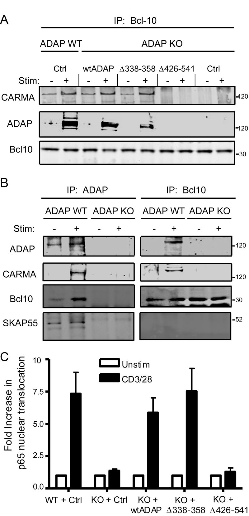
ADAP also regulates TCR-mediated activation of the transcription factor NF-κB in T cells via TCR-regulated binding of the NF-κB regulatory protein CARMA1 to aa426-541 of ADAP, and subsequent assembly of the CARMA1/Bcl10/MALT1 complex (19). However, the ADAP mutant lacking CARMA1 binding capacity (ADAPΔ426-541) can restore efficient T:APC conjugate formation (19). In line with this finding, primary ADAP-/- T cells expressing ADAPΔ426-541 showed efficient accumulation of SKAP55, suggesting that the CARMA1 binding domain within ADAP is dispensable for SKAP55 stabilization (Fig. 4). This suggests that ADAP forms physically and/or functionally distinct complexes with SKAP55 and with CARMA1 in the cell. However, it is unclear whether the ADAP/SKAP55 complex is required for NF-κB activity. To address this question, ADAP-/- T cells expressing wtADAP, ADAPΔ426-541, or ADAPΔ338-358 were stimulated and then cell lysates were prepared and subjected to immunoprecipitation for Bcl10 and Western blotting for CARMA1 and ADAP. Consistent with previous results (19), stimulation with PMA enhanced the association of CARMA1 with Bcl10 in control ADAP+/+ cells, and in ADAP-/- cells expressing wtADAP, but not in ADAP-/- cells expressing either control adenovirus or ADAPΔ426-541 (Fig. 8A). When ADAPΔ338-358 was expressed in ADAP-/- T cells, CARMA1 also associated with Bcl10 following PMA stimulation (Fig. 8A), indicating that neither the ADAP proline rich domain nor SKAP55 is required for this complex to assemble. Furthermore, ADAP was only found in the Bcl10 immunoprecipitates where the Bcl10/CARMA1 association formed (Fig. 8A).
Figure 8. The ADAP/SKAP55 interaction is not required for assembly of the CARMA1/Bcl10 complex or TCR induced NF-κB activation.
(A) Naïve DO11.10/hCAR/ADAP-/- (KO) or ADAP+/+ (WT) lymphocytes expressing the indicated ADAP constructs or the control (Ctrl) were left untreated (-) or stimulated with PMA (+) and lysed as described in Materials and Methods. Lysates were subjected to immunoprecipitation with an anti-Bcl10 antibody, and western blots were performed with antibodies to CARMA1, ADAP, or Bcl10. (B) Freshly harvested resting ADAP+/+ (WT) or ADAP-/- (KO) lymphocytes were left unstimulated or stimulated with PMA and lysed as in (A) and immunoprecipiated in parallel with antibodies to either ADAP (left panel) or Bcl-10 (right panel). Western blots were performed with antibodies to ADAP, CARMA1, Bcl10, and SKAP55 as indicated between the panels. Results are representative of three (A) or two (B) independent experiments. (C) Naïve DO11.10/hCAR/ADAP-/- (KO) or ADAP+/+ (WT) lymphocytes expressing the indicated constructs were stimulated with anti-CD3 plus anti-CD28 antibodies as described in Materials and Methods or left untreated. The samples were fixed and stained with antibodies to Thy1.1 and NF-κB (p65), and nuclei were stained with 7-AAD. Cells were analyzed on a multispectral image-scanning flow cytometer as described in Materials and Methods. Nuclear localization of p65 in unstimulated T cells was set to 1 and results show the increase in p65 nuclear translocation in stimulated relative to unstimulated Thy1.1+ cells from three independent experiments.
To assess whether the cellular pool of ADAP assembled with Bcl10/CARMA1 is separate from that found with SKAP55, identical aliquots of stimulated lysates from ADAP+/+ cells were subjected to immunoprecipitation with either anti-Bcl10 or anti-ADAP antibodies. Assembly of Bcl10, CARMA1, and ADAP was readily observed by either immunprecipitation strategy (Fig. 8B). Interestingly, while anti-ADAP immunoprecipitation showed SKAP55 association (Fig. 8B, left panels), SKAP55 was not detectable in Bcl10 immunoprecipitates (Fig. 8B, right panels). This suggests that SKAP55 and its binding domains in ADAP are not involved in TCR dependent NF-κB activation. Consistent with this model, expression of the ADAPΔ338-358 mutant rescued TCR dependent NF-κB nuclear translocation to levels similar to that observed when wtADAP is expressed (Fig. 8C). In contrast, ADAPΔ426-541, which fails to bind Bcl10 and CARMA1, did not rescue NF-κB activation (Fig. 8C). Taken together, these findings indicate that the ADAP proline rich domain and assembly of the ADAP/SKAP55 complex are not important for ADAP-dependent NF-κB activity.
Discussion
We have investigated the molecular mechanism of ADAP-dependent integrin and NF-κB activation in naïve primary murine T lymphocytes. The development and analysis of ADAP-/- mice clearly demonstrated that ADAP positively regulates T cell activation, β1 and β2 integrin dependent adhesion (23, 24) and peptide antigen-dependent T:APC conjugate formation (19, 25). However, initial analysis of ADAP function prior to the development of ADAP-/- mice utilized overexpression approaches and yielded results that were consistent with both a positive and negative function for ADAP in regulating TCR-dependent signaling (8, 9, 12, 14). Several recent investigations of ADAP function have also utilized overexpression approaches and antibody mediated TCR stimulation in Jurkat T cells (20, 29, 33) or retroviral mediated transduction and overexpression in activated primary T cells (26, 45). Given the concerns with interpreting functional effects of mutations under conditions where ADAP is overexpressed in cells, we reasoned that structure/function analysis of ADAP would be most informative under conditions where mutant ADAP constructs could be expressed in the absence of endogenous ADAP. To overcome the technical challenges of gene delivery into naïve primary murine ADAP-/- T cells, we crossed the OVAp antigen-specific DO11.10/ADAP-/- mice (25) to mice bearing the hCAR transgene (35), which allows efficient adenoviral mediated gene delivery into resting, naïve T lymphocytes.
Using this system, we show that impaired peptide antigen-dependent T:APC conjugate formation in ADAP-/- T cells (25, 46) is rescued following expression of WT ADAP to levels approaching that of endogenous ADAP. Our mutational analysis indicates that while the N-terminal 327aa of ADAP is not required for the rescue of conjugate formation, the central proline-rich domain is necessary but not sufficient for rescue. We also found that this ADAP proline-rich domain is important for optimal functional T cell activation as measured by CD69, CD25, and Bcl-xL expression following antigen stimulation. Furthermore, the C-terminal half of ADAP (aa426-819) is a second region critical for antigen-dependent integrin adhesive function. Thus, our results are consistent with recent work demonstrating a requirement for the ADAP proline-rich and C-terminal domains for TCR-induced adhesion to immobilized β1 and β2 integrin ligands (29, 33).
Several previous studies have outlined the physical and functional relationship between the T cell adapter proteins ADAP and SKAP55. A direct association between the central proline rich domain of ADAP and the SH3 domain of SKAP55 was first identified using two-hybrid screens and coimmunoprecipitation experiments with Jurkat T cells (17, 18). The link between the ADAP/SKAP55 complex and promotion of LFA-1 integrin function was demonstrated by overexpressing either protein in an antigen-specific T cell hybridoma line, or by retroviral mediated overexpression of SKAP55 in activated primary murine T cells (26). Conversely, loss of SKAP55 by siRNA mediated gene knockdown (27, 29, 33) or in SKAP55-/- mice (28) decreased LFA-1 mediated adhesion. Detailed mechanistic studies on the role of ADAP and SKAP55 in T cell activation and integrin function have recently been confounded by the observation that ADAP-/- T cells are severely deficient in SKAP55 (28, 29, 34). This loss of SKAP55 in the absence of ADAP traces to the constitutive interaction between ADAP and the SH3 domain of SKAP55, which acts to protect SKAP55 from caspase- and proteasome-mediated degradation (34). Our studies show that the SKAP55 deficiency in ADAP-/- primary murine T cells is reversible upon reintroduction of intact ADAP protein. Furthermore, we show that a 20aa region between aa338-358 in the ADAP proline rich domain is necessary and sufficient for stabilization of endogenous SKAP55. This region of murine ADAP is analogous to a 24 aa site defined in human ADAP (aa340-364) (29), and contains a 12 aa LGPPPPKPNRPP sequence that includes a canonical core PxxPxP motif capable of binding to several types of SH3 domains (47). There is high sequence identity in this region of ADAP isolated from human, mouse, monkey, dog, cow, chicken, and zebrafish, suggesting a conserved function for ADAP that is dependent on this proline-rich region.
Since cells expressing ADAP proline-rich domain mutants simultaneously fail to rescue conjugate formation and to re-express SKAP55, it is unclear whether ADAP, SKAP55, or both are required for control of antigen-dependent integrin function. Indeed, we are unaware of any experiments to date that have examined the function of SKAP55 independent of ADAP expression. In our experiments, expression of the isolated proline-rich domain of ADAP (+326-426) or of the C-terminal deletion (Δ426-819) permits significant recovery of endogenous SKAP55 expression, while still failing to rescue conjugate formation. To directly test the model that free SKAP55 expression in the absence of any ADAP interaction could be sufficient for T:APC conjugate formation, we specifically expressed SKAP55 in ADAP-/- T cells. Our results show that even in ADAP-/- T cells containing SKAP55 similar to endogenous levels, T:APC conjugate formation was not enhanced. Thus, SKAP55 expression in the absence of ADAP is not sufficient to regulate TCR signaling to integrins. The presence of the proline-rich domain of ADAP, as well as the C-terminus of ADAP, in combination with SKAP55, is necessary for optimal integrin function.
Regulation of β1 and β2 integrin function in T cells is dependent on both the activation and the membrane/synapse targeting of the small GTPase Rap1 (30, 41, 48). Recently, ADAP-/- T cells have been shown to be defective for membrane targeting of the active (GTP-bound) form of Rap1 (29), a finding consistent with the defects in integrin-mediated adhesion observed in ADAP-/- T cells. We attempted to test the role of our ADAP C-terminal mutations in controlling Rap1 membrane targeting, but were unable to obtain enough transduced cells to perform these biochemical comparisons. To test whether ADAP controls recruitment of Rap1 to the TCR signaling complex, we transduced primary wild-type or ADAP-/- cells with GFP-Rap1 and monitored the recruitment of GFP-Rap1 to anti-TCR coated beads. Using this approach, we did not detect any gross differences in the percentage of Rap1 recruited to the T cell-bead interface. This apparent discrepancy between ours and the published results may trace to the ability of Rap1 to localize or become activated on intracellular membranes (49, 50), to rapid kinetics of Rap1 activation that we were unable to capture during imaging, or to the inherent differences between soluble anti-TCR stimulation versus T cell activation against an anti-TCR coated bead, which is somewhat more similar to an APC.
Elucidation of the molecular pathways downstream of the ADAP/SKAP55 complex have implicated the involvement of the constitutive SKAP55 binding protein, RIAM (also known as PREL1), which contains a central RA-PH domain capable of binding to active Rap1 (33). RIAM has also been shown to control β1 and β2 integrin activation in T cells following TCR stimulation (51), and has been implicated in binding Ena/Vasp proteins and the regulation of actin dynamics in T cells (52). RNAi-mediated depletion of SKAP55 in Jurkat T cells results in impaired membrane localization of RIAM and Rap1 following TCR stimulation (33), consistent with the role of SKAP55 as a critical effector in TCR mediated integrin signaling (28). By contrast, ADAP membrane targeting following TCR stimulation is unaffected by SKAP55 depletion (29, 33). While we were unable to detect the ADAP/SKAP55/RIAM complex in the present study (data not shown), we were able to monitor the targeting of SKAP55 to anti-TCR coated beads. ADAP-/- cells expressing wtADAP or ADAPΔ426-819 were equally able to recruit their rescued SKAP55 to the T cell:bead interface. Interestingly, SKAP55 was also recruited even when expressed in the absence of ADAP. These previous results and our findings suggest that both ADAP and SKAP55 may contain membrane targeting information that has the capacity to recruit the proposed ADAP/SKAP55/RIAM/Rap1-GTP complex to the immune synapse following TCR activation by APCs. Alternatively, the entire complex itself may complete a quaternary structure that promotes membrane targeting and/or integrin activation.
The precise role of the ADAP C-terminus (aa426-819) in promoting integrin function remains unclear. Although the extreme C-terminal SH3-like domain (hSH3c) of ADAP has been implicated in a secondary low affinity interaction with SKAP55 that is released following TCR activation and SKAP55 phosphorylation (53, 54), this domain of ADAP is not absolutely required for enhanced integrin-mediated adhesion in mast cells (55). Indeed, we were able to detect robust TCR-independent interaction between ADAP and SKAP55 following removal of the entire ADAP C-terminus (ADAPΔ426-819), suggesting that the ADAP C terminus is not absolutely required for ADAP interaction with SKAP55. In addition, the ADAP domain between aa426-541, which is critical for interaction with CARMA1 and for NF-κB activation, is not required for T:APC conjugate formation (19) or for constitutive binding to SKAP55. However, since the ADAP C-terminus still controlled the ultimate outcome of T:APC conjugate formation and T cell activation, it is thus likely that an ∼200aa segment between murine ADAP aa542∼750 is a secondary domain that controls T:APC conjugate formation. This region contains several tyrosine residues implicated in binding SLP-76 (12, 13). Presumably, ADAP tyrosine phosphorylation in this region is responsible for its recruitment to LAT-associated SLP-76 at the plasma membrane following TCR stimulation (1, 3, 11). Although a reduction in integrin activation has been reported following treatment with a SLP-76 inhibitor peptide (56), and an ADAP construct containing mutations in the ADAP tyrosine residues implicated in binding to the SH2 domain of SLP-76 shows impaired overexpression-induced T:APC conjugate formation (45), a direct role for SLP-76 in T cell integrin function has not yet been carefully defined. The ADAP C-terminus also contains EVH1 homology motifs that have been implicated in binding members of the actin-regulatory Ena/Vasp family proteins (15, 57), which could affect T cell integrin activity. Interestingly, the helical extension of the two non-canonical hSH3 domains of ADAP has been reported to influence integrin-dependent adhesion (20). These domains bind phospholipids in vitro (21) and are predicted to influence the overall conformation of ADAP in response to oxidative stress following T cell activation (58). In addition, part of the N-terminal hSH3 domain of ADAP overlaps with the CARMA1 binding site in ADAP (19). Thus, future experiments will be required to further distinguish molecular signatures in the C-terminus of ADAP required for T:APC conjugate formation.
In addition to controlling TCR dependent integrin function, ADAP regulates TCR dependent NF-κB activation (19). This novel function for ADAP is dependent on the ability of the central aa426-541 in ADAP to bind the adapter CARMA1, which in turn allows the assembly of the CARMA1/Bcl10/Malt1 (CBM) signaling complex. This ADAP-CBM signaling complex is critical for phosphorylation and degradation of IκB, liberating NF-κB to translocate to the T cell nucleus and promote gene transcription. We previously reported that the CARMA1-binding domain in ADAP is dispensable for T:APC conjugate formation (19). In the present study we found that the CARMA1 binding function of ADAP was also not required for rescue of downstream activation markers of T cell function including CD69, CD25, and surprisingly the NF-κB regulated gene Bcl-xL. This suggests that the initial integrin-dependent adhesion during T cell priming is a critical event in determining the activation status 1-2 days following antigen challenge.
The role of the ADAP proline rich domain and the ADAP/SKAP55 complex in regulating NF-κB activation has not been previously investigated. We show here that reconstitution of ADAP-/- T cells with ADAP lacking its proline rich domain (which negates SKAP55 expression) is sufficient to rescue assembly of the CBM complex and NF-κB activation. Indeed, while ADAP immunoprecipitates from activated T cells contain Bcl10, CARMA1, and SKAP55, Bcl10 immunoprecipitates from activated T cells contain ADAP and CARMA1, but not SKAP55. This is consistent with reports that TCR induced activation of a NF-κB reporter is unaffected by RNAi mediated suppression of SKAP55 expression (59). In summary, our data support a model where ADAP coordinates two distinct and physically segregated signaling pathways following TCR stimulation. One pathway involves recruitment of the ADAP/SKAP55/RIAM/Rap1-GTP to the membrane and leads to LFA1 activation and clustering. A second pathway involves TCR-mediated protein kinase C (PKC) θ activation that promotes ADAP-dependent assembly of the CBM complex and subsequent NF-κB activation.
The relative contributions of ADAP-dependent T:APC conjugate formation and NF-κB activation toward T cell activation are currently not clear. While we found that the ADAP/CARMA1 interaction is not absolutely required for early T cell activation events in vitro, we were unable to assess antigen dependent T cell clonal expansion, which occurs 2-3 days following activation, because of the transient nature of our adenovirus expression assay. Furthermore, it is clear that these ex vivo activation assays do not accurately recapitulate the in vivo microenvironment. In particular, there are defects in clonal expansion of DO11.10 ADAP-/- T cells in response to antigen challenge in vivo that are particularly pronounced when naïve T cells are present at physiologically low precursor frequencies (25). Our results and others (28) suggest that impaired interactions of ADAP-/- T cells with APCs in vivo may lower TCR sensitivity to antigen, resulting in inefficient delivery of activation signals required for activation and clonal expansion. Thus, our finding that ADAP also controls a separate T cell activation pathway involving NF-κB suggests that impaired clonal expansion of ADAP-/- T cells in vivo in response to antigen may also be due to impaired induction of NF-κB-dependent genes critical for T cell activation and survival. The combined functions of ADAP may explain the dramatically impaired clonal expansion of ADAP-/- T cells at low precursor frequencies in vivo, despite the fact that ADAP-/- T cells still exhibit some level of T:APC conjugate formation and activation in vitro. A role for ADAP in NF-κB signaling suggests that T cells capable of forming conjugates with APCs in the absence of ADAP may still not receive the proper array of signals necessary for optimal T cell activation. Indeed, a recent report found that while ADAP-/- cytotoxic T lymphocytes (CTLs) have no defects in target cell killing, they exhibited impaired allograft-mediated rejection, consistent with the presence of underlying non-adhesion dependent activation defects in the absence of ADAP (60). Future mechanistic studies in vivo will need to distinguish between concurrent defects in both ADAP-dependent integrin-mediated adhesion and NF-κB signaling.
Acknowledgments
We thank S. Highfill and M. Schwartz for mouse genotyping and colony maintenance and Drs. C. Weaver and B. Schraven for mice and reagents.
Footnotes
Supported by National Institutes of Health grants R01AI038474 (to Y.S.), R01AI031126 (Y.S.) and R01AI056016 (E.J.P.), and National Institutes of Health grant T32DE007288 (B.J.B.). Y.S. is supported in part by the Harry Kay Chair in Biomedical Research at the University of Minnesota.
Abbreviations used in this paper: ADAP, adhesion and degranulation-promoting adapter protein; Bcl10, B-cell CLL/lymphoma 10; CARMA1, caspase-recruitment domain (CARD)-membrane-associated guanylate kinase (MAGUK) protein 1; LAT, linker for activation of T cells; Malt, mucous-associated lymphoid tissue lymphoma; PFA, paraformaldehyde; PKC, protein kinase C; RIAM, Rap1-GTP interacting adapter molecule; SKAP55, src kinase-associated phosphoprotein of 55 kDa; SLP-76, SH2 domain-containing leukocyte-specific phosphoprotein of 76 kDa
Publisher's Disclaimer: “This is an author-produced version of a manuscript accepted for publication in The Journal of Immunology (The JI). The American Association of Immunologists, Inc. (AAI), publisher of The JI, holds the copyright to this manuscript. This manuscript has not yet been copyedited or subjected to editorial proofreading by The JI; hence it may differ from the final version published in The JI (online and in print). AAI (The JI) is not liable for errors or omissions in this author-produced version of the manuscript or in any version derived from it by the United States National Institutes of Health or any other third party. The final, citable version of record can be found at www.jimmunol.org.”
References
- 1.Simeoni L, Kliche S, Lindquist J, Schraven B. Adaptors and linkers in T and B cells. Curr Opin Immunol. 2004;16:304–313. doi: 10.1016/j.coi.2004.03.001. [DOI] [PubMed] [Google Scholar]
- 2.Samelson LE. Signal transduction mediated the T cell antigen receptor: the role of adapter proteins. Annu Rev Immunol. 2002;20:371–394. doi: 10.1146/annurev.immunol.20.092601.111357. [DOI] [PubMed] [Google Scholar]
- 3.Jordan MS, Singer AL, Koretzky GA. Adaptors as central mediators of signal transduction in immune cells. Nat Immunol. 2003;4:110–116. doi: 10.1038/ni0203-110. [DOI] [PubMed] [Google Scholar]
- 4.Sommers CL, Samelson LE, Love PE. LAT: a T lymphocyte adapter protein that couples the antigen receptor to downstream signaling pathways. BioEssays. 2004;26:61–67. doi: 10.1002/bies.10384. [DOI] [PubMed] [Google Scholar]
- 5.Zhang WG, Sommers CL, Burshtyn DN, Stebbins CC, DeJarnette JB, Trible RP, Grinberg A, Tsay HC, Jacobs HM, Kessler CM, Long EO, Love PE, Samelson LE. Essential role of LAT in T cell development. Immunity. 1999;10:323–332. doi: 10.1016/s1074-7613(00)80032-1. [DOI] [PubMed] [Google Scholar]
- 6.Clements JL, Yang B, Ross-Barta SE, Eliason SL, Hrstka RF, Williamson RA, Koretzky GA. Requirement for the leukocyte-specific adapter protein SLP-76 for normal T-cell development. Science. 1998;281:416–419. doi: 10.1126/science.281.5375.416. [DOI] [PubMed] [Google Scholar]
- 7.Pivniouk V, Tsitsikov E, Swinton P, Rathbun G, Alt FW, Geha RS. Impaired viability and profound block in thymocyte development in mice lacking the adaptor protein SLP-76. Cell. 1998;94:229–238. doi: 10.1016/s0092-8674(00)81422-1. [DOI] [PubMed] [Google Scholar]
- 8.Da Silva AJ, Li ZW, De Vera C, Canto E, Findell P, Rudd CE. Cloning of a novel T-cell protein FYB that binds FYN and SH2-domain-containing leukocyte protein 76 and modulates interleukin 2 production. Proc Natl Acad Sci USA. 1997;94:7493–7498. doi: 10.1073/pnas.94.14.7493. [DOI] [PMC free article] [PubMed] [Google Scholar]
- 9.Musci MA, Hendricks-Taylor LR, Motto DG, Paskind M, Kamens J, Turck CW, Koretzky GA. Molecular cloning of SLAP-130, an SLP-76-associated substrate of the T cell antigen receptor-stimulated protein tyrosine kinases. J Biol Chem. 1997;272:11674–11677. doi: 10.1074/jbc.272.18.11674. [DOI] [PubMed] [Google Scholar]
- 10.Peterson EJ. The TCR ADAPts to integrin-mediated cell adhesion. Immunol Rev. 2003;192:113–121. doi: 10.1034/j.1600-065x.2003.00026.x. [DOI] [PubMed] [Google Scholar]
- 11.Burbach BJ, Medeiros RB, Mueller KL, Shimizu Y. T cell receptor signaling to integrins. Immunol Rev. 2007;218:65–81. doi: 10.1111/j.1600-065X.2007.00527.x. [DOI] [PubMed] [Google Scholar]
- 12.Boerth NJ, Judd BA, Koretzky GA. Functional association between SLAP-130 and SLP-76 in Jurkat T cells. J Biol Chem. 2000;275:5143–5152. doi: 10.1074/jbc.275.7.5143. [DOI] [PubMed] [Google Scholar]
- 13.Geng L, Raab M, Rudd CE. Cutting edge: SLP-76 cooperativity with FYB/FYN-T in the up-regulation of TCR-driven IL-2 transcription requires SLP-76 binding to FYB at Tyr595 and Tyr651. J Immunol. 1999;163:5753–5757. [PubMed] [Google Scholar]
- 14.Raab M, Kang H, Da Silva A, Zhu XC, Rudd CE. FYN-T-FYB-SLP-76 interactions define a T-cell receptor ζ/CD3-mediated tyrosine phosphorylation pathway that up-regulates interleukin 2 transcription in T-cells. J Biol Chem. 1999;274:21170–21179. doi: 10.1074/jbc.274.30.21170. [DOI] [PubMed] [Google Scholar]
- 15.Krause M, Sechi AS, Konradt M, Monner D, Gertler FB, Wehland J. Fyn-binding protein (Fyb)/SLP-76-associated protein (SLAP), Ena/vasodilator-stimulated phosphoprotein (VASP) proteins and the Arp2/3 complex link T cell receptor (TCR) signaling to the actin cytoskeleton. J Cell Biol. 2000;149:181–194. doi: 10.1083/jcb.149.1.181. [DOI] [PMC free article] [PubMed] [Google Scholar]
- 16.Marie-Cardine A, Bruyns E, Eckerskorn C, Kirchgessner H, Meuer SC, Schraven B. Molecular cloning of SKAP55, a novel protein that associates with the protein tyrosine kinase p59fyn in human T-lymphocytes. J Biol Chem. 1997;272:16077–16080. doi: 10.1074/jbc.272.26.16077. [DOI] [PubMed] [Google Scholar]
- 17.Marie-Cardine A, Hendricks-Taylor LR, Boerth NJ, Zhao H, Schraven B, Koretzky GA. Molecular interaction between the Fyn-associated protein SKAP55 and the SLP-76-associated phosphoprotein SLAP-130. J Biol Chem. 1998;273:25789–25795. doi: 10.1074/jbc.273.40.25789. [DOI] [PubMed] [Google Scholar]
- 18.Liu J, Kang H, Raab M, Da Silva AJ, Kraeft SK, Rudd CE. FYB (FYN binding protein) serves as a binding partner for lymphoid protein and FYN kinase substrate SKAP55 and a SKAP55-related protein in T cells. Proc Natl Acad Sci USA. 1998;95:8779–8784. doi: 10.1073/pnas.95.15.8779. [DOI] [PMC free article] [PubMed] [Google Scholar]
- 19.Medeiros RB, Burbach BJ, Mueller KL, Srivastava R, Moon JJ, Highfill S, Peterson EJ, Shimizu Y. Regulation of NF-κB activation in T cells via association of the adapter proteins ADAP and CARMA1. Science. 2007;316:754–758. doi: 10.1126/science.1137895. [DOI] [PubMed] [Google Scholar]
- 20.Heuer K, Sylvester M, Kliche S, Pusch R, Thiemke K, Schraven B, Freund C. Lipid-binding hSH3 domains in immune cell adapter proteins. J Mol Biol. 2006;361:94–104. doi: 10.1016/j.jmb.2006.06.004. [DOI] [PubMed] [Google Scholar]
- 21.Heuer K, Arbuzova A, Strauss H, Kofler M, Freund C. The helically extended SH3 domain of the T cell adaptor protein ADAP is a novel lipid interaction domain. J Mol Biol. 2005;348:1025–1035. doi: 10.1016/j.jmb.2005.02.069. [DOI] [PubMed] [Google Scholar]
- 22.Yuan M, Mogemark L, Fällman M. Fyn binding protein, Fyb, interacts with mammalian actin binding protein, mAbp1. FEBS Lett. 2005;579:2339–2347. doi: 10.1016/j.febslet.2005.03.031. [DOI] [PubMed] [Google Scholar]
- 23.Griffiths EK, Krawczyk C, Kong YY, Raab M, Hyduk SJ, Bouchard D, Chan VS, Kozieradzki I, Oliveira-dos-Santos AJ, Wakeham A, Ohashi PS, Cybulsky MI, Rudd CE, Penninger JM. Positive regulation of T cell activation and integrin adhesion by the adapter Fyb/Slap. Science. 2001;293:2260–2263. doi: 10.1126/science.1063397. [DOI] [PubMed] [Google Scholar]
- 24.Peterson EJ, Woods ML, Dmowski SA, Derimanov G, Jordan MS, Wu JN, Myung PS, Liu QH, Pribila JT, Freedman BD, Shimizu Y, Koretzky GA. Coupling of the TCR to integrin activation by SLAP-130/Fyb. Science. 2001;293:2263–2265. doi: 10.1126/science.1063486. [DOI] [PubMed] [Google Scholar]
- 25.Mueller KL, Thomas MS, Burbach BJ, Peterson EJ, Shimizu Y. Adhesion and degranulation promoting adapter protein (ADAP) positively regulates T cell sensitivity to antigen and T cell survival. J Immunol. 2007;179:3559–3569. doi: 10.4049/jimmunol.179.6.3559. [DOI] [PubMed] [Google Scholar]
- 26.Wang H, Moon EY, Azouz A, Wu X, Smith A, Schneider H, Hogg N, Rudd CE. SKAP-55 regulates integrin adhesion and formation of T cell-APC conjugates. Nat Immunol. 2003;4:366–374. doi: 10.1038/ni913. [DOI] [PubMed] [Google Scholar]
- 27.Jo EK, Wang H, Rudd CE. An essential role for SKAP-55 in LFA-1 clustering on T cells that cannot be substituted by SKAP-55R. J Exp Med. 2005;201:1733–1739. doi: 10.1084/jem.20042577. [DOI] [PMC free article] [PubMed] [Google Scholar]
- 28.Wang H, Liu H, Lu Y, Lovatt M, Wei B, Rudd CE. Functional defects of SKAP-55-deficient T cells identify a regulatory role for the adaptor in LFA-1 adhesion. Mol Cell Biol. 2007;27:6863–6875. doi: 10.1128/MCB.00556-07. [DOI] [PMC free article] [PubMed] [Google Scholar]
- 29.Kliche S, Breitling D, Togni M, Pusch R, Heuer K, Wang X, Freund C, Kasirer-Friede A, Menasche G, Koretzky GA, Schraven B. The ADAP/SKAP55 signaling module regulates T-cell receptor-mediated integrin activation through plasma membrane targeting of Rap1. Mol Cell Biol. 2006;26:7130–7144. doi: 10.1128/MCB.00331-06. [DOI] [PMC free article] [PubMed] [Google Scholar]
- 30.Katagiri K, Hattori M, Minato N, Kinashi T. Rap1 functions as a key regulator of T-cell and antigen-presenting cell interactions and modulates T-cell responses. Mol Cell Biol. 2002;22:1001–1015. doi: 10.1128/MCB.22.4.1001-1015.2002. [DOI] [PMC free article] [PubMed] [Google Scholar]
- 31.Kinashi T, Katagiri K. Regulation of lymphocyte adhesion and migration by the small GTPase Rap1 and its effector molecule, RAPL. Immunol Lett. 2004;93:1–5. doi: 10.1016/j.imlet.2004.02.008. [DOI] [PubMed] [Google Scholar]
- 32.Menasche G, Kliche S, Bezman N, Schraven B. Regulation of T-cell antigen receptor-mediated inside-out signaling by cytosolic adapter proteins and Rap1 effector molecules. Immunol Rev. 2007;218:82–91. doi: 10.1111/j.1600-065X.2007.00543.x. [DOI] [PubMed] [Google Scholar]
- 33.Menasche G, Kliche S, Chen EJ, Stradal TE, Schraven B, Koretzky G. RIAM links the ADAP/SKAP-55 signaling module to Rap1, facilitating T-cell-receptor-mediated integrin activation. Mol Cell Biol. 2007;27:4070–4081. doi: 10.1128/MCB.02011-06. [DOI] [PMC free article] [PubMed] [Google Scholar]
- 34.Huang Y, Norton DD, Precht P, Martindale JL, Burkhardt JK, Wange RL. Deficiency of ADAP/Fyb/SLAP-130 destabilizes SKAP55 in Jurkat T cells. J Biol Chem. 2005;280:23576–23583. doi: 10.1074/jbc.M413201200. [DOI] [PubMed] [Google Scholar]
- 35.Hurez V, Dzialo-Hatton R, Oliver J, Matthews RJ, Weaver CT. Efficient adenovirus-mediated gene transfer into primary T cells and thymocytes in a new coxsackie/adenovirus receptor transgenic model. BMC Immunol. 2002;3:4. doi: 10.1186/1471-2172-3-4. [DOI] [PMC free article] [PubMed] [Google Scholar]
- 36.Hurez V, Hautton RD, Oliver J, Matthews RJ, Weaver CK. Gene delivery into primary T cells: overview and characterization of a transgenic model for efficient adenoviral transduction. Immunol Res. 2002;26:131–141. doi: 10.1385/ir:26:1-3:131. [DOI] [PubMed] [Google Scholar]
- 37.Murphy KM, Heimberger AB, Loh DY. Induction by antigen of intrathymic apoptosis of CD4+CD8+TCRlo thymocytes in vivo. Science. 1990;250:1720–1723. doi: 10.1126/science.2125367. [DOI] [PubMed] [Google Scholar]
- 38.Fostel LV, Dluzniewska J, Shimizu Y, Burbach B, Peterson EJ. ADAP is dispensable for natural killer cell development and function. Int Immunol. 2006;18:1305–1314. doi: 10.1093/intimm/dxl063. [DOI] [PubMed] [Google Scholar]
- 39.Nevins JR, DeGregori J, Jakoi L, Leone G. Functional analysis of E2F transcription factor. Methods Enzymol. 1997;283:205–219. doi: 10.1016/s0076-6879(97)83017-0. [DOI] [PubMed] [Google Scholar]
- 40.Wan YY, Leon RP, Marks R, Cham CM, Schaack J, Gajewski TF, DeGregori J. Transgenic expression of the coxsackie/adenovirus receptor enables adenoviral-mediated gene delivery in naive T cells. Proc Natl Acad Sci USA. 2000;97:13784–13789. doi: 10.1073/pnas.250356297. [DOI] [PMC free article] [PubMed] [Google Scholar]
- 41.Medeiros RB, Dickey DM, Chung H, Quale AC, Nagarajan LR, Billadeau DD, Shimizu Y. Protein kinase D1 and the β1 integrin cytoplasmic domain control β1 integrin function via regulation of Rap1 activation and membrane localization. Immunity. 2005;23:213–226. doi: 10.1016/j.immuni.2005.07.006. [DOI] [PubMed] [Google Scholar]
- 42.George TC, Fanning SL, Fitzgerald-Bocarsly P, Medeiros RB, Highfill S, Shimizu Y, Hall BE, Frost K, Basiji D, Ortyn WE, Morrissey PJ, Lynch DH. Quantitative measurement of nuclear translocation events using similarity analysis of multispectral cellular images obtained in flow. J Immunol Methods. 2006;311:117–129. doi: 10.1016/j.jim.2006.01.018. [DOI] [PubMed] [Google Scholar]
- 43.Dluzniewska J, Zou L, Harmon IR, Ellingson MT, Peterson EJ. Immature hematopoietic cells display selective requirements for adhesion- and degranulation-promoting adaptor protein in development and homeostatsis. Eur J Immunol. 2007;37:3208–3219. doi: 10.1002/eji.200737094. [DOI] [PubMed] [Google Scholar]
- 44.Togni M, Swanson KD, Reimann S, Kliche S, Pearce AC, Simeoni L, Reinhold D, Wienands J, Neel BG, Schraven B, Gerber A. Regulation of in vitro and in vivo immune functions by the cytosolic adaptor protein SKAP-HOM. Mol Cell Biol. 2005;25:8052–8063. doi: 10.1128/MCB.25.18.8052-8063.2005. [DOI] [PMC free article] [PubMed] [Google Scholar]
- 45.Wang H, McCann FE, Gordan JD, Wu X, Raab M, Malik TH, Davis DM, Rudd CE. ADAP-SLP-76 binding differentially regulates supramolecular activation cluster (SMAC) formation relative to T cell-APC conjugation. J Exp Med. 2004;200:1063–1074. doi: 10.1084/jem.20040780. [DOI] [PMC free article] [PubMed] [Google Scholar]
- 46.Wu JN, Gheith S, Bezman NA, Liu QH, Fostel LV, Swanson AM, Freedman BD, Koretzky GA, Peterson EJ. Adhesion- and degranulation-promoting adapter protein is required for efficient thymocyte development and selection. J Immunol. 2006;176:6681–6689. doi: 10.4049/jimmunol.176.11.6681. [DOI] [PubMed] [Google Scholar]
- 47.Sparks AB, Rider JE, Hoffman NG, Fowlkes DM, Quillam LA, Kay BK. Distinct ligand preferences of Src homology 3 domains from Src, Yes, Abl, Cortactin, p53bp2, PLCgamma, Crk, and Grb2. Proc Natl Acad Sci USA. 1996;93:1540–1544. doi: 10.1073/pnas.93.4.1540. [DOI] [PMC free article] [PubMed] [Google Scholar]
- 48.Katagiri K, Maeda A, Shimonaka M, Kinashi T. RAPL, a Rap1-binding molecule that mediates Rap1-induced adhesion through spatial regulation of LFA-1. Nat Immunol. 2003;4:741–748. doi: 10.1038/ni950. [DOI] [PubMed] [Google Scholar]
- 49.Bivona TG, Wiener HH, Ahearn IM, Silletti J, Chiu VK, Philips MR. Rap1 up-regulation and activation on plasma membrane regulates T cell adhesion. J Cell Biol. 2004;164:461–470. doi: 10.1083/jcb.200311093. [DOI] [PMC free article] [PubMed] [Google Scholar]
- 50.Mochizuki N, Yamashita S, Kurokawa K, Ohba Y, Nagai T, Miyawaki A, Matsuda M. Spatio-temporal images of growth-factor-induced activation of Ras and Rap1. Nature. 2001;411:1065–1068. doi: 10.1038/35082594. [DOI] [PubMed] [Google Scholar]
- 51.Lafuente EM, van Puijenbroek AA, Krause M, Carman CV, Freeman GJ, Berezovskaya A, Constantine E, Springer TA, Gertler FB, Boussiotis VA. RIAM, an Ena/VASP and Profilin ligand, interacts with Rap1-GTP and mediates Rap1-induced adhesion. Dev Cell. 2004;7:585–595. doi: 10.1016/j.devcel.2004.07.021. [DOI] [PubMed] [Google Scholar]
- 52.Jenzora A, Behrendt B, Small JV, Wehland J, Stradal TE. PREL1 provides a link from Ras signalling to the actin cytoskeleton via Ena/VASP proteins. FEBS Lett. 2006;580:455–463. [PubMed] [Google Scholar]
- 53.Duke-Cohan JS, Kang H, Liu H, Rudd CE. Regulation and function of SKAP-55 non-canonical motif binding to the SH3c domain of adhesion and degranulation-promoting adaptor protein. J Biol Chem. 2006;281:13743–13750. doi: 10.1074/jbc.M508774200. [DOI] [PubMed] [Google Scholar]
- 54.Kang H, Freund C, Duke-Cohan JS, Musacchio A, Wagner G, Rudd CE. SH3 domain recognition of a proline-independent tyrosine-based RKxxYxxY motif in immune cell adaptor SKAP55. EMBO J. 2000;19:2889–2899. doi: 10.1093/emboj/19.12.2889. [DOI] [PMC free article] [PubMed] [Google Scholar]
- 55.Geng LP, Pfister S, Kraeft SK, Rudd CE. Adaptor FYB (Fyn-binding protein) regulates integrinmediated adhesion and mediator release: differential involvement of the FYB SH3 domain. Proc Natl Acad Sci USA. 2001;98:11527–11532. doi: 10.1073/pnas.191378198. [DOI] [PMC free article] [PubMed] [Google Scholar]
- 56.Jordan MS, Maltzman JS, Kliche S, Shabason J, Smith JE, Obstfeld A, Schraven B, Koretzky GA. In vivo disruption of T cell development by expression of a dominant-negative polypeptide designed to abolish the SLP-76/Gads interaction. Eur J Immunol. 2007;37:2961–2972. doi: 10.1002/eji.200636855. [DOI] [PubMed] [Google Scholar]
- 57.Coppolino MG, Krause M, Hagendorff P, Monner DA, Trimble W, Grinstein S, Wehland J, Sechi AS. Evidence for a molecular complex consisting of Fyb/SLAP, SLP-76, Nck, VASP and WASP that links the actin cytoskeleton to Fcγ receptor signalling during phagocytosis. J Cell Sci. 2001;114:4307–4318. doi: 10.1242/jcs.114.23.4307. [DOI] [PubMed] [Google Scholar]
- 58.Zimmermann J, Kuhne R, Sylvester M, Freund C. Redox-regulated conformational changes in an SH3 domain. Biochemistry. 2007;46:6971–6977. doi: 10.1021/bi700437r. [DOI] [PubMed] [Google Scholar]
- 59.Kosco KA, Cerignoli F, Williams S, Abraham RT, Mustelin T. SKAP55 modulates T cell antigen receptor-induced activation of the Ras-Erk-AP1 pathway by binding RasGRP1. Mol Immunol. 2008;45:510–522. doi: 10.1016/j.molimm.2007.05.024. [DOI] [PubMed] [Google Scholar]
- 60.Tian J, Pabst O, Romermann D, Skubich S, Forster R, Beckmann J, Chen JH, Hoffmann MW. Inactivation of T-cell receptor-mediated integrin activation prolongs allograft survival in ADAP-deficient mice. Transplantation. 2007;84:400–406. doi: 10.1097/01.tp.0000269724.06142.92. [DOI] [PubMed] [Google Scholar]


