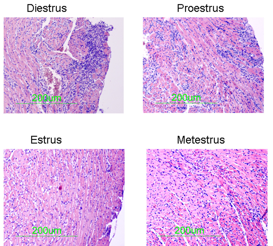
Figure 3.

Histology of TNF1.6 mice infected in different phases of the estrus cycle. Vaginal smears were performed and mice were immediately infected i.p. with 105 PFU CVB3. 7 days later, animals were killed and hearts were removed, formain fixed, paraffin embedded, sectioned and stained with hematoxylin and eosin. Photomicrographs are representative of mice in the group.
