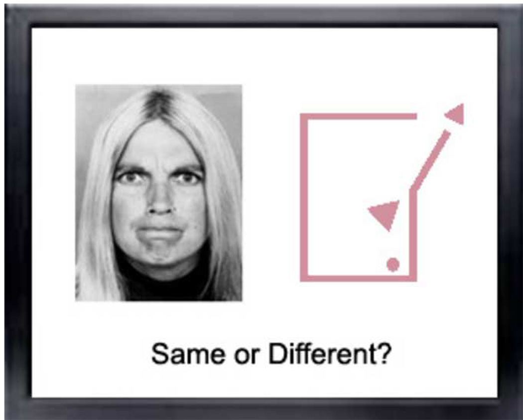
One’s brain engages in a remarkably complex series of processes when generating its pattern of neural responses to a picture of one’s mother. fMRI studies generally explicitly or implicitly measure the neural activity related to thought processes, such as attention, appraisal, manipulation of information, associations, and judgment. However, these thought processes are extremely difficult to assess independent of behavioral correlates. The dependence on behavior can be problematic as behavioral output may not fully capture the multiple dimensions of cognitive and emotional reactivity. To more fully characterize responses to particular sets of stimuli, it seems that one needs to monitor cognitive, affective, and behavioral processes simultaneously. After years of partitioning component processes, it seems that cognitive neuroscience is now engaged in trying to “piece Humpty Dumpty together again” (1).
In fMRI research of face processing in autism spectrum disorders (ASD), a host of meaningful dimensions may actually matter in interpreting results and comparing findings across studies. Examples of such dimensions are whether face stimuli are static or dynamic; contrived or naturalistic; familiar or unfamiliar; personally significant or impersonal; presented for natural viewing or guided by conspicuous attentional cueing. It would be naïve to disregard these factors by assuming that all faces are similar. Although a great deal can be learned by treating faces as a unique class of objects, we cannot forget that they are also the most ubiquitous generator of human interest and motivation, feelings and responses in our daily lives. And they are proxies to our life histories of experiences with people. This is a particularly important consideration in the work with individuals with neurodevelopmental disorders such as autism, whose mind and brain have been sculpted by years of abnormal social experiences dating back to infancy.
A study in this issue of Biological Psychiatry details how Pierce and Redcay (2) measured brain activity (using fMRI) while 6- to 12-year-old children with ASD and typical controls viewed pictures of faces. Face stimuli consisted of pictures of a familiar adult, a familiar child, a stranger adult and a stranger child. Given the established role of the fusiform gyrus (FG) in face recognition, their analysis focused primarily on this structure, but it also included highly interconnected areas such as amygdala and anterior and posterior cingulate. Their results revealed normal FG activity in the children with ASD when viewing a face of their mother or other children. This is in contrast to a host of studies involving older individuals with ASD which have consistently revealed hypoactivation of the FG (see 3 for a review). However, most of these studies involved the faces of strangers. Indeed, consistent with this literature, Pierce and Redcay did find FG abnormalities when the children with ASD viewed faces of strangers. These results led them to conclude that FG abnormalities in ASD may (1) be the result of reduced attention and interest; and (2) be the result of abnormal modulation of the FG by other structures (e.g., hypoactivation of the posterior cingulate, which is postulated to be involved in internally focused tasks such as autobiographical memory). Although the relatively small number of subjects involved in this study prevents us from seeing these results as conclusive, they raise a number of important questions that every fMRI researcher of face processing in ASD should consider in their studies:
(1) Is visual attention a confound in studies of neural responses to faces in ASD?
Past research has addressed this issue by building task constraints intended to ensure attention to stimuli (and then ascertaining comparable behavioral performance across experimental groups), or by adding a cueing device, such as a fixation cross, to the face stimuli presented. Results have been mixed. While the former typically leads to the finding of hypoactivation of the FG in individuals with ASD relative to controls, the latter reveals no differences in FG activation between the two groups (e.g., 4). Both types of studies can be criticized. The former does not fully address the possibility that individuals with ASD attend to face stimuli differently than controls (e.g., the FG hypoactivation might reflect abnormal visual attention to faces in individuals with ASD). The latter strategy may inadvertently alter the task for control subjects who are now forced to attend to a cross which, in turn, may reduce their experiences of faces as such (e.g., the lack of FG abnormalities in individuals with ASD might reflect reduced FG activation in controls). Neither of these experimental designs actually measures visual attention. In the only study to date to relate FG activation to eye-tracking measures of visual attention to faces in ASD, findings indicated that FG activation was strongly and positively correlated with the time spent fixating the eyes (5). Several behavioral studies in ASD have shown reduced eye fixation and often increased mouth fixation in spontaneous viewing of or in performance of structured tasks faces (e.g., 6). Whether a human face is primarily experienced through viewing the eyes or not is a critical question given the potentiation of social neural responses when these are mediated by eye gaze (e.g., 7). Collectively, these studies point to the need for co-registration of eye-tracking measures of visual fixation and fMRI data. This is not only to ensure that visual attention is comparable in the subject groups; it is also to ensure that significant results are not due to abnormal visual fixation patterns to face stimuli.
(2) Is motivation a confound in studies of neural responses to faces in ASD?
Pierce and Redcay’s results hinted at the involvement of structures other than the FG in face processing abnormalities in ASD. Minimally, these are amygdala as a “salience detection” system, particularly if fixation to the eyes is involved, and anterior/posterior cingulate if stimuli can be related to self or self experiences. In typical development, all of this circuitry is fully integrated, catapulted, as it were, into being by built-in and highly conserved preferential attention to eyes and to faces from the first days and weeks of life (8). These processes are disrupted in ASD from at least the age of 2 years if not earlier (9). One can postulate that the cascading effect upon the formation of social mind and brain resulting from these abnormal early experiences will be pronounced. And even though older individuals with ASD may learn to pay attention to faces, it is likely that their motivation to seek faces, that is as the result of an automatic adaptive reaction, is attenuated. Top-down expectations have been shown to modulate activation of relay structures at the earliest points of the visual stream (10). These considerations argue for the need for more inclusive, circuitry-based fMRI analyses of face processing that goes beyond the FG in ASD. It may be more revealing to focus on the process through which an end-point result is obtained. Because faces are so powerful in driving the motivation systems of the brain in typical individuals, even static, cropped, or degraded faces might suffice. In fact, a mere expectation that a face is present might be enough (11). In ASD, such representations might not be enough. Conversely, some data suggest that inherent motivation to seek stimuli other than faces, such as “digimon” characters, may hijack the specialization of the FG in individuals whose love in life is not faces, but digimon characters (12).
(3) Is the FG a brain module exclusively tied to face processing?
The findings of hypoactivation of FG have for long been considered evidence for abnormal neural processing of faces in individuals with ASD. And yet, the FG has been demonstrated to be involved in the processing of visual stimuli that are not faces. For example, we know that the FG can be “taught” to selectively light up to visual recognition of objects about which subjects developed a level of perceptual expertise (13). Does this suggest that visual perception of any class of objects can potentially become associated with the FG if the perceiver is an “expert” on the topic (e.g., bird experts, car experts)? And what mediates this phenomenon? Is it level of semantic processing (e.g., going from general – birds, to very specific – the kinds of birds), or is it that the process of becoming an expert (with the dedication, effort and self involvement required) makes that person also acquire a special, self-referential and self-identifying attitude toward the given object? Both bring us closer to the similarity in neural bases with face processing since we are all experts on faces as a class of objects, and we all feel affected by the mere presence of human faces.
In the same vein, there is evidence of strong selective FG activation during viewing of social animations involving geometric shapes (14). There is nothing face-like in geometric shapes playing human tricks on one another. How are they then related? A whole range of alternatives are possible: they both involve the attribution of social meaning to visual stimuli; they both rely on the retrieval of abstract semantic information of a social nature; they are both related to familiar, self-referential experiences; and more. What is clear is that the mental experiences they elicit are sufficiently similar so as to selectively drive the same neural structure. And neural processing of geometric shape animations of human action in ASD is, like face processing, abnormal (15).
Pierce and Redcay’s results are important in that they bring us to a higher level of scrutiny of fMRI studies of face processing in ASD. The horizons are broader than static faces of strangers and the FG. And their allusion to important developmental considerations will hopefully also prompt us to remember that our brains not only reflect what we are now; our brains also reflect what we have been.
Figure 1.
Footnotes
Publisher's Disclaimer: This is a PDF file of an unedited manuscript that has been accepted for publication. As a service to our customers we are providing this early version of the manuscript. The manuscript will undergo copyediting, typesetting, and review of the resulting proof before it is published in its final citable form. Please note that during the production process errors may be discovered which could affect the content, and all legal disclaimers that apply to the journal pertain.
Dr. Klin reported no biomedical financial interests or potential conflicts of interest.
REFERENCES
- 1.Hobson P. The cradle of thought. London: Macmillan; 2002. [Google Scholar]
- 2.Pierce K, Redcay E. Fusiform function in children with as ASD is a matter of “who”. Biol Psychiatry. 2008 doi: 10.1016/j.biopsych.2008.05.013. this issue. [DOI] [PMC free article] [PubMed] [Google Scholar]
- 3.Schultz RT. Developmental deficits in social perception in autism: the role of the amygdala and fusiform face area. Int J of Dev Neurosci. 2005;23:125–141. doi: 10.1016/j.ijdevneu.2004.12.012. [DOI] [PubMed] [Google Scholar]
- 4.Hadjikhani N, Joseph RM, Snyder J, Chabris CF, Clark J, Steele S, et al. Activation of the fusiform gyrus when individuals with autism spectrum disorder view faces. Neuroimage. 2004;22:1141–1150. doi: 10.1016/j.neuroimage.2004.03.025. [DOI] [PubMed] [Google Scholar]
- 5.Dalton KM, Nacewicz BM, Johnstone T, Schaefer HS, Gernsbacher MA, Goldsmith HH, et al. Gaze fixation and the neural circuitry of face processing in autism. Nat Neurosci. 2005;8:519–526. doi: 10.1038/nn1421. [DOI] [PMC free article] [PubMed] [Google Scholar]
- 6.Klin A, Jones W, Schultz R, Volkmar F, Cohen DJ. Visual fixation patterns during viewing of naturalistic social situations as predictors of social competence in individuals with autism. Arch Gen Psychiatry. 2002;59:809–816. doi: 10.1001/archpsyc.59.9.809. [DOI] [PubMed] [Google Scholar]
- 7.Kampe KKW, Frith CD, Dolan RJ, Frith U. Reward value of attractiveness and gaze. Nature. 2001;413:589–590. doi: 10.1038/35098149. [DOI] [PubMed] [Google Scholar]
- 8.Farroni T, Csibra G, Simion F, Johnson MH. Eye contact detection in humans from birth. Proc Natl Acad Sci USA. 2002;99:9602–9605. doi: 10.1073/pnas.152159999. [DOI] [PMC free article] [PubMed] [Google Scholar]
- 9.Jones W, Carr K, Klin A. Absence of preferential looking to the eyes of approaching adults predicts level of social disability in 2-year-olds with autism. Arch Gen Psychiatry. 2008 doi: 10.1001/archpsyc.65.8.946. [DOI] [PubMed] [Google Scholar]
- 10.Treue S. Visual attention: the where, what, how and why of saliency. Curr Opin Neurobiol. 2003;13:428–432. doi: 10.1016/s0959-4388(03)00105-3. [DOI] [PubMed] [Google Scholar]
- 11.Cox D, Meyers E, Sinha P. Contextually evoked object-specific responses in human visual cortex. Science. 2004;304:115–117. doi: 10.1126/science.1093110. [DOI] [PubMed] [Google Scholar]
- 12.Grelotti DJ, Klin A, Volkmar FR, Gauthier I, Skudlarski P, Cohen DJ, et al. fMRI activation of the fusiform gyrus and amygdala to cartoon characters but not to faces in a boy with autism. Neuropsychologia. 2005;43:373–385. doi: 10.1016/j.neuropsychologia.2004.06.015. [DOI] [PubMed] [Google Scholar]
- 13.Gauthier I, Tarr MJ, Anderson AW, Skudlarski P, Gore JC. Activation of the middle fusiform ‘face area’ increases with expertise in recognizing novel objects. Nat Neurosci. 1999;2:568–573. doi: 10.1038/9224. [DOI] [PubMed] [Google Scholar]
- 14.Schultz RT, Grelotti D, Klin A, Kleinman J, van der Gaag C, Marois R, et al. The role of the fusiform face area in social cognition: Implications for the pathobiology of autism. Philos Trans R Soc Lond B Biol Sci. 2003;358:415–427. doi: 10.1098/rstb.2002.1208. [DOI] [PMC free article] [PubMed] [Google Scholar]
- 15.Castelli F, Frith C, Happé F, Frith U. Autism, Asperger syndrome and brain mechanisms for the attribution of mental states to animated shapes. Brain. 2002;125:1839–1849. doi: 10.1093/brain/awf189. [DOI] [PubMed] [Google Scholar]


