Abstract
Interaction of Activation Induced cytidine Deaminase (AID) with Replication Protein A (RPA) has been proposed to promote AID access to transcribed double strand (ds) DNA during IgH Class Switch Recombination (CSR). Mouse AID (mAID) interaction with RPA and transcription-dependent dsDNA deamination in vitro requires protein kinase A (PKA) phosphorylation at Serine-38 (S38), and normal mAID CSR activity depends on S38. However, zebrafish AID (zAID) catalyzes robust CSR in mouse cells despite lacking an S38-equivalent PKA site. Here, we show that aspartate 44 (D44) in zAID provides similar in vitro and in vivo functionality as mAID S38-phosphorylation. Moreover, introduction of a PKA site into a zAID D44 mutant made it PKA-dependent for in vitro activities and restored normal CSR activity. Based on these findings, we generated mAID mutants that similarly function independent of S38-phosphorylation. Comparison of bony fish versus amphibian and mammalian AIDs suggests evolutionary divergence from constitutive to PKA-regulated RPA/AID interaction.
INTRODUCTION
B cells undergo two different forms of immunoglobulin (Ig) gene alteration following antigen activation. Somatic hypermutation (SHM) introduces point mutations into Ig light chain and heavy chain (IgH) variable region exons, allowing generation of antibodies with increased affinity (Di Noia and Neuberger, 2007; Odegard and Schatz, 2006). IgH Class switch recombination (CSR) replaces the IgH Cμ exons, which encode the first IgH constant region (CH) expressed, with one of several downstream sets of CH exons, which encode different antibody classes (Chaudhuri et al., 2007; Honjo et al., 2002). Long repetitive switch (S) regions precede each set of CH exons. CSR involves introduction of DNA double strand breaks (DSBs) into the donor Sμ and into a downstream acceptor S region, followed by joining of the two S regions (Chaudhuri et al., 2007). Both CSR and SHM require Activation Induced cytidine Deaminase (AID) (Muramatsu et al., 2000; Revy et al., 2000). AID initiates CSR and SHM by catalyzing deamination of cytidines in S regions and variable region exons, respectively, in a process that requires transcription of targeted sequences (Chaudhuri et al., 2007; Goodman et al., 2007). Subsequently, deaminated cytidines are processed by subversion of normal repair pathways to generate variable region point mutations or S region mutations and DSBs (Di Noia and Neuberger, 2007).
Purified AID has cytidine deaminase activity on single strand (ss) but not double strand (ds) DNA (Bransteitter et al., 2003; Chaudhuri et al., 2003; Dickerson et al., 2003; Ramiro et al., 2003; Sohail et al., 2003). Several mechanisms have been implicated in AID access of duplex DNA in vivo (Chaudhuri et al., 2003). When transcribed in physiologic orientation, but not inverted orientation, mammalian S regions generate ssDNA within R loops (Tian and Alt, 2000; Yu et al., 2003). In this regard, biochemical and genetic studies indicated that R-loop formation in transcribed mammalian S regions can enhance AID access (Shinkura et al., 2003; Chaudhuri et al., 2003). However, R loop formation cannot fully explain AID access to S regions, since they still support reduced CSR when transcribed in non-physiologic orientation. Also, R-loops cannot explain AID access to transcribed variable region exons, which do not form R-loops. In the latter context, biochemical assays revealed that RPA, a trimeric ssDNA binding protein involved in replication and repair (Wold, 1997), promotes efficient AID deamination of in vitro transcribed sequences rich in SHM motifs ("SHM substrates") that do not form R-loops (Chaudhuri et al., 2004). While AID and SHM are present in tetrapods (bony fish); CSR first occurs evolutionarily in amphibians, (e.g., Xenopus), suggesting CSR evolved after SHM (Stavnezer and Amemiya, 2004). Transcribed Xenopus Sμ (XSμ) does not form R-loops but can replace mouse Sγ1 to effect CSR (Zarrin et al., 2004). XSμ CSR junctions and AID deamination sites occurred within a region dense in AGCT sequences, a canonical SHM motif (Zarrin et al., 2004). Thus, AID CSR activities may have evolved from AID SHM functions (Barreto et al., 2005; Wakae et al., 2006). As mammalian S regions are rich in SHM motifs, AID/RPA targeting may function in mammalian CSR (Zarrin et al., 2004).
A portion of mouse B cell AID (mAID) is phosphorylated at Serine-38 (Basu et al., 2005; McBride et al., 2006), and mAID interaction with RPA is dependent upon S38 phosphorylation (Basu et al., 2005). The mAID S38 site is part of a RRX(S/T) cAMP-dependent protein kinase A (PKA) consensus motif (Basu et al., 2005). Correspondingly, mAID can be phosphorylated by PKA in vitro at S38, and co-expression of PKA with mAID in fibroblasts enhances S38 phosphorylation (Basu et al., 2005; Basu et al., 2007; McBride et al., 2006; Pasqualucci et al., 2006). In vitro phosphorylation of non-phosphorylated mAID by PKA conferred ability to bind RPA and to mediate transcription-dependent dsDNA deamination (Basu et al., 2005). In addition, mutation of mAID S38 to alanine (mAIDS38A) had no effect on ssDNA catalytic activity, but abolished AID phosphorylation by PKA and, correspondingly, impaired ability of AID to interact with RPA and deaminate transcribed SHM substrates (Basu et al., 2005). Moreover, mAIDS38A had significantly reduced ability to catalyze CSR when expressed in AID-deficient B cells (Basu et al., 2005; Basu et al., 2007; McBride et al., 2006; Pasqualucci et al., 2006), consistent with AID phosphorylation at S38 and RPA interaction augmenting AID function in CSR. Finally, mAIDS38A showed greatly reduced ability to carry out gene conversion (GCV) and SHM in chicken DT40 cells (Chatterji et al., 2007).
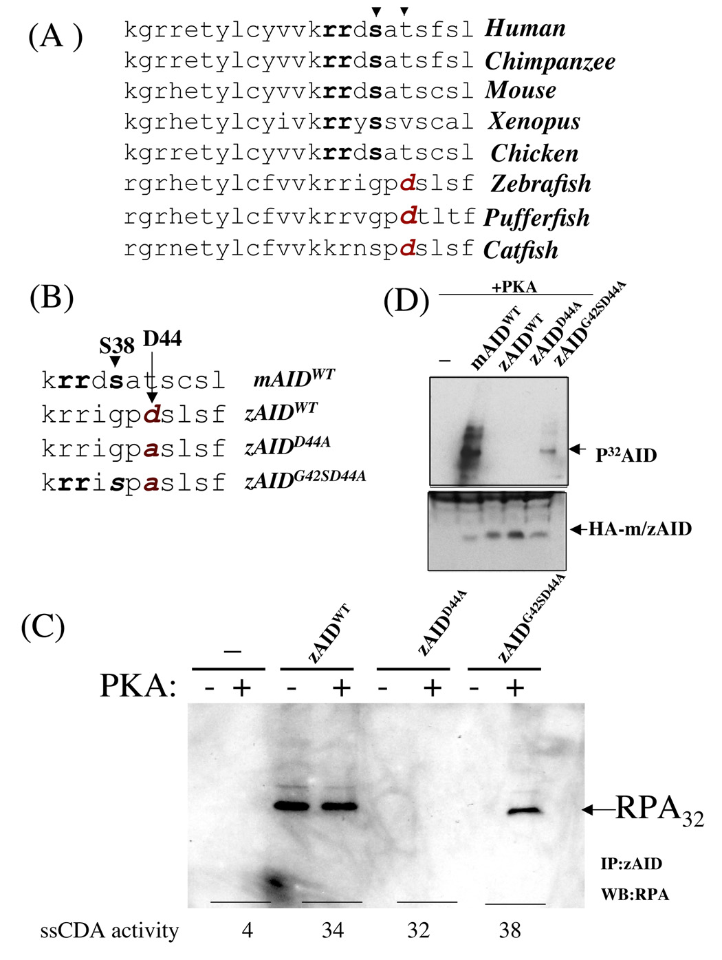
AID has a PKA-consensus S38 phosphorylation site in all analyzed tetrapods (mouse, human, Xenopus), but not in any analyzed teleost (zebrafish, fugu, catfish), the latter of which undergo SHM but not CSR (Fig. 1A). Yet, zebrafish AID (zAID), despite lacking a clear counterpart of the mammalian AID S38 PKA site, catalyzes robust CSR in mouse B cells (Barreto et al., 2005; Wakae et al., 2006). These findings were viewed as a challenge to the model that S38 phosphorylation and RPA interaction play roles in physiological AID function (Shinkura et al., 2007; Pham et al., 2008). The mechanism by which zAID "bypasses" PKA phosphorylation has remained speculative (Pan-Hammarstrom et al., 2007), with one possibility being that zAID might interact with RPA via phosphorylation at a different site or via a phosphorylation-independent mechanism. In the latter context, AID from all analyzed teleost species has a conserved aspartate residue (D44 in zebrafish) in the region that harbors tetrapod S38 (Fig. 1A) (Barreto et al., 2005; Wakae et al., 2006), suggesting D44 might be a phosphorylation mimic in teleost AID (Basu et al., 2005). In this context, only catfish AID has a serine residue corresponding to mAID S38 (at position S41); but this residue is not found in a consensus PKA site (Fig. 1A). In this study, we demonstrate zAID D44 functionally mimics S38 phosphorylation in mAID by allowing constitutive association with RPA. In addition, we have engineered a zAID that interacts with RPA and deaminates transcribed dsDNA in a PKA phosphorylation-dependent fashion and a mAID that similarly functions in a phosphorylation-independent fashion.
Figure 1. Zebrafish AID interacts with RPA in vitro.
(A) Phylogenetic comparison of AID from Homo sapiens (human), Pan troglodytes (chimpanzee), Mus musculus (mouse), Xenopus laevis (amphibian), Gallus gallus (chicken), Danio rerio (zebrafish), Takifugu rubripes (pufferfish) and Ictalurus punctatus (catfish). Arrows indicate Serine 38 in mammals, birds, and amphibian AID and aspartate 44 in fish AID. The conserved PKA site in tetrapods is indicated in bold and the conserved aspartic acid in telosts in red (B) The zAIDWT, zAIDD44A, and zAIDG42S,D44A are represented with the mutated residues indicated (C) Mutant tagged-zAID proteins were expressed in 293T cells and nuclear extracts obtained were treated (+) or not treated (−) with PKA and immunoprecipitated. The immunoprecipitates were washed and assayed for zAID-RPA interactions by western blotting with anti-RPA antibodies. Presence of AID in the immunoprecipitation reaction was estimated by ssDNA deamination as described (Chaudhuri et al., 2003) and presented at the bottom of panel C. (D) Flag/HA tagged mAID and Flag-HA tagged (zAIDWT, zAIDD44A, and zAIDG42SD44A were expressed in 293 cells and partially purified by immunoprecipitation, incubated with PKA in the presence of γ-32P-ATP, and analyzed by SDS-PAGE followed by autoradiography (top panel). Presence of Flag/HA epitope-tagged mouse AID or zAID proteins in immunoprecipitates was determined via western blotting with anti-HA antibodies(bottom panel).
RESULTS
Binding of zAID to RPA Without Phosphorylation
To test potential functions of the conserved D44 residue in zAID, we expressed N-terminal flag-tagged wild-type zAID (zAIDWT) and zAID in which D44 had been mutated to alanine (zAIDD44A) (Fig. 1B) in HEK293 cells. Subsequently, we partially purified zAIDWT and zAIDD44A from HEK293T nuclear extracts, and immunoprecipitated the partially purified proteins via anti-Flag antibodies in the presence of recombinant RPA plus or minus recombinant PKA (Fig. 1C). First, we confirmed that immunoprecipitates had similar amounts of active AID, as evidenced by ssDNA cytidine deamination activity (Fig. 1C, bottom). Interaction of RPA with zAID then was assessed via SDS-PAGE followed by western blotting with anti-RPA antibodies. Notably, we observed constitutive zAID interaction with RPA whether or not the zAID was pre-incubated with recombinant PKA (Fig. 1C; Sup. Fig. 1 and 2A). In contrast, we did not find readily detectable interaction of zAIDD44A with RPA, either in the presence or absence of PKA and ATP (Fig. 1C; Supp. Fig. 1, 2A). As a control, mutation of adjacent residues, including glycine 42 to serine (zAIDG42S) or proline 43 to alanine (zAIDP43), did not appear to alter zAID binding to RPA, indicating the specificity of D44 for this activity (Supp. Fig. 2A). Finally, following ectopic expression in B cells, mAIDwt and zAIDwt interact with RPA, whereas mAIDS38A and zAIDD44A do not (Supp. Fig. 2D).
We next generated and expressed in HEK293 cells a second-site mutant of zAIDD44A in which glycine 42 was mutated to serine to form the zAIDG42S,D44A double mutant (Fig.1B). The G42S mutation reconstitutes a PKA phosphorylation site at a position corresponding to mAID S38 (Fig. 1B). Immunoprecipitated zAIDG42S,D44A retained ssDNA deamination activity similar to that of zAIDWT or zAIDD44A (Fig. 1C, lower panel). When partially purified zAIDWT, zAIDD44A or zAIDG42SD44A was incubated with γ-32P ATP in the presence of recombinant PKA and analyzed via SDS-PAGE, we observed that zAIDG42S,D44A clearly was phosphorylated by PKA; whereas zAIDwt and zAIDD44A were not detectably phosphorylated (Fig. 1D, mAIDWT is shown as a control; Supp. Figs 3A,B). In these experiments, zAIDG42S,D44A appeared less phosphorylated than the mAIDWT control protein, potentially reflecting roles for other mAID sequences in enhancing PKA phosphorylation. Strikingly, while untreated zAIDG42S,D44A failed to show RPA interaction, PKA phosphorylation of this double mutant led to clear RPA interaction (Fig 1C; Supp. Figs. 1 and 2A).
RPA-Dependent zAID dsDNA Deamination Activity
Because zAID constitutively associates with RPA and zAIDD44A lacks ability to associate with RPA (Fig. 1C), we asked whether zAID, in the presence of RPA, also showed constitutive ability to deaminate transcribed dsDNA SHM substrates in vitro and whether such ability was dependent on integrity of D44. As expected, partially purified (from HEK-293 cells) mAIDwt, mAIDS38A, zAIDwt, and zAIDD44A proteins demonstrated similar ability to deaminate ssDNA, again confirming the respective mutations do not affect catalytic activity (Fig. 2A). We then assayed each protein, following pre-incubation with recombinant PKA and ATP, for ability to deaminate a T7 RNA polymerase-transcribed dsDNA SHM substrate in the presence of RPA (Chaudhuri et al., 2004; see Supp. Fig. 4 for schematic of the assay). Both the mAIDWT and zAIDWT were active in these assays, although zAIDWT appeared to have somewhat less activity (Fig. 2B). However, similar to mAIDS38A versus mAIDWT, zAIDD44A, despite having normal catalytic activity on ssDNA, showed greatly impaired transcription-dependent dsDNA deamination activity relative to zAIDWT (Fig. 2B).
Figure 2. zAID deaminates transcribed RGYW-rich dsDNA in a RPA-dependent manner.
(A and B) mAIDWT, mAIDS38A, zAIDWT and zAIDD44A were expressed in 293T cells and partially purified (through DEAE cellulose and glycerol gradient). The proteins were assayed for ssDNA deamination activity without PKA addition (A) or RPA-dependent dsDNA deamination activity after PKA addition (B). For (A) and (B), values represent the mean of three experiments and error bars represent standard deviation from the mean. (C) Indicated wildtype and mutant AID proteins were expressed in 293T cells, partially purified as above, standardized for ssDNA deamination activity, and then 50 ug of each partially purified AID protein, as present in the peak fraction of the glycerol gradient, was incubated with or without 50 units of PKA for 30 mins at 37°C, followed by assay for RPA-dependent dsDNA deamination activity. Cytidine deamination activity for each protein was adjusted by subtracting the background value obtained with processed from cells not transfected with AID. Deamination acivity is plotted as a % of total counts added. Background in individual assays varied from 0.3–1.5%. For (A), (B) and (C), values represent the mean from three experiments and the error bar represent the standard deviation from the mean.
We next compared activities of partially purified zAIDWT, zAIDD44A and zAIDG42S,D44A proteins in the transcription-dependent dsDNA cytidine deamination assay, before and after PKA treatment. The partially purified AID proteins used for these assays again had similar ssDNA deamination activities (Supp. Fig. 5A) and were used in roughly similar amounts in each assay (Supp. Fig. 5B). The presence or absence of PKA had no affect on zAIDWT dsDNA deamination activity (Fig. 2C) or the lack of this activity observed for zAIDD44A (Fig. 2C). In contrast, pre-incubation with PKA led to a striking increase in dsDNA deamination activity of zAIDG42S,D44A, to levels substantially above the background observed in the absence of treatment (Fig.2C). In these assays, the activity of PKA treated zAIDG42S,D44A was not fully restored to that of zAIDWT (Fig. 1C; D). However, these findings make the clear and important qualitative point that the second site G42S mutation simultaneously allowed the AIDD44A to serve as a substrate for PKA phosphorylation, to acquire PKA-dependent ability to bind RPA, and to acquire PKA-dependent dsDNA deamination activity.
Rescue of Impaired CSR of the zAIDD44A Protein by a Second Site Mutation
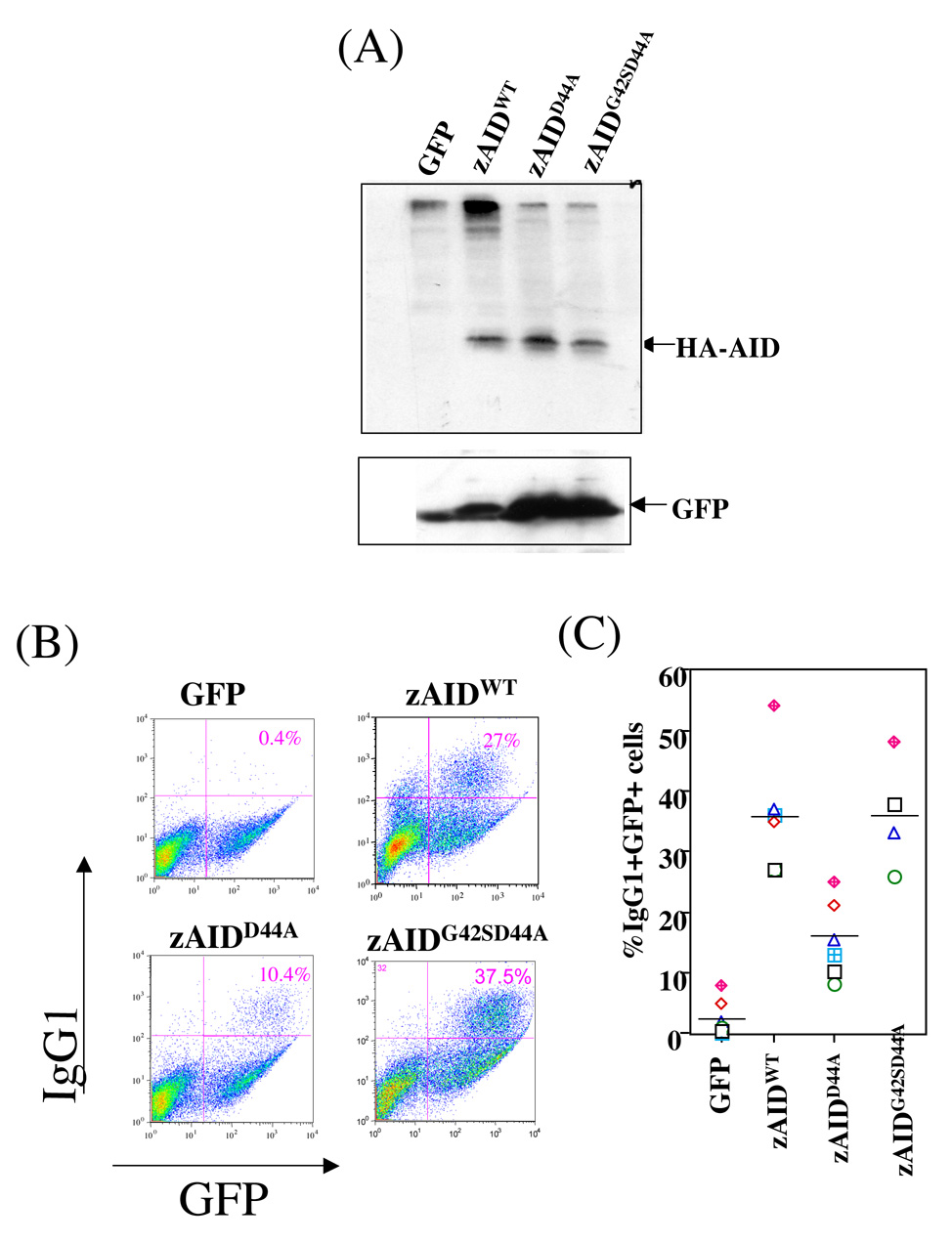
To assess effects of different AID mutations on IgH CSR, we retrovirally introduced HA-tagged zAIDWT, HA-tagged zAIDD44A or HA-tagged zAIDG42,D44A into anti-CD40 plus IL-4 stimulated AID-deficient mouse B cells and assayed ability of introduced AID proteins to rescue class switching to IgG1 (Fagarasan et al., 2001). HA-tagged zAID proteins were employed because there are no available antibodies that react well with zAID in total cell extracts. Retrovirally-expressed WT and mutant zAID proteins were expressed at roughly similar levels in B cells (Fig. 3A, Suppl. Fig. 2C) and in HEK293 cells (data not shown). As expected, introduction of zAIDWT resulted in a substantial rescue of class-switching to IgG1, to levels similar to those obtained with mAIDWT (Fig. 3B,C; data not shown). In contrast, CSR levels obtained via introduction of zAIDD44A were, on average, about 40–45% those of zAIDWT (based on 6 independent experiments), indicating that D44 integrity is required for optimal zAID CSR activity (Fig. 3B, C; Supp. Fig. 2B). This high residual level of CSR supported by zAIDD44A, along with it apparently robust activity in GCV and SHM in DT40 chicken cells (Chatterji et al., 2007) supports the notion that zAID also can access transcribed DNA via RPA-independent mechanisms (see Supplementary Discussion). Strikingly, the zAIDD44A,G42S double mutant showed significantly increased CSR activity compared with zAIDD44A, with levels that were, on average, similar to zAIDWT (Fig. 3B, C); Supp. Fig. 2B). A zAIDG42S mutant had CSR activity similar to that of zAIDWT, consistent with the AIDG42S mutation functioning to rescue the AIDD44A mutation as opposed to simply activating overall CSR activity of AIDWT (Supp. Fig. 2B). We conclude that introduction of the second-site G42S mutation into the zAIDD44A mutant restored essentially normal ability of the zAIDD44A mutant to catalyze CSR in vivo under assayed conditions.
Figure 3. CSR Activity of zAID Mutants.
(A,B) AID-deficient B cells stimulated with anti-CD40 and IL4 were infected with retrovirus expressing tagged zAIDwt or zAIDD44A or zAIDG42S, D44A, expression of HA-tagged analyzed by loading 50ug of total B cell extracts on a SDS-PAGE followed by western blotting using anti-HA antibodies (A) and the level of CSR to IgG1 was evaluated flow cytometry (B) as described (Basu et al., 2005). ((C) The percentage of GFP-positive cells that had undergone CSR to IgG1 from multiple experiments is indicated plotted. Individual sets of experiment are represented by different symbols. In (C), the mean of all the experiments of a particular zAID protein is represented by a line.
Phospho-mimetic mutation of mouse AID Ser-38 phosphorylation
We confirmed prior studies that showed a mAID mutant in which S38 was replaced with aspartic acid, to potentially provide a mimetic of constitutive phosphorylation, failed to generate normal CSR activity (McBride et al., 2006; Shinkura et al., 2007; data not shown). However, only positive results are clearly interpretable in such a study, as any amino acid replacement may negatively alter protein structure and function by various effects. Based on our finding that zAID employs D44 as a mimetic of mAID S38 phosphorylation, we predicted that replacement of mouse mAID T40 (which corresponds in position to zAID D44; Fig 4A) might generate a mAID protein that functions independently of S38 phosphorylation, and rescue activities lost in the context of the mAID S38A mutation. A purified mAIDS38A,T40D double mutant protein had similar ssDNA deamination activity as AIDWT, confirming retention of basic catalytic activity (Fig. 4B). Strikingly, the mAIDS38AT40D protein bound RPA (Supp. Fig. 7A, B) and catalyzed deamination of T7 polymerase-transcribed dsDNA SHM substrates even more robustly that PKA phosphorylated mAIDWT (Fig. 4C), with both activities occurring independent of PKA phosphorylation (Fig. 4C; Supp. Fig. 7B). In agreement with restored biochemical activities, retrovirally introduced mAIDS38AT40D supported CSR in mouse B cells at levels similar to those of mAIDWT (Fig. 4D; Supp. Fig.7C, D). Finally, a mAIDS38G/T40D mutant also had CSR similar to that of mAIDWT (data not shown). Taken together, these findings indicate that the mAID T40D mutation serves as a mimetic of mAID phosphorylation on S38. Thus, using an evolutionary approach, we have identified a dominantly active mutant of mAID in the context of S38 phosphorylation.
Figure 4. Mouse AID “phospho-mimetic” mutants.
(A) The mAIDWT, mAIDS38A and mAIDS38AT40D are represented with the mutated residues indicated. (B) Partially purified AID protein indicated were assayed for ssDNA deamination activities. (C) The RPA- and transcription-dependent dsDNA deamination activities of AIDWT and AIDS38AT40D proteins were analyzed in presence (+) or absence (−) of PKA as described (Basu et al., 2005). (D) CSR activities of mAIDWT, mAIDS38A and mAIDS38At40D mutants in ex vivo complementation assay. Assays are presented in the same format as corresponding assays in Fig. 2, 3 and Supp. Fig.6). In (B), (C) and (D), values represent the mean from three experiments and the error bar represent the standard deviation from the mean.
DISCUSSION
The model that mAID CSR activity is augmented via S38 phosphorylation was challenged based on observations that zAID lacks a PKA site equivalent to S38, but still supports CSR in AID-deficient mouse B cells (Shinkura et al., 2007; Pham et al., 2008). However, we now show that the D44 residue in zAID mimics S38 phosphorylation in mAID. Thus, constitutive zAID association with RPA, constitutive zAID activity in the in vitro transcription-dependent dsDNA deamination assay, and normal zAID CSR activity in B cells depend fully on integrity of the zAID D44 residue, as all are impaired in zAIDD44A even though the mutant protein retains normal catalytic activity on ssDNA. Remarkably, the zAIDD44A,G42S double mutant, in which we have reconstituted a PKA site within zAIDD44A, can be phoshorylated by PKA and, moreover, has PKA-dependent RPA-association, PKA-dependent dsDNA deamination activity, and in vivo CSR activity equivalent to that of zAIDWT. The linkage of this second-site zAIDD44A mutation to simultaneous restoration of all these biochemical and in vivo activities provides compelling evidence for their linkage. Furthermore, based on our zAID findings, we generated a mAIDS38A,T40D mutant that functions as a constitutively S38 phosphorylated mimetic in both biochemical and CSR assays. Together, our current mAID and zAID studies strongly support a significant role for S38 phosphorylation and RPA association with respect to AID function. Moreover, our ability to generate mAID mutants that mimic "constitutive" AID activation via S38 phosphorylation should allow "knock-in" approaches to firmly elucidate roles of this form of AID regulation in vivo.
Bony fish undergo SHM but not CSR; whereas amphibians and higher tetrapods undergo both processes. Notably, Xenopus S regions do not form R loops like S regions of higher tetrapods, and appear to rely on abundant SHM motifs to target AID induced DSBs. Thus, CSR may have evolved from SHM through an evolutionary process that, in large part, required just the evolution of S regions as specialized AID targets for generating DSBs (Chaudhuri et al., 2007). DSBs are a dangerous form of cellular DNA damage, leading to cell death or oncogenic deletions and translocations if not properly repaired (Povirk, 2006). Moreover, modest increases in AID levels have disproportionately greater effects on AID mutator activities, such as promoting translocations, than on CSR per se (Dorsett et al. 2008; Teng et al. 2008). Therefore, evolution of CSR might have selected for evolutionary fixation of post-translational control mechanisms to more tightly regulate AID activity. In this regard, AID regulation via S38 phosphorylation in tetrapods may have evolved from constitutively active AID in bony fish; or, alternatively, the two AID may have evolved from a common AID ancestor, as potentially suggested by both a classical PKA site and an adjacent aspartate in a Dogfish (elasmobranch) AID EST (Conticello et al., 2006). The importance of fine-tuning AID activity via S38 phosphorylation is supported by the growing list of post-transcriptional AID regulatory mechanisms, which also include control of AID mRNA stability by miRNA155 (Dorsett et al. 2008; Teng et al. 2008) and regulation of AID levels via nuclear transport and proteosomal degradation (Aoufouchi et al. 2008; McBride et al. 2004).
MATERIALS AND METHODS
In vitro AID phosphorylation
Approximately 500ug of 293T cell extract containing N-terminal Flag/HA epitope-tagged zAID was incubated with 50 units of recombinant PKA (Sigma) for 1h at 30°C in a buffer containing 20mM Tris.HCl (pH. 7.5), 80mM NaCl, 1mM DTT and 10mM MgCl2 and 1mMATP. Extracts were immunoprecipitated with αFLAG and αAID antibodies and assayed by Western blotting as described (Basu et al., 2005).
Antibodies and reagents
Sources of antibodies were: anti-RPA32 from Santa Cruz Biotechnology; anti-GFP from Clontech; anti-HA and anti-Flag from Sigma and anti-AID as described (Chaudhuri et al., 2003). Anti-AID antibodies were used as described (Chaudhuri et al., 2004; Chaudhuri et al., 2003).
Protein preparation and deamination assays
Ectopically expressed mAID or N-terminal Flag/HA epitope tagged zAID were purified from 293 cells (Chaudhuri et al., 2004; Chaudhuri et al., 2003). Deamination assays on ssDNA or dsDNA containing RGYW motifs were as described (Chaudhuri et al., 2004; Chaudhuri et al., 2003)
Recombinant Retroviral production and infection of B cells
Mutant AIDs were generated by mutation of AID ORF cloned in pcDNA3.0 (Invitrogen) using site directed mutagenesis (Stratagene). Mutant AID ORFs were subcloned in pMX-IRES-GFP with an N-terminal Flag/HA epitope tag (Fagarasan et al., 2001). The plasmid was transfected into virus packaging cell line (phoenix cells, Orbigen) using the Trans-It reagent (Mirus). The virus was mixed with pre-activated B cells (with anti-CD40 and IL4), which were assayed for CSR after 72 hours as described (Basu et al., 2005).
Flow Cytometric analysis
The antibodies PE-Cy5 conjugated anti-mouse CD45R (B220) (eBioscience) and PE antimouse IgG1 (A85-1) (BD Pharmingen) were used for staining B cells for flow cytometric analyses performed with a BD FACScaibur.
AID immunoprecipitation assay for in vitro AID phosphorylation and RPA-interaction
Nuclear extracts from mouse or zebrafish Flag-HA epitope tagged AID cDNA-transfected 293 cells or B cells were prepared as described (Chaudhuri et al., 2004; Chaudhuri et al., 2003). 500ug of extract was incubated with 50 units of purified PKA (sigma) and [γ-32P] ATP for 1h at 30°C, incubated with protein A-sepharose for 6h at 4°C in buffer A (20mM Tris, pH 7.5, 1mM DTT, 10 µM ZnCl2, 0.5mM EDTA, 500 mM NaCl and 10% glycerol) and immunoprecipitated with a mix of anti-Flag (sigma) and anti-AID antibodies (Chaudhuri et al., 2004; Chaudhuri et al., 2003). Immunoprecipitates were washed with buffer A and analyzed by SDS-gel electrophoresis followed by autoradiography. To determine RPA interaction, immunoprecipitates were incubated (in Buffer A+150 mM NaCl) with whole 293 cell extracts. An additional 50 units of PKA also was added to each reaction in presence of 10 mM MgCl2. The immunoprecipitate was washed in the same buffer and analyzed by western blotting using α-RPA32 antibodies. The presence of AID in the immunoprecipitation reactions was analyzed in some experiments by ssDNA deamination activity and in others by western blotting using αHA antibodies or αAID antibodies as indicated.
Supplementary Material
ACKNOWLEDGEMENTS
We thank David Schatz, Jayanta Chaudhuri, Andrew Franklin, Jing Wang, Vivian Choi, Abhishek Datta, and Ting Ting Zhang for evaluation of the manuscript. We thank Tasuku Honjo for AID-deficient mice and Vasco Barretto and Michel Nussenszweig for a retroviral construct expressing wild-type zebrafish AID. Tiffany Borjeson and Lisa Aquaviva provided technical assistant in maintenance of AID-deficient mice. U.B. was a fellow of the Irvington Institute of Immunology program of the Cancer Research Institute and is currently a Special Fellow of the Leukemia and Lymphoma Society of America. This work was supported by an NIH grant to F.W.A. F.W.A is an Investigator of the Howard Hughes Medical Institute.
Footnotes
Publisher's Disclaimer: This is a PDF file of an unedited manuscript that has been accepted for publication. As a service to our customers we are providing this early version of the manuscript. The manuscript will undergo copyediting, typesetting, and review of the resulting proof before it is published in its final citable form. Please note that during the production process errors may be discovered which could affect the content, and all legal disclaimers that apply to the journal pertain.
REFERENCES
- Aoufouchi S, Faili A, Zober C, D'Orlando O, Weller S, Weill JC, Reynaud CA. Proteasomal degradation restricts the nuclear lifespan of AID. J Exp Med. 2008;205:1357–1368. doi: 10.1084/jem.20070950. [DOI] [PMC free article] [PubMed] [Google Scholar]
- Barreto VM, Pan-Hammarstrom Q, Zhao Y, Hammarstrom L, Misulovin Z, Nussenzweig MC. AID from bony fish catalyzes class switch recombination. J Exp Med. 2005;202:733–738. doi: 10.1084/jem.20051378. [DOI] [PMC free article] [PubMed] [Google Scholar]
- Basu U, Chaudhuri J, Alpert C, Dutt S, Ranganath S, Li G, Schrum JP, Manis JP, Alt FW. The AID antibody diversification enzyme is regulated by protein kinase A phosphorylation. Nature. 2005;438:508–511. doi: 10.1038/nature04255. [DOI] [PubMed] [Google Scholar]
- Basu U, Chaudhuri J, Phan RT, Datta A, Alt FW. Regulation of activation induced deaminase via phosphorylation. Adv Exp Med Biol. 2007;596:129–137. doi: 10.1007/0-387-46530-8_11. [DOI] [PubMed] [Google Scholar]
- Bransteitter R, Pham P, Scharff MD, Goodman MF. Activation-induced cytidine deaminase deaminates deoxycytidine on single-stranded DNA but requires the action of RNase. Proc Natl Acad Sci U S A. 2003;100:4102–4107. doi: 10.1073/pnas.0730835100. [DOI] [PMC free article] [PubMed] [Google Scholar]
- Chatterji M, Unniraman S, McBride KM, Schatz DG. Role of activation-induced deaminase protein kinase a phosphorylation sites in Ig gene conversion and somatic hypermutation. J Immunol. 2007;179:5274–5280. doi: 10.4049/jimmunol.179.8.5274. [DOI] [PubMed] [Google Scholar]
- Chaudhuri J, Basu U, Zarrin A, Yan C, Franco S, Perlot T, Vuong B, Wang J, Phan RT, Datta A, et al. Evolution of the immunoglobulin heavy chain class switch recombination mechanism. Adv Immunol. 2007;94:157–214. doi: 10.1016/S0065-2776(06)94006-1. [DOI] [PubMed] [Google Scholar]
- Chaudhuri J, Khuong C, Alt FW. Replication protein A interacts with AID to promote deamination of somatic hypermutation targets. Nature. 2004;430:992–998. doi: 10.1038/nature02821. [DOI] [PubMed] [Google Scholar]
- Chaudhuri J, Tian M, Khuong C, Chua K, Pinaud E, Alt FW. Transcription-targeted DNA deamination by the AID antibody diversification enzyme. Nature. 2003;422:726–730. doi: 10.1038/nature01574. [DOI] [PubMed] [Google Scholar]
- Conticello SG, Thomas CJ, Petersen-Mahrt SK, Neuberger MS. Evolution of the AID/APOBEC family of polynucleotide (deoxy)cytidine deaminases. Mol Biol Evol. 2005;22(2):367–377. doi: 10.1093/molbev/msi026. [DOI] [PubMed] [Google Scholar]
- Di Noia JM, Neuberger MS. Molecular mechanisms of antibody somatic hypermutation. Annu Rev Biochem. 2007;76:1–22. doi: 10.1146/annurev.biochem.76.061705.090740. [DOI] [PubMed] [Google Scholar]
- Dickerson SK, Market E, Besmer E, Papavasiliou FN. AID Mediates Hypermutation by Deaminating Single Stranded DNA. J Exp Med. 2003;197:1291–1296. doi: 10.1084/jem.20030481. [DOI] [PMC free article] [PubMed] [Google Scholar]
- Dorsett Y, McBride KM, Jankovic M, Gazumyan A, Thai TH, Robbiani DF, Di Virgilio M, San-Martin BR, Heidkamp G, Schwickert TA, et al. MicroRNA-155 suppresses activation-induced cytidine deaminase-mediated Myc-Igh translocation. Immunity. 2008;28:630–638. doi: 10.1016/j.immuni.2008.04.002. [DOI] [PMC free article] [PubMed] [Google Scholar]
- Fagarasan S, Kinoshita K, Muramatsu M, Ikuta K, Honjo T. In situ class switching and differentiation to IgA-producing cells in the gut lamina propria. Nature. 2001;413:639–643. doi: 10.1038/35098100. [DOI] [PubMed] [Google Scholar]
- Goodman MF, Scharff MD, Romesberg FE. AID-initiated purposeful mutations in immunoglobulin genes. Adv Immunol. 2007;94:127–155. doi: 10.1016/S0065-2776(06)94005-X. [DOI] [PubMed] [Google Scholar]
- Honjo T, Kinoshita K, Muramatsu M. Molecular Mechanism of Class Switch Recombination: Linkage with Somatic Hypermutation. Annu Rev Immunol. 2002;20:165–196. doi: 10.1146/annurev.immunol.20.090501.112049. [DOI] [PubMed] [Google Scholar]
- McBride KM, Barreto V, Ramiro AR, Stavropoulos P, Nussenzweig MC. Somatic hypermutation is limited by CRM1-dependent nuclear export of activation-induced deaminase. J Exp Med. 2004;199:1235–1244. doi: 10.1084/jem.20040373. [DOI] [PMC free article] [PubMed] [Google Scholar]
- McBride KM, Gazumyan A, Woo EM, Barreto VM, Robbiani DF, Chait BT, Nussenzweig MC. Regulation of hypermutation by activation-induced cytidine deaminase phosphorylation. Proc Natl Acad Sci U S A. 2006;103:8798–8803. doi: 10.1073/pnas.0603272103. [DOI] [PMC free article] [PubMed] [Google Scholar]
- Muramatsu M, Kinoshita K, Fagarasan S, Yamada S, Shinkai Y, Honjo T. Class switch recombination and hypermutation require activation-induced cytidine deaminase (AID), a potential RNA editing enzyme. Cell. 2000;102:553–563. doi: 10.1016/s0092-8674(00)00078-7. [DOI] [PubMed] [Google Scholar]
- Odegard VH, Schatz DG. Targeting of somatic hypermutation. Nat Rev Immunol. 2006;6:573–583. doi: 10.1038/nri1896. [DOI] [PubMed] [Google Scholar]
- Pan-Hammarstrom Q, Zhao Y, Hammarstrom L. Class switch recombination: a comparison between mouse and human. Adv Immunol. 2007;93:1–61. doi: 10.1016/S0065-2776(06)93001-6. [DOI] [PubMed] [Google Scholar]
- Pasqualucci L, Kitaura Y, Gu H, Dalla-Favera R. PKA-mediated phosphorylation regulates the function of activation-induced deaminase (AID) in B cells. Proc Natl Acad Sci U S A. 2006;103:394–400. doi: 10.1073/pnas.0509969103. [DOI] [PMC free article] [PubMed] [Google Scholar]
- Pham P, Smolka MB, Calabrese P, Landolph A, Zhang K, Zhou H, Goodman MF. Impact of phosphorylation and phosphorylation-null mutants on the activity and deamination specificity of activation-induced cytidine deaminase. J Biol Chem. 2008;283:17428–17439. doi: 10.1074/jbc.M802121200. [DOI] [PMC free article] [PubMed] [Google Scholar]
- Povirk LF. Biochemical mechanisms of chromosomal translocations resulting from DNA double-strand breaks. DNA Repair (Amst) 2006;5:1199–1212. doi: 10.1016/j.dnarep.2006.05.016. [DOI] [PubMed] [Google Scholar]
- Ramiro AR, Stavropoulos P, Jankovic M, Nussenzweig MC. Transcription enhances AID-mediated cytidine deamination by exposing single-stranded DNA on the nontemplate strand. Nat Immunol. 2003;4:452–456. doi: 10.1038/ni920. [DOI] [PubMed] [Google Scholar]
- Revy P, Muto T, Levy Y, Geissmann F, Plebani A, Sanal O, Catalan N, Forveille M, Dufourcq-Labelouse R, Gennery A, et al. Activation-induced cytidine deaminase (AID) deficiency causes the autosomal recessive form of the Hyper-IgM syndrome (HIGM2) Cell. 2000;102:565–575. doi: 10.1016/s0092-8674(00)00079-9. [DOI] [PubMed] [Google Scholar]
- Shinkura R, Okazaki IM, Muto T, Begum NA, Honjo T. Regulation of AID function in vivo. Adv Exp Med Biol. 2007;596:71–81. doi: 10.1007/0-387-46530-8_7. [DOI] [PubMed] [Google Scholar]
- Shinkura R, Tian M, Smith M, Chua K, Fujiwara Y, Alt FW. The influence of transcriptional orientation on endogenous switch region function. Nat Immunol. 2003;4:435–441. doi: 10.1038/ni918. [DOI] [PubMed] [Google Scholar]
- Stavnezer J, Amemiya CT. Evolution of isotype switching. Semin Immunol. 2004;16:257–275. doi: 10.1016/j.smim.2004.08.005. [DOI] [PubMed] [Google Scholar]
- Sohail A, Klapacz J, Samaranayake M, Ullah A, Bhagwat AS. Human activation-induced cytidine deaminase causes transcription-dependent, strand-biased C to U deaminations. Nucleic Acids Res. 2003;31:2990–2994. doi: 10.1093/nar/gkg464. [DOI] [PMC free article] [PubMed] [Google Scholar]
- Teng G, Hakimpour P, Landgraf P, Rice A, Tuschl T, Casellas R, Papavasiliou FN. MicroRNA-155 is a negative regulator of activation-induced cytidine deaminase. Immunity. 2008;28:621–629. doi: 10.1016/j.immuni.2008.03.015. [DOI] [PMC free article] [PubMed] [Google Scholar]
- Tian M, Alt FW. Transcription-induced cleavage of immunoglobulin switch regions by nucleotide excision repair nucleases in vitro. J Biol Chem. 2000;275:24163–24172. doi: 10.1074/jbc.M003343200. [DOI] [PubMed] [Google Scholar]
- Wakae K, Magor BG, Saunders H, Nagaoka H, Kawamura A, Kinoshita K, Honjo T, Muramatsu M. Evolution of class switch recombination function in fish activation-induced cytidine deaminase, AID. Int Immunol. 2006;18:41–47. doi: 10.1093/intimm/dxh347. [DOI] [PubMed] [Google Scholar]
- Wold MS. Replication protein A: a heterotrimeric, single-stranded DNA-binding protein required for eukaryotic DNA metabolism. Annu Rev Biochem. 1997;66:61–92. doi: 10.1146/annurev.biochem.66.1.61. [DOI] [PubMed] [Google Scholar]
- Yu K, Chedin F, Hsieh CL, Wilson TE, Lieber MR. R-loops at immunoglobulin class switch regions in the chromosomes of stimulated B cells. Nat Immunol. 2003;4:442–451. doi: 10.1038/ni919. [DOI] [PubMed] [Google Scholar]
- Zarrin AA, Alt FW, Chaudhuri J, Stokes N, Kaushal D, Du Pasquier L, Tian M. An evolutionarily conserved target motif for immunoglobulin class-switch recombination. Nat Immunol. 2004;5:1275–1281. doi: 10.1038/ni1137. [DOI] [PubMed] [Google Scholar]
Associated Data
This section collects any data citations, data availability statements, or supplementary materials included in this article.


